

 PDF
PDF ePub
ePub Citation
 Print
Print


Abstract
Purpose
To report a rare case of Aspergillus endophthalmitis as a preceding symptom of central nervous system (CNS) lymphoma.
Case summary
A 66-year-old female was admitted to our clinic with mental change for 3 days. Seven months earlier, she had been diagnosed with retinal vasculitis in an ophthalmology clinic because of blurred vision in both eyes and was administered steroid therapy. Three months earlier, because of progressive symptoms, vitreous fluid culture had been performed and showed Aspergillus endophthalmitis. She was treated with intravitreous voriconazole injection and oral voriconazole. Initial brain magnetic resonance imaging (MRI) and cerebrospinal fluid study was normal. Two months later, a second MRI showed multiple enhancing lesions, which were aggravated on the third MRI at admission to our clinic. Although brain biopsy was not performed due to the poor condition of the patient, CNS lymphoma was suspected based on the neuroimaging. After steroid pulse therapy and whole brain radiation, follow-up neurologic examination showed improved mental state, and follow-up MRI showed remarkable shrinkage of multiple lesions.
Conclusions
As Aspergillus endophthalmitis is an opportunistic infection in those with an immune-compromised state and the orbit is near the central nervous system, the clinician should be alert to concomitant disorders in CNS. For a prompt and accurate diagnosis of CNS disorder, early evaluation of neurologic symptoms beyond symptoms of endophthalmitis and neuroimaging is essential.
Go to : 
References
1. Weishaar PD, Flynn HW Jr, Murray TG, et al. Endogenous aspergillus endophthalmitis. Clinical features and treatment outcomes. Ophthalmology. 1998; 105:57–65.
2. Chatzimichalis A, Massard G, Kessler R, et al. Bronchopulmonary aspergilloma: a reappraisal. Ann Thorac Surg. 1998; 65:927–9.

3. Machado Od Ode O, Gonçalves R, Fernandes EM, et al. Bilateral aspergillus endophthalmitis in a patient with chronic lymphocytic leukaemia. Br J Ophthalmol. 2003; 87:1429–30.
4. Denning DW. Invasive aspergillosis. Clin Infect Dis. 1998; 26:781–803. quiz 804-5.
5. Bodey GP, Vartivarian S. Aspergillosis. Eur J Clin Microbiol Infect Dis. 1989; 8:413–37.
6. Nadkarni T, Goel A. Aspergilloma of the brain: an overview. J Postgrad Med. 2005; 51(Suppl 1):S37–41.
7. Mohile NA, Abrey LE. Primary central nervous system lymphoma. Neurol Clin. 2007; 25:1193–207. xi.
Go to :
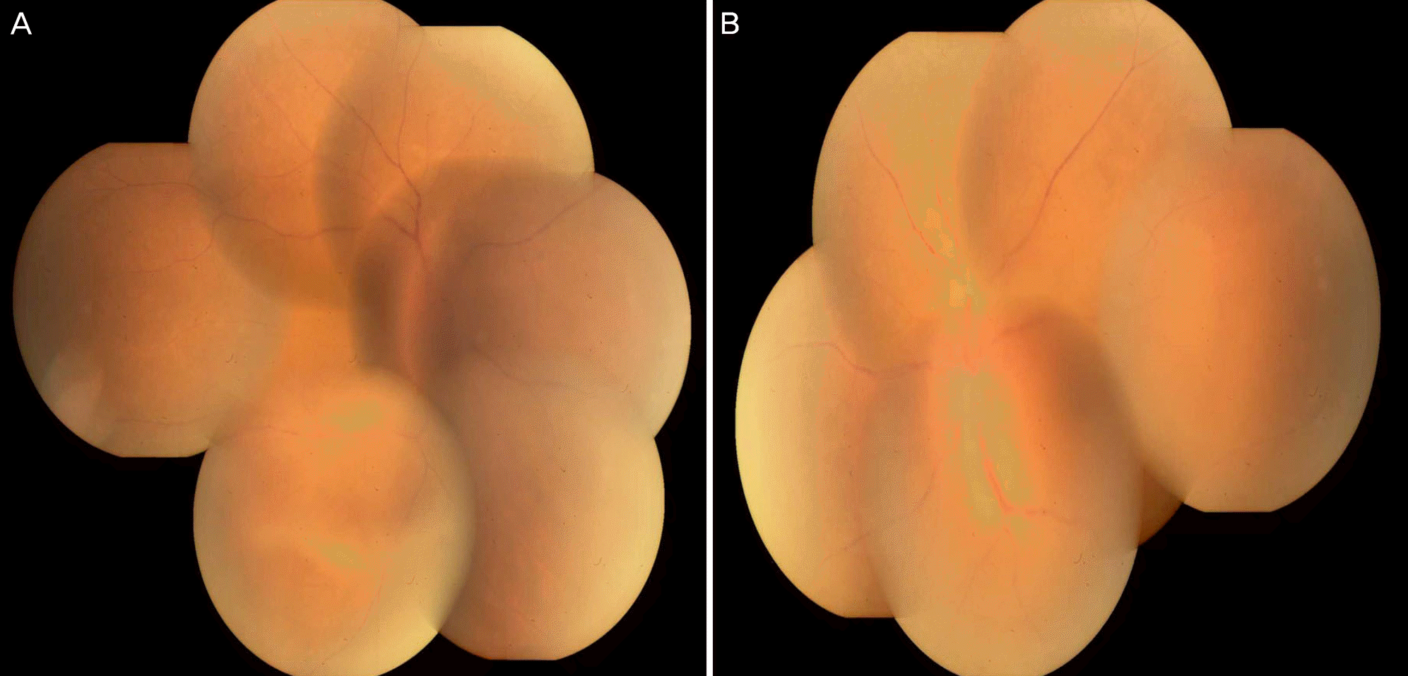 | Figure 1.Fundus photograph. Bilateral vitreous opacity with whitish infiltration inferiorly and venous beading is demonstrated. Both optic disc swelling and blurred margin are also observed. Right eye (A) and left eye (B). |
 | Figure 2.Fluorescein angiograph. Filling defect and early leakage from bilateral optic discs is shown. Both vein and artery beading are observed. Multiple hyperfluorescent dots represent diffuse capillary leakage. Right eye (A) and left eye (B). |
 | Figure 3.Brain magnetic resonance imaging (MRI). (A, C, E) gadolinium (Gd) T1-weighted imaging (T1WI). (B, D, F) T2 fluid-attennated inversion recovery images. Brain MRI, taken 2 months before admission to our Department of Neurology, shows multiple enhancing lesions in Gd-enhanced T1WI in the right frontal cortex and the left basal ganglia (A), with surrounding edema noted in T2 fluid-attenuated inversion recovery images (B). Follow-up MRI at admission shows enlargement of Gd-enhancing lesions in the frontal lobe and basal ganglia (C). Surrounding edema is also aggravated (D). MRI after completion of radiation therapy reveals remarkable shrinkage of the multiple Gd-enhancing lesions (E) and edema (F). |
 XML Download