

 PDF
PDF ePub
ePub Citation
 Print
Print


Abstract
Purpose
To investigate the quantification of metamorphopsia with a novel method that uses Monpack One (Metrovision, Perenchies, France) and to compare the relationship between metamorphopsia score and spectral-domain optical coherence tomography (SD-OCT) findings in patients with epiretinal membrane (ERM).
Methods
This study included 37 eyes of 35 patients with idiopathic ERM. We examined the patients using SD-OCT and quantified the degree of metamorphopsia using the Monpack One. On the topographic map of the Early Treatment Diabetic Retinopathy (ETDRS) grid, central retinal thickness at the fovea (1 mm), and parafovea (3 mm) was measured with the SD-OCT software. The correlation between these factors was analyzed. We repeated the metamorphopsia test twice in 22 eyes of 11 healthy subjects in order to calculate intraclass correlation coefficients (ICCs) and evaluate the reproducibility and reliability of the new metamorphopsia test.
Results
On the ETDRS grid, the retinal thickness (µm) of the central, superior, inferior, nasal, and temporal subfields was 495 ± 102, 428 ± 98, 454 ± 78, 434 ± 83, and 463 ± 95, respectively. The mean total metamorphopsia score was 24.8 ± 13.9, while those for the superior, inferior, nasal, and temporal subfields were 14.7 ± 9.1, 15.1 ± 8.6, 15.9 ± 8.9, and 14.6 ± 8.6, respectively. Linear regression analysis revealed that total metamorphopsia score was significantly related to central retinal thickness (p = 0.01). Moreover, each subfield of parafoveal retinal thickness positively correlated with metamorphopsia subfield score (p < 0.01 −0.023). The ICCs for the metamorphopsia tests of the healthy individuals showed almost perfect repeatability (>0.9) in all subfields.
References
1. Seo S, Lim HW, Shin YU, et al. Morphologic and functional evaluation before and after vitrectomy in idiopathic epiretinal membrane patients ssing microperimetry. J Korean Ophthalmol Soc. 2013; 54:893–901.
2. Bouwens MD, Van Meurs JC. Sine Amsler Charts: a new method for the follow-up of metamorphopsia in patients undergoing macular pucker surgery. Graefes Arch Clin Exp Ophthalmol. 2003; 241:89–93.

3. Wong JG, Sachdev N, Beaumont PE, Chang AA. Visual outcomes following vitrectomy and peeling of epiretinal membrane. Clin Exp Ophthalmol. 2005; 33:373–8.
4. Matsumoto C, Arimura E, Okuyama S, et al. Quantification of metamorphopsia in patients with epiretinal membranes. Invest Ophthalmol Vis Sci. 2003; 44:4012–6.
5. van Velthoven ME, Faber DJ, Verbraak FD, et al. Recent developments in optical coherence tomography for imaging the retina. Prog Retin Eye Res. 2007; 26:57–77.
6. Ko TH, Fujimoto JG, Schuman JS, et al. Comparison of ultrahigh- and standard-resolution optical coherence tomography for imaging macular pathology. Ophthalmology. 2005; 112:1922.e1–15.
7. Schmidt-Erfurth U, Leitgeb RA, Michels S, et al. Three-dimensional ultrahigh-resolution optical coherence tomography of macular diseases. Invest Ophthalmol Vis Sci. 2005; 46:3393–402.
8. Klein R, Klein BE, Wang Q, Moss SE. The epidemiology of epiretinal membranes. Trans Am Ophthalmol Soc. 1994; 92:403–25. discussion 425-30.
9. Gastaud P, Bétis F, Rouhette H, Hofman P. Ultrastructural findings of epimacular membrane and detached posterior hyaloid in vitreomacular traction syndrome. J Fr Ophtalmol. 2000; 23:587–93.
10. Lakshminarayanan V, Aziz S, Enoch JM. Quantification of meta-morphopsia using hyperacuity techniques. Optom Vis Sci. 1991; 68:942–5.
11. Shinoda K, Ishida S, Kawashima S, et al. A new method for quantification of metamorphopsia in patients with epiretinal membrane. Jpn J Ophthalmol. 2000; 44:424–7.
12. Watanabe A, Arimoto S, Nishi O. Correlation between meta-morphopsia and epiretinal membrane optical coherence tomography findings. Ophthalmology. 2009; 116:1788–93.
13. Liu X, Ling Y, Huang J, Zheng X. Optic coherence tomography of idiopathic macular epiretinal membranes. Yan Ke Xue Bao. 2002; 18:14–9.
14. Michalewski J, Michalewska Z, Cisiecki S, Nawrocki J. Morphologically functional correlations of macular pathology connected with epiretinal membrane formation in spectral optical coherence tomography (SOCT). Graefes Arch Clin Exp Ophthalmol. 2007; 245:1623–31.
15. Amsler M. Earliest symptoms of diseases of the macula. Br J Ophthalmol. 1953; 37:521–37.
16. Matsumoto C, Tsuboi S, Okuyama S, et al. Quantification of metamorphopsia. Method of evaluation. Rinsho Ganka. 1990; 44:271–4.
17. Talbot R, Goldberg I, Kelly P. Evaluating the accuracy of the visual field index for the Humphrey Visual Field Analyzer in patients with mild to moderate glaucoma. Am J Ophthalmol. 2013; 156:1272–6.
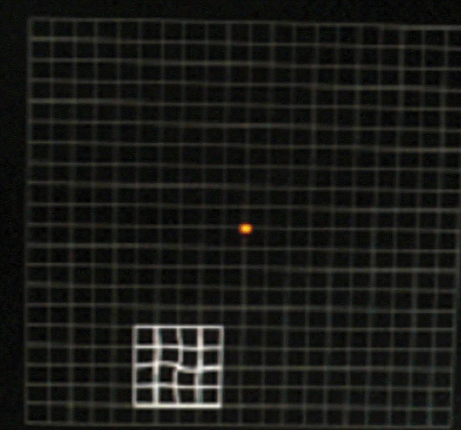
Figure 2.
The metamorphopsia test software consists of tests in 21 different locations positioned every four degrees over the macular field. RE = right eye; LE = left eye.
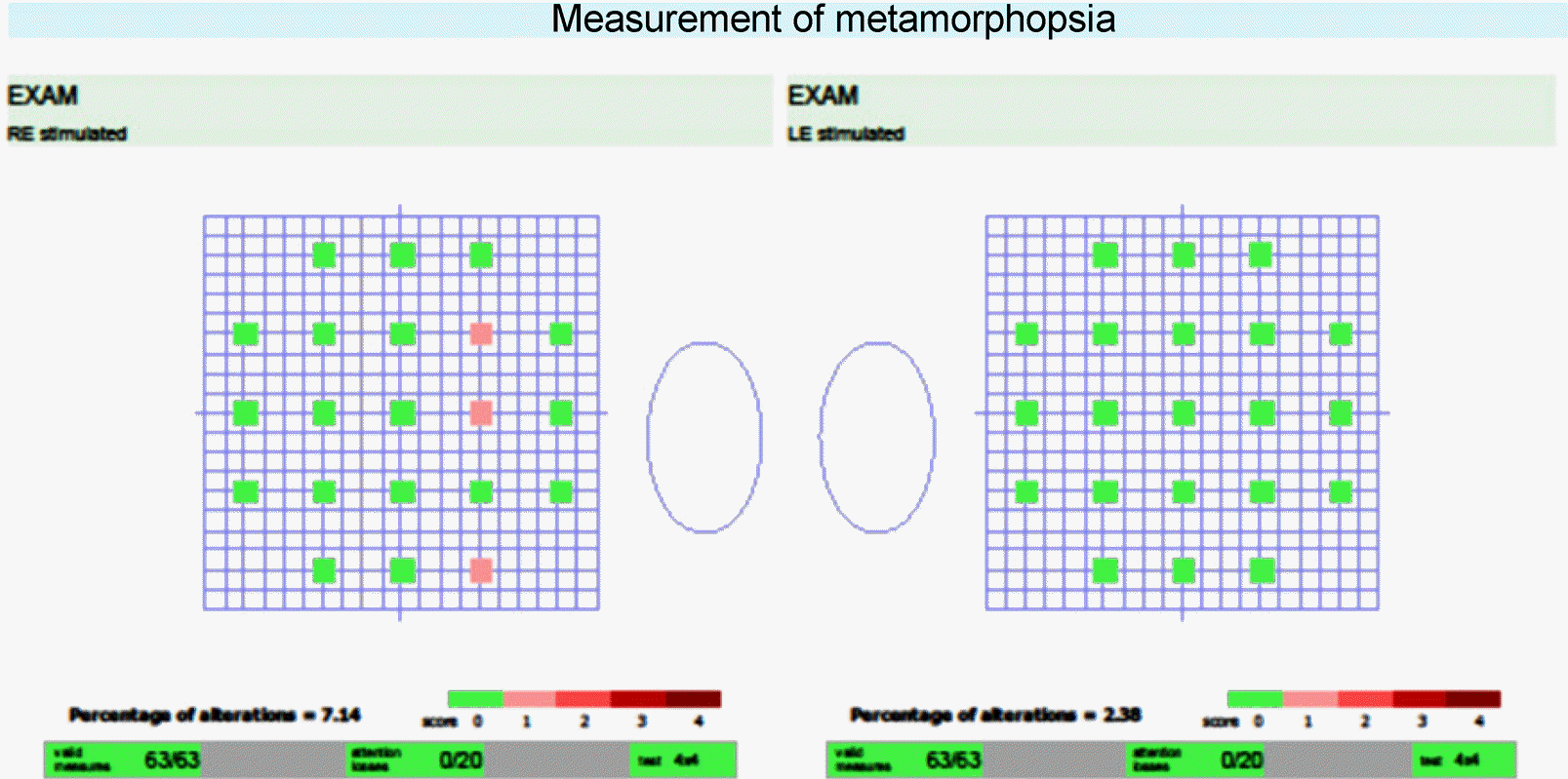
Figure 3.
Correlation between total metamorphopsia score and CRT (μm). The CRT (μm) and total metamorphopsia score were significantly positively correlated (r = 0.635, p = 0.001). p-value was determined using linear regression analysis. CRT = central retinal thickness.
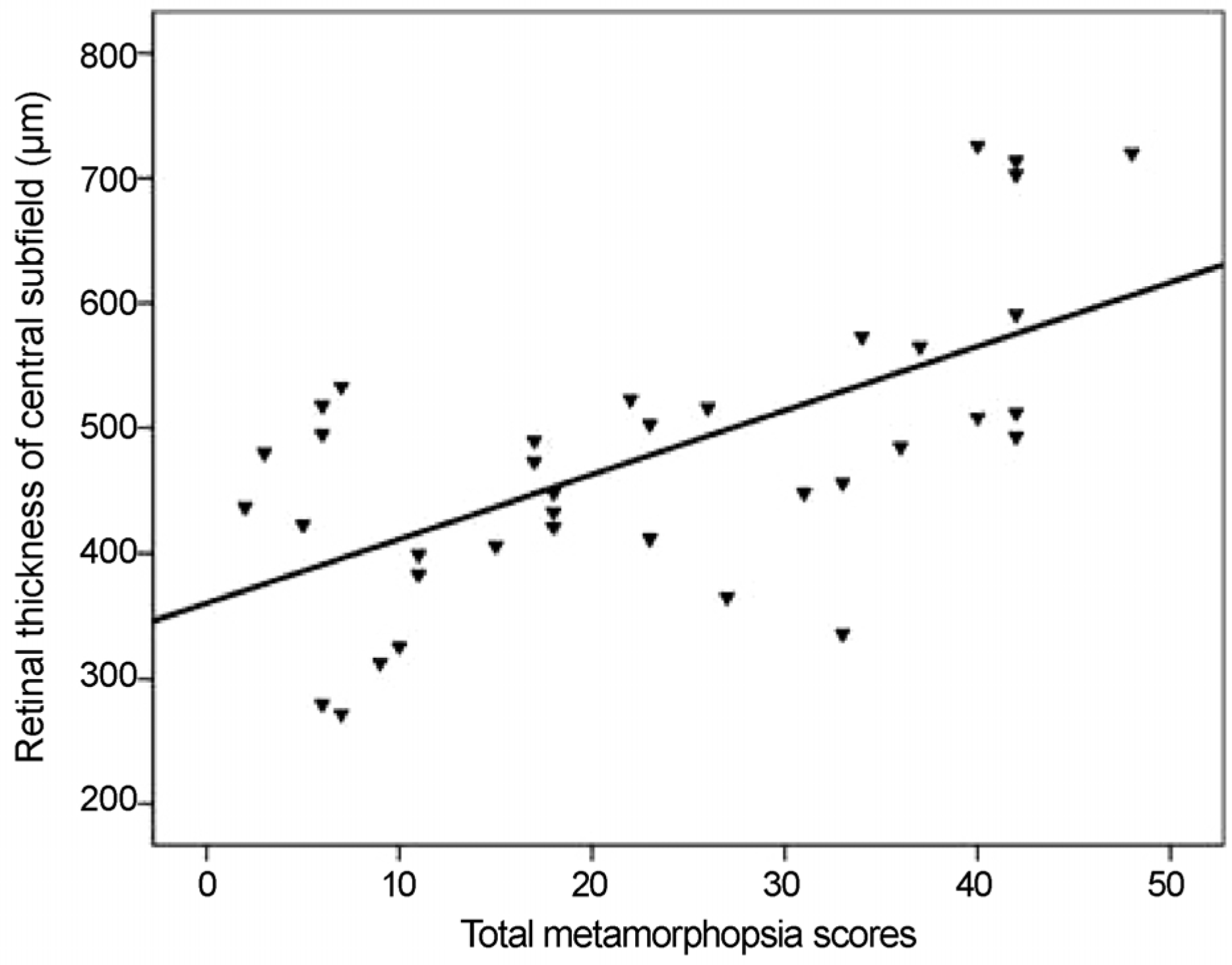
Figure 4.
Relationship between metamorphopsia score and retinal thickness in subjects with epiretinal membrane. Correlation between metamorphopsia score and retinal thickness (μm) in the (A) inferior subfield, (B) superior subfield, (C) temporal subfield, and (D) nasal subfield. (A) Significant positive correlation was found between retinal thickness of the inferior subfield and metamorphopsia score of the superior field (r = 0.469, p = 0.003). (B) Significant positive correlation between retinal thickness of the superior subfield and metamorphopsia score of the inferior field (r = 0.574, p < 0.001). (C) Significant positive correlation between retinal thickness of the temporal subfield and metamorphopsia score of the nasal field (r = 0.630, p = 0.001). (D) Significant positive correlation between retinal thickness of the nasal subfield and metamorphopsia score of the temporal field (r = 0.373, p = 0.001). p-value was determined using linear regression analysis.
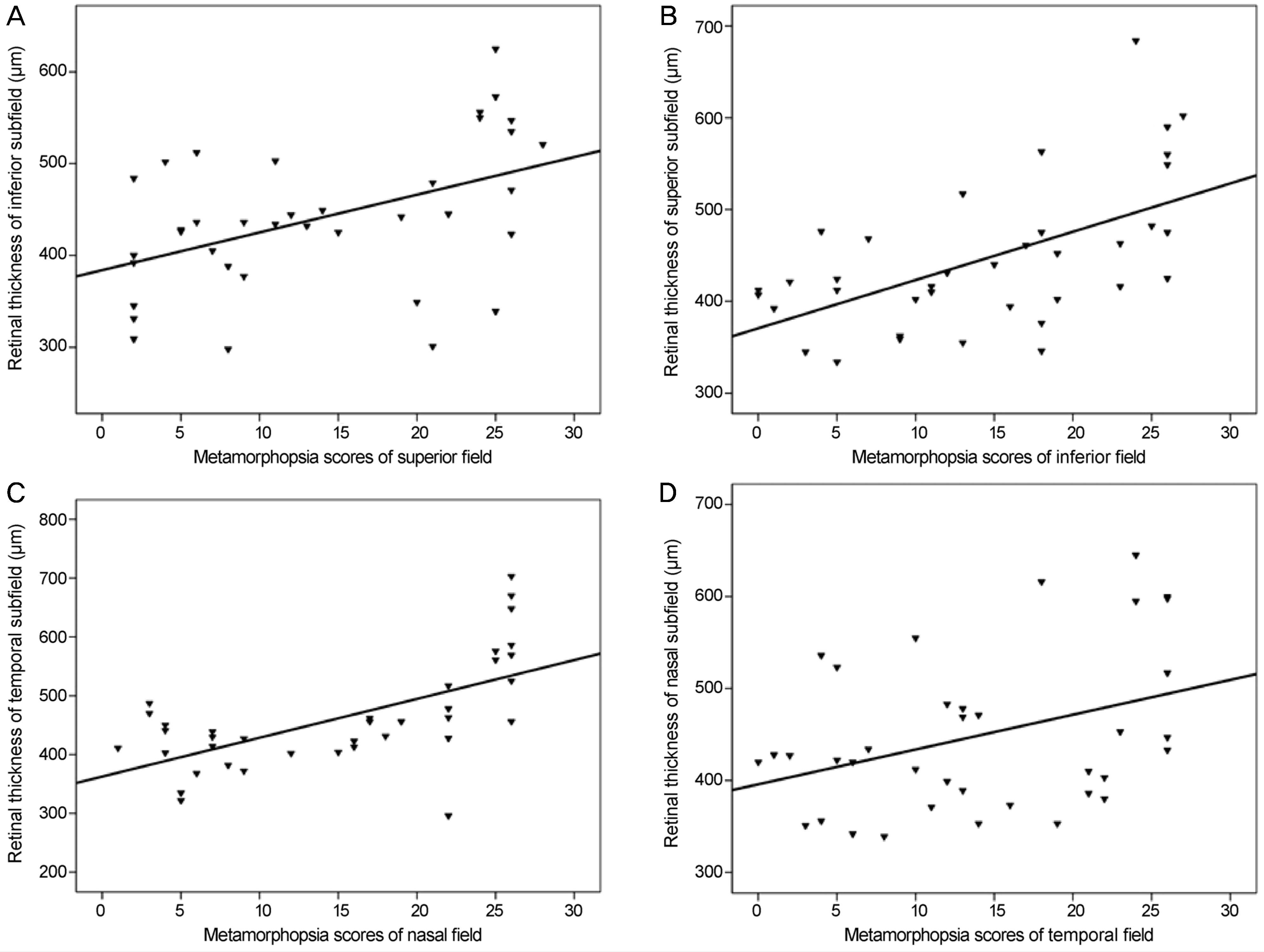
Figure 5.
Clinical manifestations of a patient with epiretinal membrane. (A) Fundus photograph of a 49-year-old female with epiretinal membrane on the right eye. (B) Horizontal section scan showing epiretinal membrane. (C) Colored topographic map showing increased retinal thickening in the superior field. (D) Numeric value of ETDRS grid also showing increased superior thickening. (E) Metamorphopsia test results showed a higher score in the inferior than superior field. ETDRS = Early Treatment Diabetic Retinopathy.
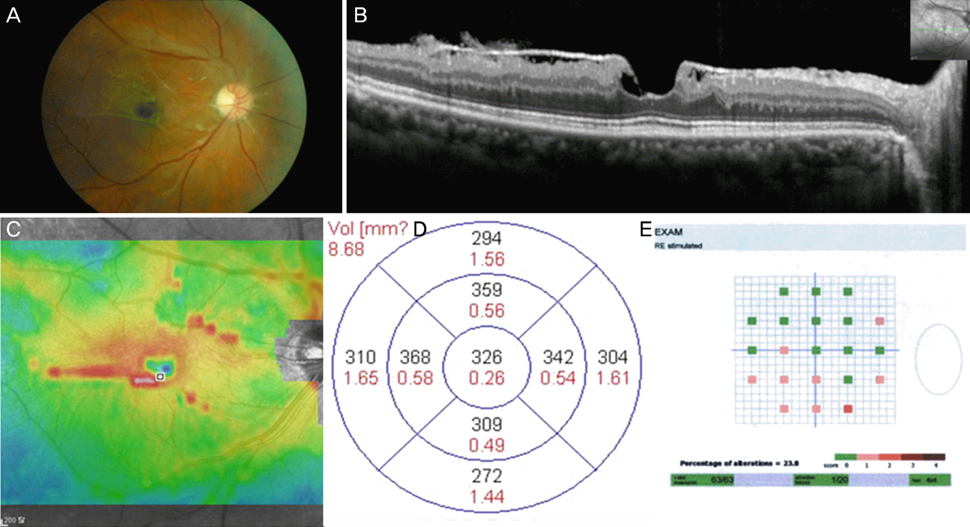
Table 1.
Baseline characteristics of patients with idiopathic epiretinal membrane
 XML Download