

 PDF
PDF ePub
ePub Citation
 Print
Print


Abstract
Purpose
To investigate clinical manifestations and prognosis of traumatic wound dehiscence after penetrating keratoplasty (PKP).
Methods
This is a retrospective study of patients with traumatic wound dehiscence after penetrating keratoplasty performed between January 2004 and July 2014. All patients underwent primary repair of wound dehiscence. Main outcome measurements included pre- and post-injury best corrected visual acuity (BCVA), mechanism of injury, indication of PKP, time interval from PKP to injury, time interval from injury to primary repair, extent of dehiscence, type of suture, presence of suture, prolapse of intraocular tissue, secondary operation, BCVA and graft state at 6 months after primary repair.
Results
The incidence of traumatic wound dehiscence after PKP was 3.96% (12/303). Mean post-injury BCVA and BCVA at 6 months after primary repair (log MAR) were 2.58 ± 0.95 and 2.50 ± 1.05, respectively, and visual acuity did not show significant improvement (p = 1.000). After primary repair, graft failure developed in 9 patients (75%) and evisceration in 2 patients; re-PKP was performed in 3 patients. Pars plana vitrectomy was performed in 1 patient due to retinal detachment. Visual prognosis was poor in patients with wound dehiscence greater than 180° than those with wound dehiscence less than 180° (3.24 ± 0.13 vs. 1.97 ± 1.11, p = 0.030), and in patients with iris prolapse than those without iris prolapse (3.17 ± 0.16 vs. 1.56 ± 1.05, p = 0.048).
Go to : 
References
1. Choi SH, Lee YW, Kim HM, et al. Epidemiologic studies of keratoplsty in Korea. J Korean Ophthalmol Soc. 2006; 47:538–47.
2. Kawashima M, Kawakita T, Shimmura S, et al. Characteristics of traumatic globe rupture after keratoplasty. Ophthalmology. 2009; 116:2072–6.

3. Kartal B, Kandemir B, Set T, et al. Traumatic wound dehiscence after penetrating keratoplasty. Ulus Travma Acil Cerrahi Derg. 2014; 20:181–8.
4. Jafarinasab MR, Feizi S, Esfandiari H, et al. Traumatic wound dehiscence following corneal transplantation. J Ophthalmic Vis Res. 2012; 7:214–8.
5. Brown SI, Tragakis MP. Wound dehiscence with keratoplasty: complication of the continuous-suture technique. Am J Ophthalmol. 1971; 72:115–6.
6. Farley MK, Pettit TH. Traumatic wound dehiscence after penetrating keratoplasty. Am J Ophthalmol. 1987; 104:44–9.
7. Friedman AH. Late traumatic wound rupture following successful partial penetrating keratoplasty. Am J Ophthalmol. 1973; 75:117–20.
8. Raber IM, Arentsen JJ, Laibson PR. Traumatic wound dehiscence after penetrating keratoplasty. Arch Ophthalmol. 1980; 98:1407–9.
9. Topping TM, Stark WJ, Maumenee E, Kenyon KR. Traumatic wound dehiscence following penetrating keratoplasty. Br J Ophthalmol. 1982; 66:174–8.
10. Kim KS, Myong YW. Traumatic wound dehiscence after penetrating keratoplasty. J Korean Ophthalmol Soc. 1999; 40:2438–42.
11. Hollander DA, Giaconi JA, Holland GN, et al. Graft failure after penetrating keratoplasty in eyes with Ahmed valves. Am J Ophthalmol. 2010; 150:169–78.
12. Elder MJ, Stack RR. Globe rupture following penetrating keratoplasty: how often, why, and what can we do to prevent it? Cornea. 2004; 23:776–80.
13. Cherry PM. Rupture of the globe. Arch Ophthalmol. 1972; 88:498–507.
14. Agrawal V, Wagh M, Krishnamachary M, et al. Traumatic wound dehiscence after penetrating keratoplasty. Cornea. 1995; 14:601–3.
15. Calkins JL, Hochheimer BF, Stark WJ. Corneal wound healing: holographic stress-test analysis. Invest Ophthalmol Vis Sci. 1981; 21:322–34.
16. Rohrbach JM, Weidle EG, Steuhl KP, et al. Traumatic wound dehiscence after penetrating keratoplasty. Acta Ophthalmol Scand. 1996; 74:501–5.
17. Renucci AM, Marangon FB, Culbertson WW. Wound dehiscence after penetrating keratoplasty: clinical characteristics of 51 cases treated at Bascom Palmer Eye Institute. Cornea. 2006; 25:524–9.
18. Das S, Whiting M, Taylor HR. Corneal wound dehiscence after penetrating keratoplasty. Cornea. 2007; 26:526–9.
19. Lam FC, Rahman MQ, Ramaesh K. Traumatic wound dehiscence after penetrating keratoplasty-a cause for concern. Eye (Lond). 2007; 21:1146–50.
20. Sari ES, Koytak A, Kubaloglu A, et al. Traumatic wound dehiscence after deep anterior lamellar keratoplasty. Am J Ophthalmol. 2013; 156:767–72.
21. Binder PS, Abel R Jr, Polack FM, Kaufman HE. Keratoplasty wound separations. Am J Ophthalmol. 1975; 80:109–15.
22. Tseng SH, Lin SC, Chen FK. Traumatic wound dehiscence after penetrating keratoplasty: clinical features and outcome in 21 cases. Cornea. 1999; 18:553–8.
23. Kim EC, Kim MS. Three cases of corneal perforation caused by noncontact tonometry. Cornea. 2008; 27:1191–4.
24. Williams MA, Gawley SD, Jackson AJ, Frazer DG. Traumatic graft dehiscence after penetrating keratoplasty. Ophthalmology. 2008; 115:276–8.e1.
25. Bowman RJ, Yorston D, Aitchison TC, et al. Traumatic wound rupture after penetrating keratoplasty in Africa. Br J Ophthalmol. 1999; 83:530–4.
26. Hiratsuka Y, Sasaki S, Nakatani S, Murakami A. Traumatic wound dehiscence after penetrating keratoplasty. Jpn J Ophthalmol. 2007; 51:146–7.
27. Nagra PK, Hammersmith KM, Rapuano CJ, et al. Wound dehiscence after penetrating keratoplasty. Cornea. 2006; 25:132–5.
28. Rehany U, Rumelt S. Ocular trauma following penetrating keratoplasty: incidence, outcome, and postoperative recommendations. Arch Ophthalmol. 1998; 116:1282–6.
Go to :
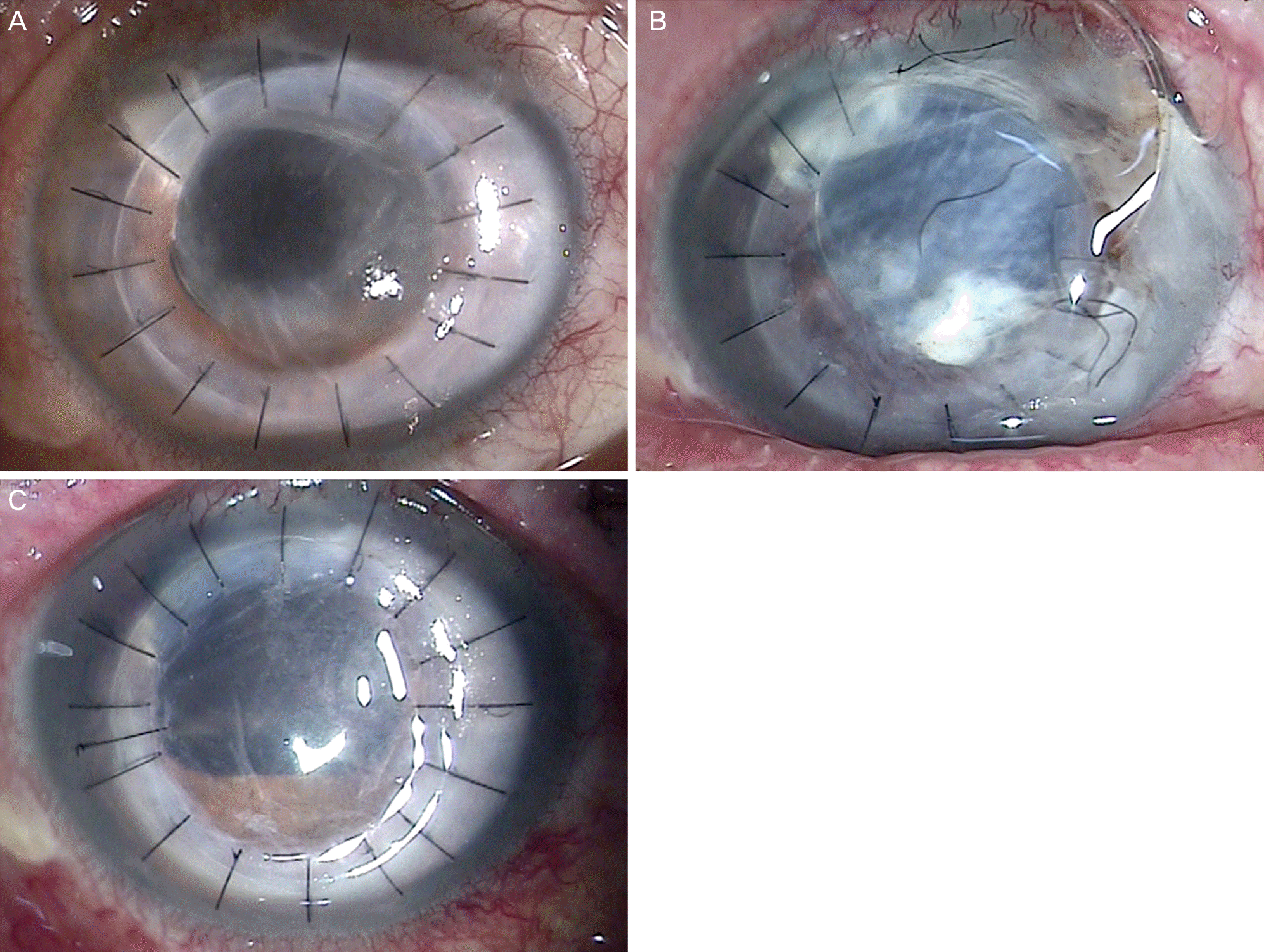 | Figure 1.Slit lamp photography of a 71-year-old male. (A) Good graft state after penetrating keratoplasty. (B) Traumatic wound dehiscence with intraocular lens prolapse after being struck by a door. (C) Post operative state of primary repair. |
Table 1.
Clinical characteristics of patients before primary repair
Table 2.
Clinical characteristics of patients after primary repair
Table 3.
Comparison of visual acuity (log MAR)
| Group | Postoperative BCVA | p-value* |
|---|---|---|
| Age ≤ 55 | 2.68 ± 0.78 | 0.394 |
| Age > 55 | 2.06 ± 1.32 | |
| Suture (+) | 2.02 ± 1.08 | 0.268 |
| Suture (-) | 2.84 ± 0.96 | |
| Iris prolapsed (+) | 3.17 ± 0.16 | 0.048 |
| Iris prolapsed (-) | 1.56 ± 1.05 | |
| Extent of dehiscence < 180° | 3.24 ± 0.13 | 0.030 |
| Extent of dehiscence ≥ 180° | 1.97 ± 1.11 | |
| Time interval from PKP and injury ≤ 100 months | 2.13 ± 1.22 | 0.394 |
| Time interval from PKP and injury > 100 months | 2.86 ± 0.78 | |
| Time interval from injury and repair ≤ 10 hours | 2.88 ± 0.71 | 0.432 |
| Time interval from injury and repair > 10 hours | 1.96 ± 1.28 |
 XML Download