

 PDF
PDF ePub
ePub Citation
 Print
Print


Abstract
Purpose
To present the clinical outcomes of small incision lenticule extraction (SMILE) including visual quality analysis in Korean patients with myopia
Methods
The medical records of 228 eyes of 116 patients who underwent SMILE in HanGil Eye Hospital LASIK Center from May 2014 to Feb 2015 and were followed-up for at least 3 months was analyzed retrospectively. The patients were followed up at 1 day, 1 week, 1 month, and 3 months after the operation. Refractive value, visual acuity, intraocular pressure, and visual quality were measured at each visit
Results
Preoperatively, uncorrected distant visual acuity was 0.01 ± 0.02 in log MAR, spherical equivalent was −5.03 ± 1.72 diopters, intraocular pressure was 15.85 ± 2.85 mm Hg, and the objective scattering index (OSI) value was 0.68 ± 0.49. The postoperative uncorrected distant visual acuity was 0.13 ± 0.10, 0.05 ± 0.08, 0.04 ± 0.09, and 0.02 ± 0.04 and OSI was 2.16 ± 1.89, 1.25 ± 0.64, 1.14 ± 0.69, and 0.81 ± 0.36 at 1 day, 1 week, 1 month, and 3 months after the operation, respectively. The postoperative intraocular pressure was 12.55 ± 3.74 mm Hg, 13.03 ± 4.35 mm Hg, 11.65 ± 2.49 mm Hg at 1 week, 1 month, and 3 months after the operation. The efficacy of refractive surgery 3 months after the operation was 0.97 ± 0.11, the safety was 0.99 ± 0.10, and predictability was 99.56% and 100.00% at the range of ±0.5 diopters and ± 1.0 diopter, respectively.
Go to : 
References
1. Moshirfar M, McCaughey MV, Reinstein DZ, et al. Small-incision lenticule extraction. J Cataract Refract Surg. 2015; 41:652–65.

2. Sandoval HP, de Castro LE, Vroman DT, Solomon KD. Refractive Surgery Survey 2004. J Cataract Refract Surg. 2005; 31:221–33.
3. Kim HJ, Cho SH, Kim JH, Joo CK. Risk factors and clinical evaluation for corneal ectasia after LASIK. J Korean Ophthalmol Soc. 2005; 46:589–96.
4. Khoueir Z, Haddad NM, Saad A, et al. Traumatic flap dislocation 10 years after LASIK. Case report and literature review. J Fr Ophtalmol. 2013; 36:82–6.
5. Sekundo W, Kunert KS, Blum M. Small incision corneal refractive surgery using the small incision lenticule extraction (SMILE) procedure for the correction of myopia and myopic astigmatism: results of a 6 month prospective study. Br J Ophthalmol. 2011; 95:335–9.
6. Shah R, Shah S, Sengupta S. Results of small incision lenticule extraction: all-in-one femtosecond laser refractive surgery. J Cataract Refract Surg. 2011; 37:127–37.
7. Lin F, Xu Y, Yang Y. Comparison of the visual results after SMILE and femtosecond laser-assisted LASIK for myopia. J Refract Surg. 2014; 30:248–54.
8. Vestergaard A, Ivarsen AR, Asp S, Hjortdal JØ. Small-incision lenticule extraction for moderate to high myopia: predictability, safety, and patient satisfaction. J Cataract Refract Surg. 2012; 38:2003–10.
9. Sekundo W, Gertnere J, Bertelmann T, Solomatin I. One-year refractive results, contrast sensitivity, high-order aberrations and complications after myopic small-incision lenticule extraction (ReLEx SMILE). Graefes Arch Clin Exp Ophthalmol. 2014; 252:837–43.
10. Vestergaard AH, Grauslund J, Ivarsen AR, Hjortdal JØ. Central corneal sublayer pachymetry and biomechanical properties after refractive femtosecond lenticule extraction. J Refract Surg. 2014; 30:102–8.
11. Wu D, Wang Y, Zhang L, et al. Corneal biomechanical effects: small-incision lenticule extraction versus femtosecond laser-assisted laser in situ keratomileusis. J Cataract Refract Surg. 2014; 40:954–62.
12. Kamiya K, Shimizu K, Igarashi A, et al. Intraindividual comparison of changes in corneal biomechanical parameters after femtosecond lenticule extraction and small-incision lenticule extraction. J Cataract Refract Surg. 2014; 40:963–70.
13. Wei S, Wang Y. Comparison of corneal sensitivity between FS-LASIK and femtosecond lenticule extraction (ReLEx flex) or small-incision lenticule extraction (ReLEx smile) for myopic eyes. Graefes Arch Clin Exp Ophthalmol. 2013; 251:1645–54.
14. Reinstein DZ, Archer TJ, Gobbe M, Bartoli E. Corneal sensitivity after small-incision lenticule extraction and laser in situ keratomileusis. J Cataract Refract Surg. 2015; 41:1580–7.
15. Kim JR, Kim HS, Mun SJ, Chung YT. Outcomes of small incision lenticule extraction: mild to moderate myopia versus high myopia. J Korean Ophthalmol Soc. 2014; 55:963–8.
16. Hjortdal JØ, Vestergaard AH, Ivarsen A, et al. Predictors for the outcome of small-incision lenticule extraction for myopia. J Refract Surg. 2012; 28:865–71.
17. Zhao J, Yao P, Li M, et al. The morphology of corneal cap and its relation to refractive outcomes in femtosecond laser small incision lenticule extraction (SMILE) with anterior segment optical coherence tomography observation. PloS One. 2013; 8:e70208.
18. Chan TC, Ng AL, Cheng GP, et al. Vector analysis of astigmatic correction after small-incision lenticule extraction and femtosecond-assisted LASIK for low to moderate myopic astigmatism. Br J Ophthalmol. 2016; 100:553–9.
19. Ivarsen A, Hjortdal J. Correction of myopic astigmatism with small incision lenticule extraction. J Refract Surg. 2014; 30:240–7.
20. Vestergaard A, Ivarsen A, Asp S, Hjortdal JØ. Femtosecond (FS) laser vision correction procedure for moderate to high myopia: a prospective study of ReLEx([R])flex and comparison with a retrospective study of FS-laser in situ keratomileusis. Acta Ophthalmol. 2013; 91:355–62.
21. Agca A, Ozgurhan EB, Yildirim Y, et al. Corneal backscatter analysis by in vivo confocal microscopy: fellow eye comparison of small incision lenticule extraction and femtosecond laser-assisted LASIK. J Ophthalmol. 2014; 2014:265012.
22. Miao H, He L, Shen Y, et al. Optical quality and intraocular scattering after femtosecond laser small incision lenticule extraction. J Refract Surg. 2014; 30:296–302.
23. Jung BJ, Oh TH, Chung SK. Eight-year follow-up of laser epithelial keratomileusis for correcting moderate and high myopia. J Korean Ophthalmol Soc. 2012; 53:1438–44.
24. Jung HG, Lim TH. The recovery of optical quality after laser vision correction. Korean J Ophthalmol. 2013; 27:249–55.
25. Ivarsen A, Asp S, Hjortdal J. Safety and complications of more than 1500 small-incision lenticule extraction procedures. Ophthalmology. 2014; 121:822–8.
26. Kim BK, Mun SJ, Lee DG, Chung YT. A case of suction loss during SMILE and a switch to LASIK. J Korean Ophthalmol Soc. 2015; 56:1274–7.
27. Ivarsen A, Hjortdal JØ. Topography-guided photorefractive keratectomy for irregular astigmatism after small incision lenticule extraction. J Refract Surg. 2014; 30:429–32.
28. Vestergaard AH. Past and present of corneal refractive surgery: a retrospective study of long-term results after photorefractive keratectomy and a prospective study of refractive lenticule extraction. Acta Ophthalmol. 2014; 92 Thesis. 2:1–21.
Go to :
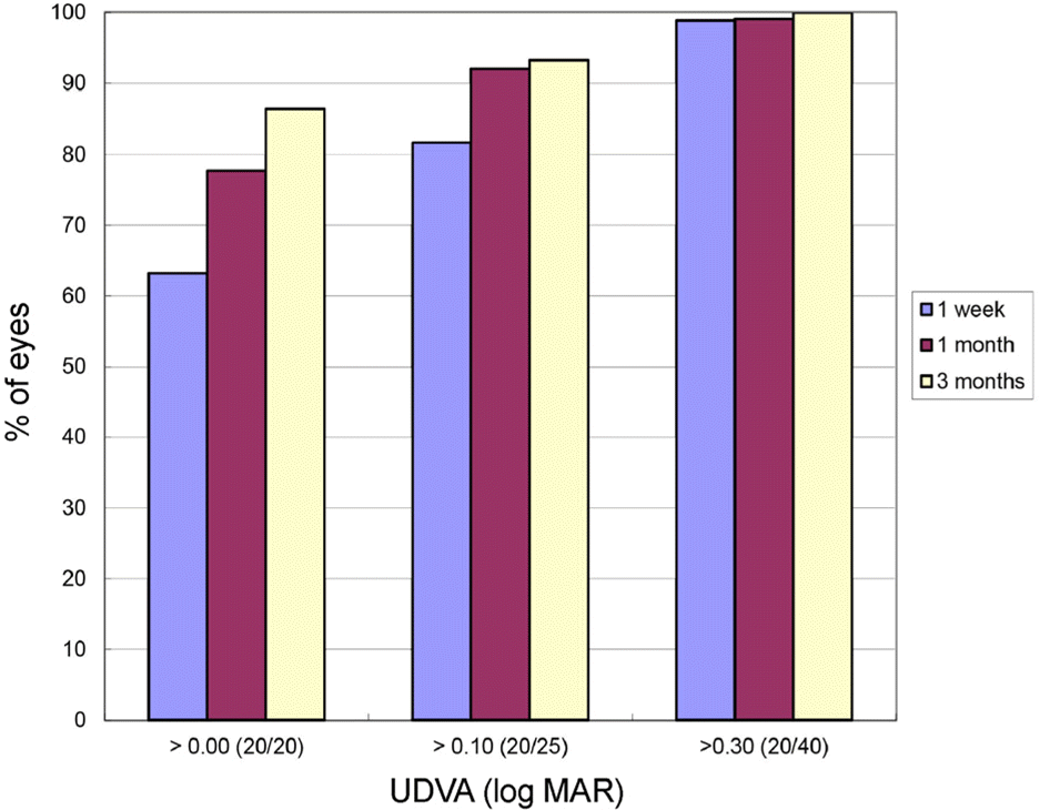 | Figure 1.Postoperative cumulative uncorrected distant visual acuity. Visual acuity was expressed in log MAR. UDVA = uncorrected distant visual acuity. |
Table 1.
Preoperative demographic data of the patients
| Parameter | Value |
|---|---|
| Age (year) | 26.84 ± 6.44 |
| Gender (male/female) | 56/60 |
| S.E. (D) | −5.03 ± 1.72 |
| CDVA (log MAR) | 0.01 ± 0.02 |
| K1* (D) | 42.51 ± 2.86 |
| K2* (D) | 43.91 ± 1.35 |
| Corneal thickness (μm) | 566.17 ± 45.17 |
| Pupil diameter (mm) | 7.13 ± 4.57 |
| OSI | 0.68 ± 0.49 |
| MTF | 36.91 ± 10.06 |
| Sterhl ratio | 0.19 ± 0.06 |
| IOP (mm Hg) | 15.85 ± 2.85 |
Table 2.
Postoperative refractive value and visual acuity
Table 3.
Postoperative visual quality and intraocular pressure
| Period |
Visual quality* |
IOP (mm Hg) | ||
|---|---|---|---|---|
| OSI† | MTF | Sterhl ratio | ||
| Preoperative | 0.67 ± 0.49 | 36.91 ± 10.06 | 0.19 ± 0.06 | 15.82 ± 2.85 |
| 1 day | 2.16 ± 1.89 (p < 0.001)† | 28.84 ± 12.6 | 0.15 ± 0.06 | − |
| 1 week | 1.25 ± 0.65 (p < 0.001)† | 32.02 ±13.31 | 0.17 ± 0.11 | 12.55 ± 3.74 |
| 1 month | 1.14 ± 0.69 (p < 0.001)† | 31.5 ± 11.53 | 0.16 ± 0.60 | 13.03 ± 4.35 |
| 3 months | 0.81 ± 0.36 (p = 0.146)† | 31.61 ± 11.44 | 0.16 ± 0.05 | 11.65 ± 2.49 |
 XML Download