Abstract
Purpose
We report a case of a scleral perforation during inferior rectus recession in congenital fibrosis of extraocular muscles and the management of this perforation with a scleral patch graft.
Case summary
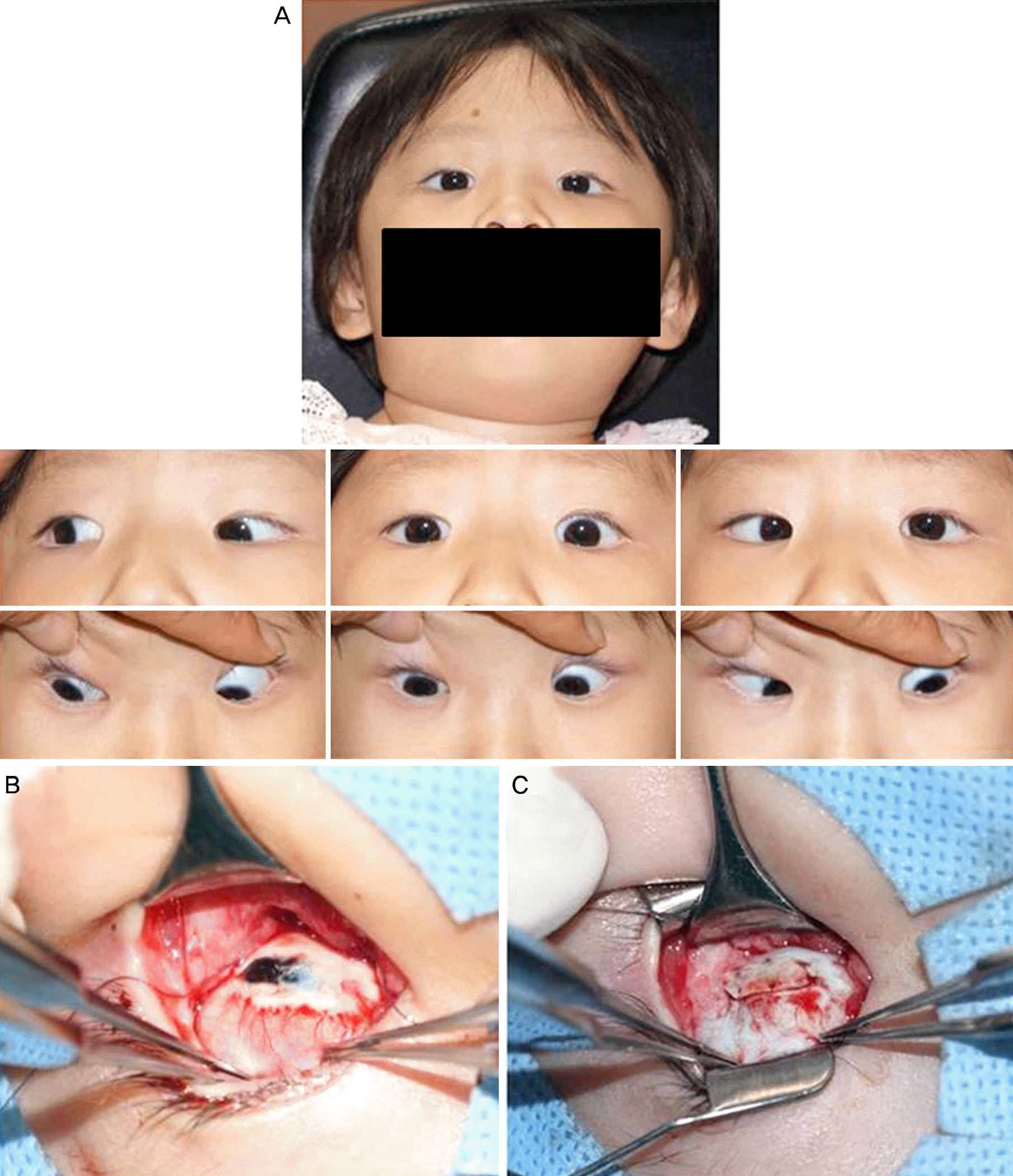
A 20-month-old female with bilateral ptosis, absence of elevation and a chin-up position was diagnosed with congenital fibrosis of extraocular muscles. Because severe esotropia in the downward gaze was observed, we first performed esotropia surgery. After 1 year, she underwent a bilateral ptosis correction. We decided to perform bilateral inferior rectus recession due to an abnormal head posture and the absence of elevation. Because the inferior rectus muscles were extremely tight and adhered to the sclera, hooking and isolating these muscles during surgery was difficult. After muscle suture placement, a portion of the sclera that contacted the left inferior rectus was chipped off as this muscle was disinserted with blunt Westcott scissors. A scleral perforation was observed, thus, we placed a scleral patch graft using the donor sclera and finished the bilateral inferior rectus recession. No abnormal findings for the vitreous or retina were detected. At 8 months after surgery, the patient exhibited exotropia of 12 prism diopters in her primary gaze. Her abnormal head posture nearly disappeared.
Go to : 
References
1. Awad AH, Mullaney PB, Al-Hazmi A, et al. Recognized globe perforation during strabismus surgery: incidence, risk factors, and sequelae. J AAPOS. 2000; 4:150–3.

4. Yazdani A, Traboulsi EI. Classification and surgical management of patients with familial and sporadic forms of congenital fibrosis of the extraocular muscles. Ophthalmology. 2004; 111:1035–42.

5. Heidary G, Engle EC, Hunter DG. Congenital fibrosis of the extraocular muscles. Semin Ophthalmol. 2008; 23:3–8.

6. Kim JH, Hwang JM. Hypoplastic oculomotor nerve and absent abducens nerve in congenital fibrosis syndrome and synergistic divergence with magnetic resonance imaging. Ophthalmology. 2005; 112:728–32.

7. Choi SR, Baek SH, Kim US. Dissociated vertical deviation in congenital fibrosis of the extraocular muscles. Graefes Arch Clin Exp Ophthalmol. 2013; 251:1007–8.

8. Apt L, Axelrod RN. Generalized fibrosis of the extraocular muscles. Am J Ophthalmol. 1978; 85:822–9.

9. Morris RJ, Rosen PH, Fells P. Incidence of inadvertent globe perforation during strabismus surgery. Br J Ophthalmol. 1990; 74:490–3.

10. Park K, Hong S, Chung W, et al. Inadvertent scleral perforation after strabismus surgery: incidence and association with refractive error. Can J Ophthalmol. 2008; 43:669–72.

11. MacEwen CJ, Gregson RMC. Manual of strabismus surgery, 1st ed. Oxford: Butterworth-Heinemann. 2001; 171–84.
Go to : 
 | Figure 1.The clinical manifestation and intraoperative findings. (A) Eye movement prior to bilateral inferior rectus muscle recession. The patient exhibits downward eye fixation, an absence of elevation and limited abduction in the left eye. (B) A scleral perforation with uveal prolapse is observed (3 × 3 mm). (C) A scleral patch graft is immediately placed. |


 PDF
PDF ePub
ePub Citation
 Print
Print


 XML Download