

 PDF
PDF ePub
ePub Citation
 Print
Print


Abstract
Purpose
To report a case of steroid-induced glaucoma in a child who was treated with systemic steroids for a long period due to graft-versus-host disease.
Case summary
A 10-year-old male was referred to our ophthalmologic clinic for examination of papilledema due to persistent headache and nausea. He was diagnosed as aplastic anemia 8 years prior and took approximately 4,000 mg of oral prednisolone for 8 years from April 2007 to April 2015 for treatment of lung graft-versus-host disease after hematopoietic stem cell transplantation. His best corrected visual acuity was 0.8 (decimal) in the right eye, 0.5 in the left eye and intraocular pressure (IOP) measured using a Goldmann applanation tonometer was 42 mm Hg in the right eye and 43 mm Hg in the left eye. His cup-to-disc ratio was 0.8 in the right eye and 0.7 in the left eye. Additionally, superior and inferior neuroretinal rim thinning was present in both eyes. Despite using IOP-lowering agents, IOP was not controlled. However, after trabeculectomy with mitomycin C in both eyes, IOP became normalized.
References
1. David DS, Berkowitz JS. Ocular effects of topical and systemic corticosteroids. Lancet. 1969; 2:149–51.

2. Bernstein HN, Mills DW, Becker B. Steroid-induced elevation of intraocular pressure. Arch Ophthalmol. 1963; 70:15–8.
3. Kersey JP, Broadway DC. Corticosteroid-induced glaucoma: a review of the literature. Eye (Lond). 2006; 20:407–16.
4. Biedner BZ, David R, Grudsky A, Sachs U. Intraocular pressure response to corticosteroids in children. Br J Ophthalmol. 1980; 64:430–1.
5. de Queiroz Mendonca C, de Souza CP Jr, Martins-Filho PR, et al. Steroid-induced ocular hypertensive response in children and adolescents with acute lymphoblastic leukemia and non-Hodgkin lymphoma. Pediatr Blood Cancer. 2014; 61:2083–5.
6. Hayasaka Y, Hayasaka S, Matsukura H. Ocular findings in Japanese children with nephrotic syndrome receiving prolonged corticosteroid therapy. Ophthalmologica. 2006; 220:181–5.
7. Armaly MF. Statistical attributes of the steroid hypertensive response in the clinically normal eye. I. The demonstration of three levels of response. Invest Ophthalmol. 1965; 4:187–97.
8. Godel V, Feiler-Ofry V, Stein R. Systemic steroids and ocular fluid dynamics. I. Analysis of the sample as a whole. Influence of dosage and duration of therapy. Acta Ophthalmol (Copenh). 1972; 50:655–63.
9. Garbe E, LeLorier J, Boivin JF, Suissa S. Risk of ocular hypertension or open-angle glaucoma in elderly patients on oral glucocorticoids. Lancet. 1997; 350:979–82.
10. Mitchell P, Cumming RG, Mackey DA. Inhaled corticosteroids, family history, and risk of glaucoma. Ophthalmology. 1999; 106:2301–6.
11. Cadera W, Pachtman MA, Cantor LB, et al. Filtering surgery in childhood glaucoma. Ophthalmic Surg. 1984; 15:319–22.
12. Tham CC, Ng JS, Li RT, et al. Intraocular pressure profile of a child on a systemic corticosteroid. Am J Ophthalmol. 2004; 137:198–201.
13. Lee SW, Jin KH, Lee SC, et al. Cataract and glaucoma in Korean children with chronic glomerulonephritis receiving systemic corticosteroid treatment. Acta Ophthalmol. 2010; 88:e344–5.
14. Brito PN, Silva SE, Cotta JS, Falcão-Reis F. Severe ocular hypertension secondary to systemic corticosteroid treatment in a child with nephrotic syndrome. Clin Ophthalmol. 2012; 6:1675–9.
15. Yamashita T, Kodama Y, Tanaka M, et al. Steroid-induced glaucoma in children with acute lymphoblastic leukemia: a possible complication. J Glaucoma. 2010; 19:188–90.
Figure 1.
Fundus photographs of the patient. (A, B) Normal fundus photographs with a normal cup to disc ratio 5 years ago, (C, D) glaucomatous cupping of the optic disc with a cup-to-disc ratio of 0.8 in the right eye and 0.7 in the left eye, and superior and inferior neuro-retinal rim thinning in both eyes.
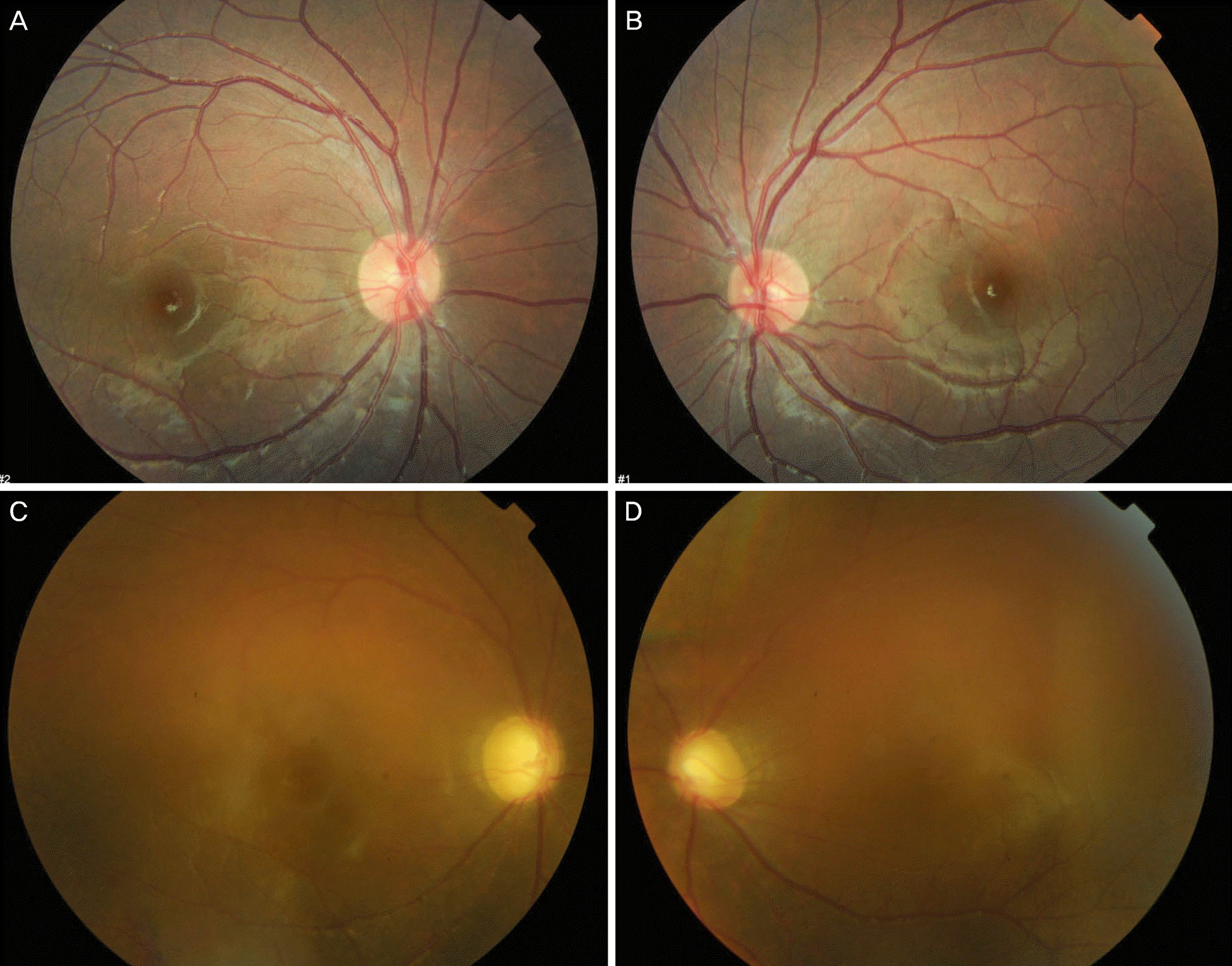
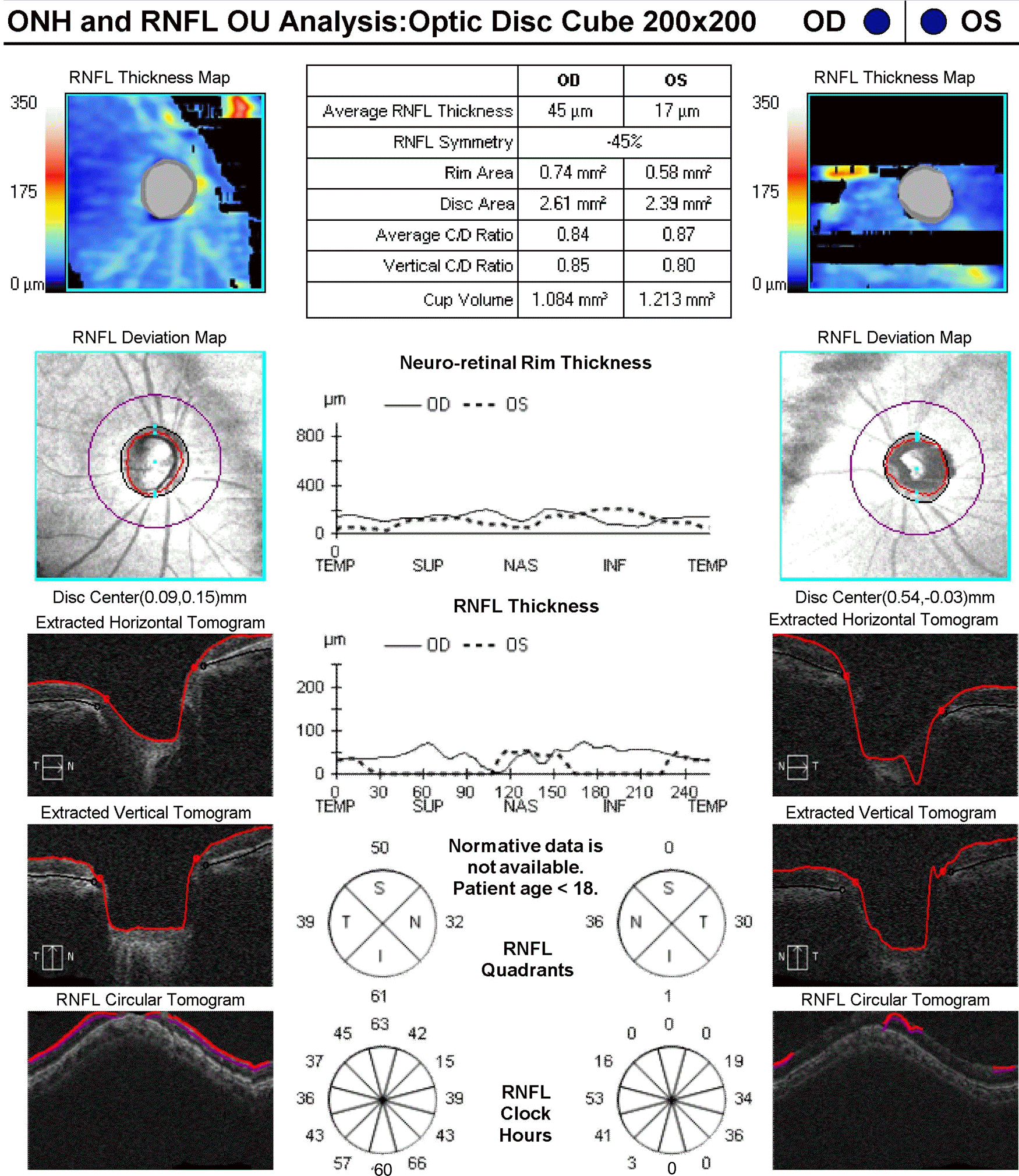
Figure 2.
Neuro-retinal rim thinning and decreased retinal nerve fiber layer in both eyes measured by optical coherent tomography. ONH = optic nerve head; RNFL = retinal nerve fiber layer; OU = oculus unitas; OD = oculus dexter; OS = oculus sinister; TEMP = temporal; SUP = superior; NAS = nasal; INF = inferior; S = superior; T = temporal; I = inferior; N = nasal.
 XML Download