Abstract
Purpose
To report a case of ocular surface squamous cell carcinoma with intraorbital extension in a patient with renal transplantation and long-term immunosuppressive therapy.
Case summary
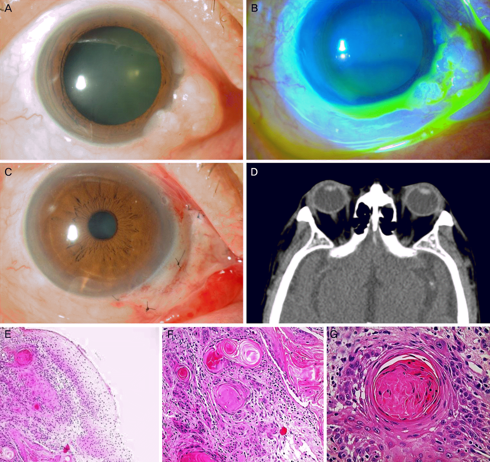
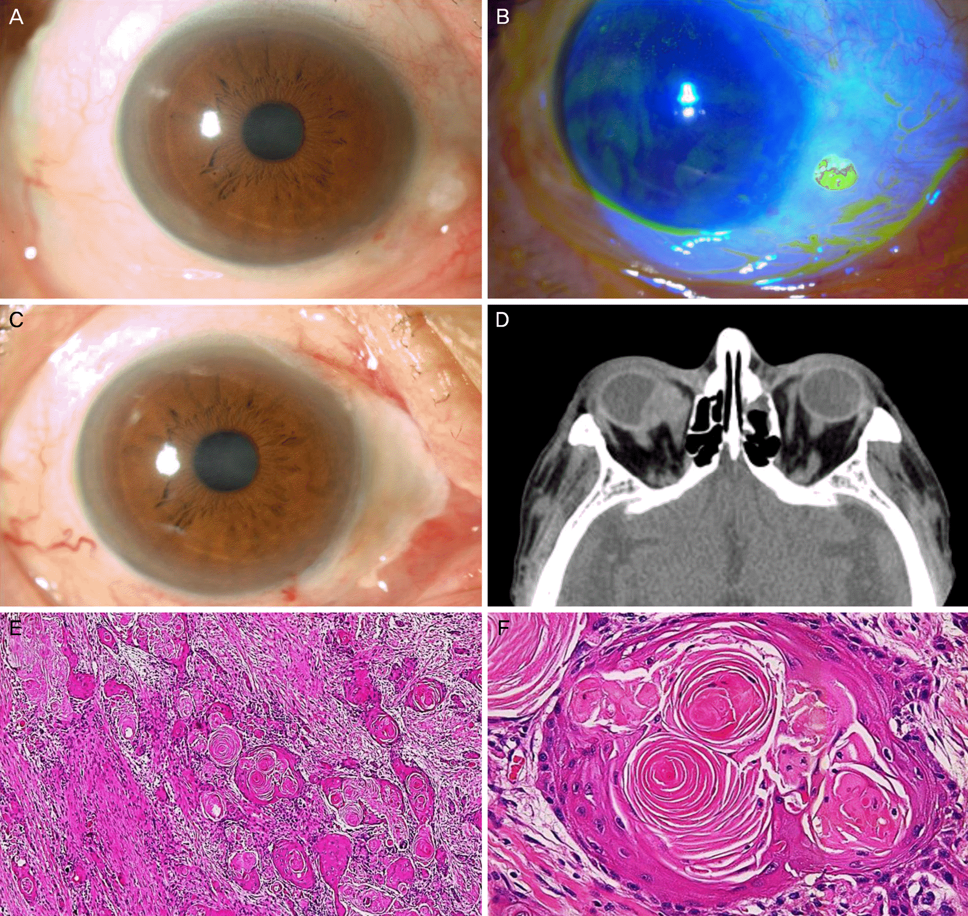
A 59-year-old Korean male presented with a whitish mass in the medial limbus and conjunctiva of the right eye. The patient had undergone renal transplantation 17 years prior due to lupus nephritis and was on systemic immunosuppression with daily prednisolone (10 mg), tacrolimus (5 mg), and mycophenolate sodium (720 mg). The complete excision of the mass was performed and mitomycin C application and amniotic membrane transplantation on the excised area were combined. Histopathological examination revealed the mass was squamous cell carcinoma. There were no abnormal findings on the orbit computed tomography (CT). The patient was additionally treated with topical interferon alpha 2b 6 months postoperatively. One year later, a mass recurred at the same site in the right eye. The complete excision of the mass, mitomycin C application, cryotherapy, and amniotic membrane transplantation were performed. Orbit CT showed a 1.9 cm-sized intraorbital mass involving the medial rectus of the right eye. The orbital exenteration was performed and the intraorbital mass was histologically proven to be squamous cell carcinoma.
References
2. Basti S, Macsai MS. Ocular surface squamous neoplasia: a review. Cornea. 2003; 22:687–704.
3. Gichuhi S, Ohnuma S, Sagoo MS, Burton MJ. Pathophysiology of ocular surface squamous neoplasia. Exp Eye Res. 2014; 129:172–82.

4. Tsatsos M, Karp CL. Modern management of ocular surface squamous neoplasia. Expert Rev Ophthalmol. 2013; 8:287–95.

5. Euvrard S, Kanitakis J, Claudy A. Skin cancers after organ transplantation. N Engl J Med. 2003; 348:1681–91.

6. Shields CL, Ramasubramanian A, Mellen PL, Shields JA. Conjunctival squamous cell carcinoma arising in immunosuppressed patients (organ transplant, human immunodeficiency virus infection). Ophthalmology. 2011; 118:2133–7.e1.

7. Lindholm A, Ohlman S, Albrechtsen D, et al. The impact of acute rejection episodes on long-term graft function and outcome in 1347 primary renal transplants treated by cyclosporine regimens. Transplantation. 1993; 56:307–15.
8. Penn I. Occurrence of cancers in immunosuppressed organ transplant recipients. Clin Transpl. 1998:147–58.
9. Penn I. The incidence of malignancies in transplant recipients. Transplant Proc. 1975; 7:323–6.
10. Vajdic CM, van Leeuwen MT, McDonald SP, et al. Increased incidence of squamous cell carcinoma of eye after kidney transplantation. J Natl Cancer Inst. 2007; 99:1340–2.

11. Porges Y, Groisman GM. Prevalence of HIV with conjunctival squamous cell neoplasia in an African provincial hospital. Cornea. 2003; 22:1–4.

13. Schreiber RD, Old LJ, Smyth MJ. Cancer immunoediting: integrating immunity's roles in cancer suppression and promotion. Science. 2011; 331:1565–70.

Figure 1.
Clinical, radiological, and histological findings of the tumor at first appearance. (A, B) Anterior segment photography showed papillary mass with surface keratinization and irregular margin involving the inferomedial conjunctiva and corneal limbus. (C) Anterior segment photography taken 10 days after excision. The mass was completely removed. (D) Orbit CT showed no abnormal findings in the intraorbital area. (E-G) Histology of the excised mass revealing squamous cell carcinoma of well-differentiated type. Horn pearls and abundant keratinization were observed (Hematoxylin-eosin ×100, ×200, and ×400). CT = computed tomography.

Figure 2.
Clinical, radiological, and histological findings of the tumor at recurrence. (A, B) Anterior segment photography taken 3 months after the first excision showed that small mass recurred in the inferomedial conjunctiva and had epithelial defect on the surface. (C) Anterior segment photography taken 8 days after the second excision. (D) Orbit CT revealed a 1.9 cm-diameter high density mass involving the medial side of the eyeball and medial rectus muscle in the right eye. (E, F) Histology of the intraorbital mass after orbital exenteration demonstrated squamous cell carcinoma composed of nests and cords of tumor cells with marked dysplasia. Tumor was well-differentiated with horn pearls and abundant keratinization (Hematoxylin-eosin ×40 and ×400). CT = computed tomography.


 PDF
PDF ePub
ePub Citation
 Print
Print


 XML Download