

 PDF
PDF ePub
ePub Citation
 Print
Print


Abstract
Purpose
Nestin, a marker of neural stem cells, is expressed in Müller cells during retinal development. However, the role of nestin in retinal vascular development is not well established. Thus, we investigated the expression of nestin in developmental mouse retina and identified which retinal cells are related to the expression of nestin during the retinal vascular development.
Methods
Eyes were enucleated from C57BL/6 mice on postnatal day (P) 4, P8, P12, P16 and P26. Immunofluorescence was used to evaluate nestin expression in relation to endothelial cells (isolectin B4), pericytes (neural/glial antigen 2) and astrocytes (glial fibrillary acidic protein).
Results
Nestin was strongly expressed from the ganglion cell layer to retinoblast layer at P4. At P8, P12 and P16, the expression of nestin was observed from the upper border of the ganglion cell layer, and vertically penetrating to outer nuclear layer. At P26, the expression of nestin was decreased and confined to the ganglion cell layer and inner nuclear layer. Interestingly, there was strong vascular shape expression of nestin at all stages. The superficial, deep and intermediate vascular plexus was completely merged with nestin expression at P4, P8, P12 and P16. In addition, the nestin expression merged with pericytes but not with astrocytes.
References
1. Guillemin K, Krasnow MA. The hypoxic response: huffing and HIFing. Cell. 1997; 89:9–12.

2. Muñoz B, West SK, Rubin GS, et al. Causes of blindness and visual impairment in a population of older Americans: The Salisbury Eye Evaluation Study. Arch Ophthalmol. 2000; 118:819–25.
3. Yoon KC, Mun GH, Kim SD, et al. Prevalence of eye diseases in South Korea: data from the Korea National Health and Nutrition Examination Survey 2008-2009. Korean J Ophthalmol. 2011; 25:421–33.
4. Jee D, Lee WK, Kang S. Prevalence and risk factors for diabetic retinopathy: the Korea National Health and Nutrition Examination Survey 2008-2011. Invest Ophthalmol Vis Sci. 2013; 54:6827–33.
5. Park SJ, Lee JH, Woo SJ, et al. Age-related macular degeneration: prevalence and risk factors from Korean National Health and Nutrition Examination Survey, 2008 through 2011. Ophthalmology. 2014; 121:1756–65.
6. Chan-Ling T, Gock B, Stone J. The effect of oxygen on vasoformative cell division. Evidence that ‘physiological hypoxia’ is the stimulus for normal retinal vasculogenesis. Invest Ophthalmol Vis Sci. 1995; 36:1201–14.
7. Stone J, Itin A, Alon T, et al. Development of retinal vasculature is mediated by hypoxia-induced vascular endothelial growth factor (VEGF) expression by neuroglia. J Neurosci. 1995; 15(7 Pt 1):4738–47.
8. Dorrell MI, Aguilar E, Friedlander M. Retinal vascular development is mediated by endothelial filopodia, a preexisting astrocytic template and specific R-cadherin adhesion. Invest Ophthalmol Vis Sci. 2002; 43:3500–10.
9. Kim JH, Kim JH, Yu YS, et al. Recruitment of pericytes and astrocytes is closely related to the formation of tight junction in developing retinal vessels. J Neurosci Res. 2009; 87:653–9.
10. Lendahl U, Zimmerman LB, McKay RD. CNS stem cells express a new class of intermediate filament protein. Cell. 1990; 60:585–95.
11. Hockfield S, McKay RD. Identification of major cell classes in the developing mammalian nervous system. J Neurosci. 1985; 5:3310–28.
12. Terling C, Rass A, Mitsiadis TA, et al. Expression of the intermediate filament nestin during rodent tooth development. Int J Dev Biol. 1995; 39:947–56.
13. Kachinsky AM, Dominov JA, Miller JB. Myogenesis and the intermediate filament protein, nestin. Dev Biol. 1994; 165:216–28.
14. Sejersen T, Lendahl U. Transient expression of the intermediate filament nestin during skeletal muscle development. J Cell Sci. 1993; 106(Pt 4):1291–300.
15. Suzuki S, Namiki J, Shibata S, et al. The neural stem/progenitor cell marker nestin is expressed in proliferative endothelial cells, but not in mature vasculature. J Histochem Cytochem. 2010; 58:721–30.
16. Yamahatsu K, Matsuda Y, Ishiwata T, et al. Nestin as a novel therapeutic target for pancreatic cancer via tumor angiogenesis. Int J Oncol. 2012; 40:1345–57.
17. Dorrell MI, Friedlander M. Mechanisms of endothelial cell guidance and vascular patterning in the developing mouse retina. Prog Retin Eye Res. 2006; 25:277–95.
18. Lee JH, Park HS, Shin JM, et al. Nestin expressing progenitor cells during establishment of the neural retina and its vasculature. Anat Cell Biol. 2012; 45:38–46.
19. Wohl SG, Schmeer CW, Kretz A, et al. Optic nerve lesion increases cell proliferation and nestin expression in the adult mouse eye in vivo. Exp Neurol. 2009; 219:175–86.
20. Steinert PM, Chou YH, Prahlad V, et al. A high molecular weight intermediate filament-associated protein in BHK-21 cells is nestin, a type VI intermediate filament protein. Limited co-assembly in vitro to form heteropolymers with type III vimentin and type IV alpha-internexin. J Biol Chem. 1999; 274:9881–90.
21. Marvin MJ, Dahlstrand J, Lendahl U, McKay RD. A rod end deletion in the intermediate filament protein nestin alters its subcellular localization in neuroepithelial cells of transgenic mice. J Cell Sci. 1998; 111(Pt 14):1951–61.
22. Liang ZW, Wang Z, Chen H, et al. Nestin-mediated cytoskeletal remodeling in endothelial cells: novel mechanistic insight into VEGF-induced cell migration in angiogenesis. Am J Physiol Cell Physiol. 2015; 308:C349–58.
Figure 1.
Retinal vascular development in normal mouse retina. On P4, superficial vascular plexus originating from the optic nerve head extended radically, which reached to the mid-peripheral retina (A) and extended to far periphery on P8 (B). P12 shows the vertical sprouting vessels toward the deep vascular plexus, which is secondary angiogenesis (C). Further maturation of vessels, especially intermediate vascular plexuses, occurred during P16 to P26 (C). Scale bar means 50 μm. P = postnatal day; GCL = ganglion cell layer; RBL = retinoblast layer; INL = inner nuclear layer; ONL = outer nuclear layer.
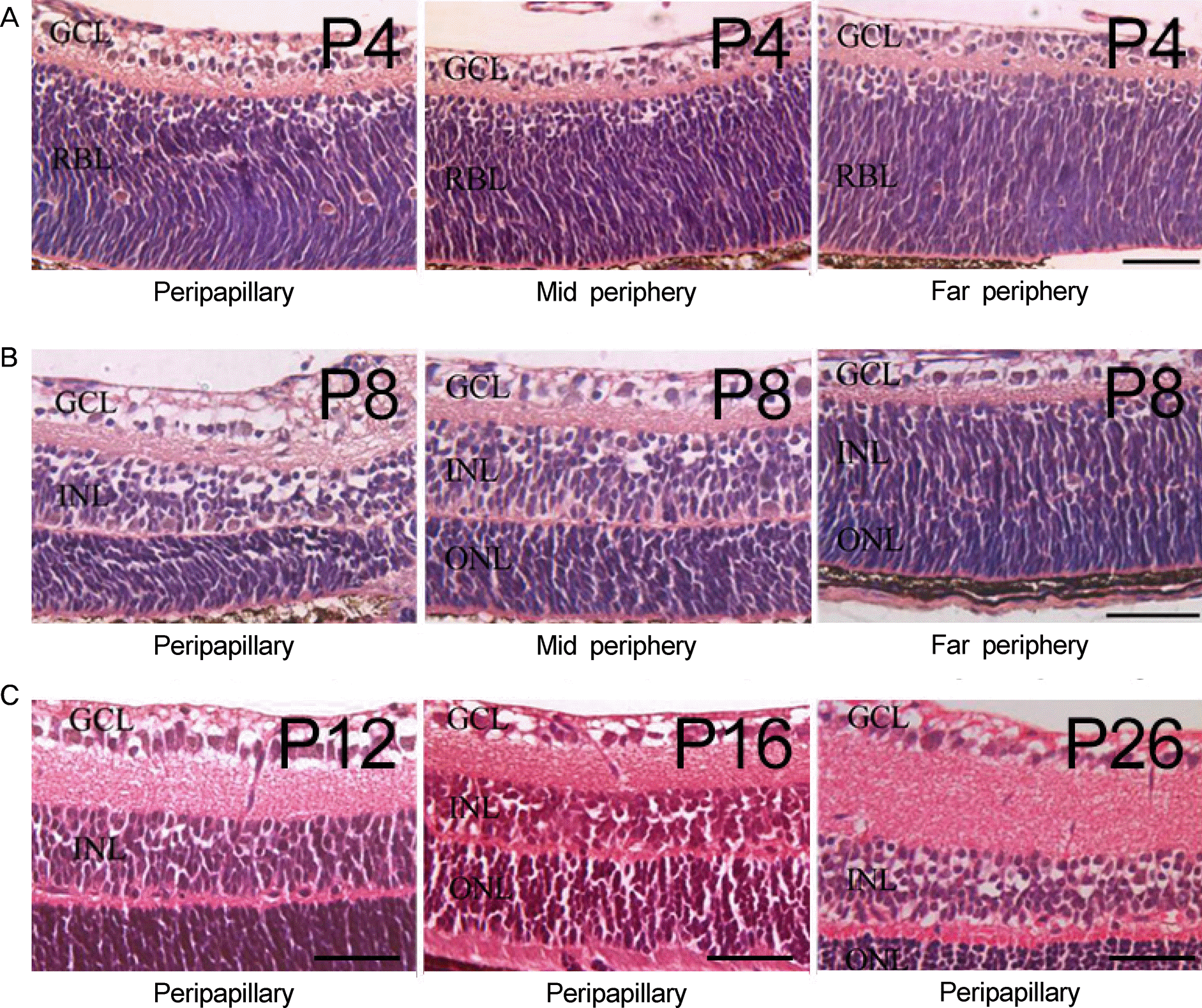
Figure 2.
The nestin expression merged with the expression of IB4 in developing mouse retina. At P4, nestin (red) expression is observed as a fibrous bundle penetrating the retina vertically, from the upper border of the ganglion cell layer to outer nuclear layer (A). This pattern of expression decreased as the mouse grew and finally confined to GCL and INL at P26 (B-E). The other observed pattern of nestin expression is an intracytoplasmic aggregation showing vascular shape at all stages. From P4 to P16, this pattern of nestin expression is completely merged with expression of IB4 (green) at the location of superficial, intermediate and deep vascular plexuses (A-D: arrowheads). At P26, intracytoplasmic expression of nestin is partially merged with expression of IB4 (E: arrowheads). Scale bar means 20 μm. IB4 = isolectin B4; DAPI = 4’,6-diamidino-2-phenylindole; GCL = ganglion cell layer; RBL = retinoblast layer; INL = inner nuclear layer; ONL = outer nuclear layer; P = postnatal day.
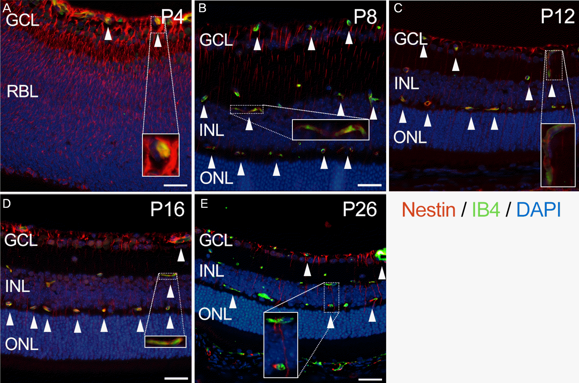
Figure 3.
The nestin expression merged with the expression of NG2 in developing mouse retina. NG2 (green) expression is observed at the level of retinal vascular plexuses and partially merged with expression of nestin (red) from P4 to P26 (A-E: arrowheads). Scale bar means 20 μm. NG2 = neural/glial antigen 2; DAPI = 4’,6-diamidino-2-phenylindole; GCL = ganglion cell layer; RBL = retinoblast layer; INL = inner nuclear layer; ONL = outer nuclear layer; P = postnatal day.
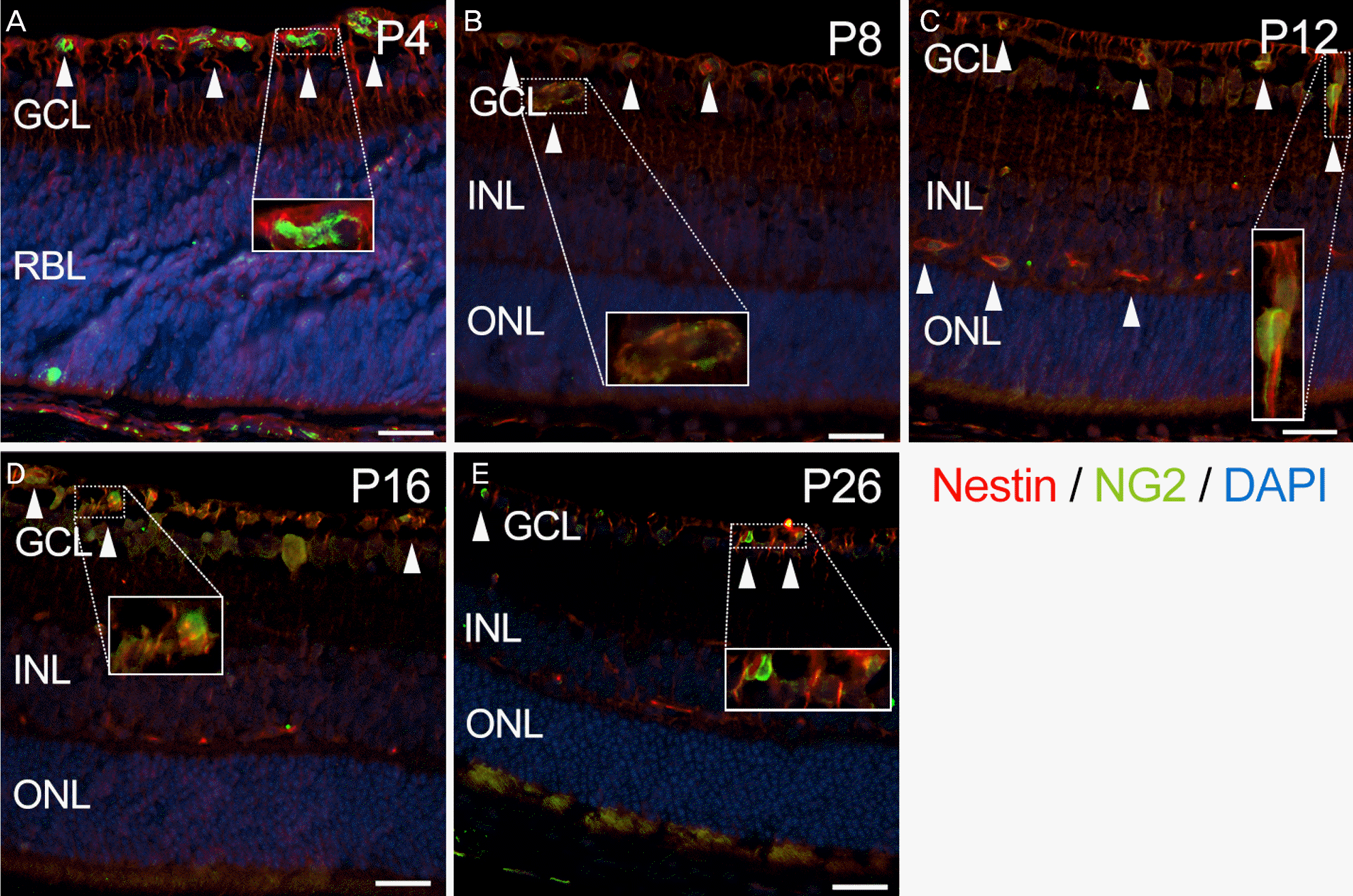
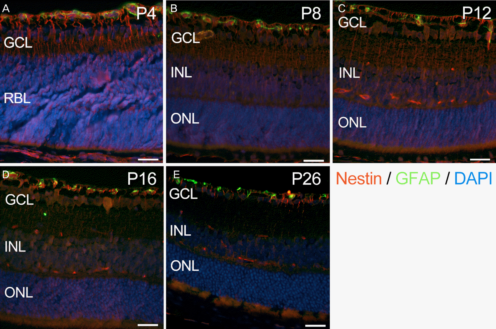
Figure 4.
The nestin expression (red) merged with the expression of GFAP (green) in developing mouse retina. Expression of GFAP is confined to GCL and not merged with expression of nestin at any stage (A-E). Scale bar means 20 μm. GFAP = glial fibrillary acidic protein; DAPI = 4’,6-diamidino-2-phenylindole; GCL = ganglion cell layer; RBL = retinoblast layer; INL = inner nuclear layer; ONL = outer nuclear layer; P = postnatal day.
 XML Download