Abstract
Purpose
To report serial cases of patients who received surgical treatment for tube erosion after Ahmed valve implantation.
Methods
In this retrospective and longitudinal study, surgical outcomes of reconstruction for tube erosion after Ahmed valve implantation were evaluated. Tube erosion occurred in 7 of 125 eyes (121 patients) at 60.5 ± 67.5 months (2 to 196 months). To prevent recurrence of tube erosion, the tube was repositioned backward in 4 eyes. Scleral allograft was used in all cases. Partial thickness scleral flap, collagen matrix implant, or bovine pericardium was used to cover the exposed tube in selected cases. In eyes with large conjunctival defects or severe adhesions between surrounding conjunctiva and episclera, rotational conjunctival flap, conjunctival autograft, or amniotic membrane graft was used to reconstruct the ocular surface.
Results
Three of 7 eyes (42.9%) with tube erosion were successfully repaired by the first surgical treatment. No recurrence of tube erosion was found after the second and the fourth surgical procedure in 3 and 1 eyes, respectively. There was no case of explantation of the Ahmed valve in our series. Tube erosion recurred in 1 of the 4 eyes in which tube-repositioning was required and in 3 of the 5 eyes in which a partial thickness scleral flap was made to cover a tube. One eye experienced recurrent tube erosion in a relatively short-term interval and was repaired successfully using both collagen matrix implantation and amniotic membrane transplantation.
Go to : 
References
1. Joseph NH, Sherwood MB, Trantas G, et al. A one-piece drainage system for glaucoma surgery. Trans Ophthalmol Soc U K. 1986; 105(Pt 6):657–64.
2. Taglia DP, Perkins TW, Gangnon R, et al. Comparison of the Ahmed glaucoma valve, the Krupin eye valve with Disk, and the double-plate Molteno implant. J Glaucoma. 2002; 11:347–53.

3. Huang MC, Netland PA, Coleman AL, et al. Intermediate-term clinical experience with the Ahmed Glaucoma Valve implant. Am J Ophthalmol. 1999; 127:27–33.
4. Ayyala RS, Zurakowski D, Smith JA, et al. A clinical study of the Ahmed glaucoma valve implant in advanced glaucoma. Ophthalmology. 1998; 105:1968–76.
5. Wilson MR, Mendis U, Paliwal A, Haynatzka V. Long-term follow-up of primary glaucoma surgery with Ahmed glaucoma valve implant versus trabeculectomy. Am J Ophthalmol. 2003; 136:464–70.

6. Lee D, Kim HM, Kim T. Amniotic membrane transplantation for tube erosion after Ahmed glaucoma valve implant surgery: two cases. J Korean Ophthalmol Soc. 2003; 44:2950–4.
7. Geffen N, Buys YM, Smith M, et al. Conjunctival complications related to Ahmed glaucoma valve insertion. J Glaucoma. 2014; 23:109–14.

8. Han YS, Cho YS, Ahn JH. Scleral allograft with autologous buccal mucosal transplantation for tube erosion ocurred after Ahmed glaucoma valve implant surgery: three cases. J Korean Ophthalmol Soc. 2001; 42:401–6.
9. Lee ES, Kang SY, Kim NR, et al. Split-thickness hinged scleral flap in the management of exposed tubing of a glaucoma drainage device. J Glaucoma. 2011; 20:319–21.

10. Chen PP, Palmberg PF. Needling revision of glaucoma drainage device filtering blebs. Ophthalmology. 1997; 104:1004–10.

11. Huddleston SM, Feldman RM, Budenz DL, et al. Aqueous shunt exposure: a retrospective review of repair outcome. J Glaucoma. 2013; 22:433–8.
12. Stewart WC, Kristoffersen CJ, Demos CM, et al. Incidence of conjunctival exposure following drainage device implantation in patients with glaucoma. Eur J Ophthalmol. 2010; 20:124–30.

13. Low SA, Rootman DB, Rootman DS, Trope GE. Repair of eroded glaucoma drainage devices: mid-term outcomes. J Glaucoma. 2012; 21:619–22.
14. Al-Torbak AA, Al-Shahwan S, Al-Jadaan I, et al. Endophthalmitis associated with the Ahmed glaucoma valve implant. Br J Ophthalmol. 2005; 89:454–8.

15. Gedde SJ, Scott IU, Tabandeh H, et al. Late endophthalmitis associated with glaucoma drainage implants. Ophthalmology. 2001; 108:1323–7.

16. Bayraktar Z, Kapran Z, Bayraktar S, et al. Delayed-onset streptococcus pyogenes endophthalmitis following Ahmed glaucoma valve implantation. Jpn J Ophthalmol. 2005; 49:315–7.

17. Gutiérrez-Diaz E, Montero-Rodríguez M, Mencía-Gutiérrez E, et al. Long-term persistence of fascia lata patch graft in glaucoma drainage device surgery. Eur J Ophthalmol. 2005; 15:412–4.
18. Raviv T, Greenfield DS, Liebmann JM, et al. Pericardial patch grafts in glaucoma implant surgery. J Glaucoma. 1998; 7:27–32.

19. Tanji TM, Lundy DC, Minckler DS, et al. Fascia lata patch graft in glaucoma tube surgery. Ophthalmology. 1996; 103:1309–12.
20. Singh M, Chew PT, Tan D. Corneal patch graft repair of exposed glaucoma drainage implants. Cornea. 2008; 27:1171–3.

Go to : 
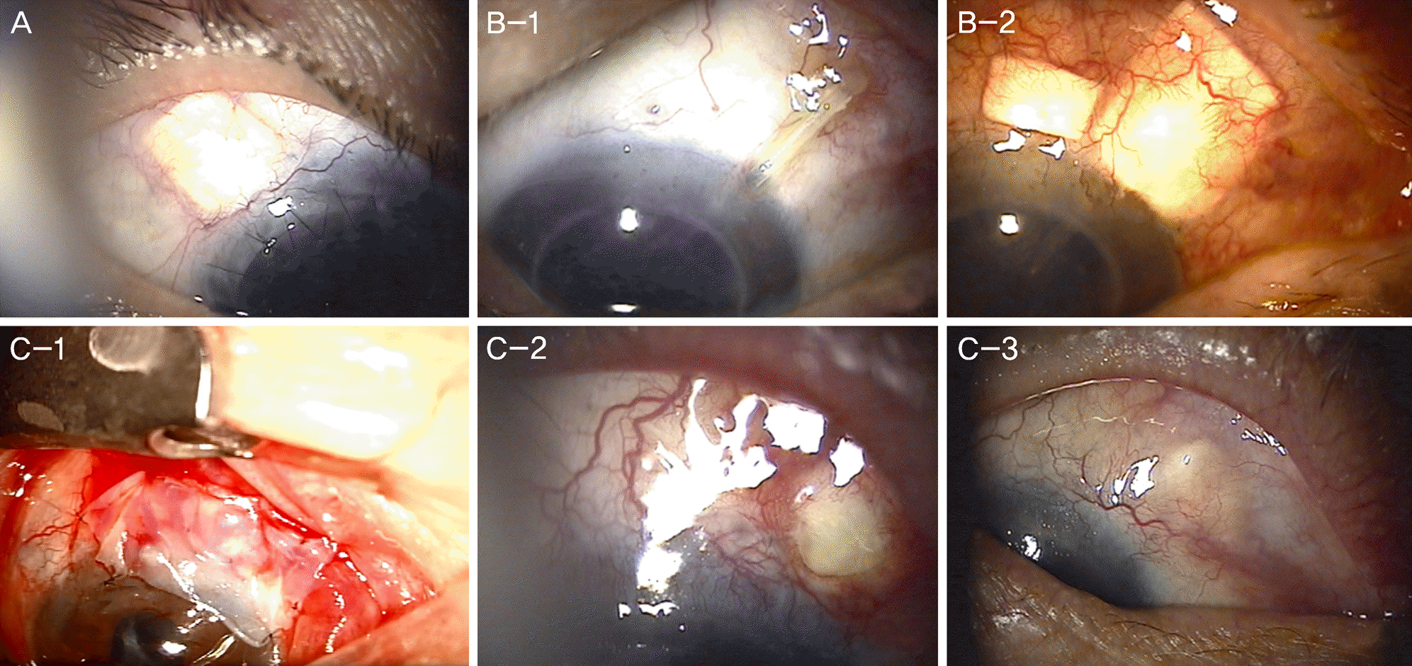
 | Figure 1.Surgical repair of tube erosion in patients with Ahmed valve implantation. (A) Repair of tube exposure using bovine pericardial graft in case 3. Implanted pericardium and overlying conjunctiva are well maintained at 4 months after the reconstruction. (B) Preoperative and postoperative findings using scleral allograft for tube exposure in case 6. (B-1) Exposed tube and scleral fixation suture are noted at 196 months after Ahmed valve implantation. (B-2) Exposed tube is repaired using scleral allograft and partial thickness scleral flap combined with amniotic membrane transplantation. Exposed suture knot is covered with scleral allograft and the conjunctiva. There is no exposure noted at 9 months after the reconstruction. (C) Combined methods to repair recurrent conjunctival erosions and exposure of scleral allograft using collagen matrix implantation and amniotic membrane transplantation in case 7. (C-1) Intraoperative findings during the 3rd amniotic membrane transplantation. Amniotic membrane is covered with epithelial side down over implanted scleral allograft and amniotic membrane inlay graft. (C-2) The scleral allograft is exposed again at 1 month after the 3rd amniotic membrane transplantation. (C-3) The conjunctiva over the previously exposed site is well maintained at 7 months after the 4th amniotic membrane transplantation combined with collagen matrix implantation. |
Table 1.
Characteristics of the patients with tube erosion
Table 2.
Clinical features of the patients who underwent surgical treatment for tube erosion
| Case No. | Sex/age (years) | Diagnosis | Intraocular operation history | Exposure time after surgery (months) |
Method for the first repair |
Additional procedure for re-exposure*r | Follow up after the first reconstruction (months) | ||
|---|---|---|---|---|---|---|---|---|---|
| Tube reposit ioning | Partial thickness scleral flap | Materials for ocular surface repair | |||||||
| 1 | F/61 | NVG | None | 80 | Done | (-) | Advancement flap AM overlay | N/A | 33 |
| 2 | F/58 | NVG | Cataract | 38 | (-) | (-) | Rotational flap | Tube repositioning, partial thickness scleral flap, scleral allograft, conjunctival autograft | 113 |
| 3 | F/38 | ICE syndrome | DMEK,PKP, cataract | 78 | (-) | Done | Advancement flap | Tube repositioning, bovine pericardium, advancement flap | 35 |
| 4 | M/68 | NVG | Trabeculectomy | 26 | (-) | Done | Rotational flap | N/A | 8 |
| 5 | F/64 | CACG | Cataract, trabeculectomy | 4 | Done | Done | Advancement flap, AM overlay | Scleral allograft, AM inlay + overlay | 12 |
| 6 | F/53 | Secondary glaucoma | PKP, cataract | 196 | (-) | Done | AM inlay + overlay | N/A | 9 |
| 7 | M/67† | NVG | Cataract, removal of implanted Ahmed valve | 2 | (-) | Done | AM inlay + overlay | Collagen matrix implant, AM inlay + overlay | 13 |


 PDF
PDF ePub
ePub Citation
 Print
Print


 XML Download