Abstract
Purpose
To investigate the long-term results of transscleral fixation of posterior chamber intraocular lens (IOL) for unstable pos-terior capsular supporting structure.
Methods
We performed a retrospective review of 67 patients (67 eyes) with unstable posterior capsular supporting structure who underwent transscleral fixation at Soonchunhyang University Bucheon Hospital from March 2005 to January 2013. Transscleral fixation without scleral flap was performed by a single surgeon. We analyzed the causes of transscleral fixation and compared postoperative best-corrected visual acuity (BCVA) and spherical diopter.
Results
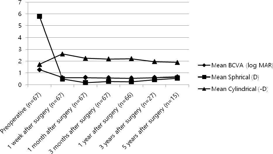
Among the 67 eyes of 67 patients, the causes of transscleral fixation included IOL subluxation (33 cases, 49.2%), IOL dislocation (11 cases, 16.4%), intraoperative posterior capsule rupture (8 cases, 11.9%), aphakia associated with previous intra-ocular surgery (7 cases, 10.4%), crystalline lens disorder with zonular dialysis (4 cases, 5.9%) and IOL opacity (4 cases, 5.9%). The mean BCVA before surgery was 1.26 ± 0.94 (log MAR) and the visual acuity improved to 0.59 ± 0.71, 0.60 ± 0.69, 0.58 ±0.70, 0.55 ± 0.70, 0.60 ± 0.58 and 0.66 ± 0.70 (1 week, 1 month, 3 months, 1 year, 3 years and 5 years, respectively, after the surgery; p < 0.05).
Conclusions
Posterior chamber IOL transscleral fixation in unstable posterior capsular supporting structure is effective for in-creasing visual acuity and spherical diopter. Specifically, the most improvement was observed at one month after surgery. Transscleral fixation is an adequate surgical procedure for fast improvement of visual acuity with long-term effects.
Go to : 
References
1. Malbran ES, Malbran E Jr, Negri I. Lens guide suture for transport and fixation in secondary IOL implantation after intracapsular extraction. Int Ophthalmol. 1986; 9:151–60.

2. Holt DG, Young J, Stagg B, Ambati BK. Anterior chamber intra-ocular lens, sutured posterior chamber intraocular lens, or glued in-traocular lens: where do we stand? Curr Opin Ophthalmol 2012; 23. 62–7.
3. Hannush SB. Sutured posterior chamber intraocular lenses: in-dications and procedure. Curr Opin Ophthalmol. 2000; 11:233–40.

4. Hu BV, Shin DH, Gibbs KA, Hong YJ. Implantation of posterior chamber lens in the absence of capsular and zonular support. Arch Ophthalmol. 1988; 106:416–20.

6. Stark WJ, Goodman G, Goodman D, Gottsch J. Posterior chamber intraocular lens implantation in the absence of posterior capsular support. Ophthalmic Surg. 1988; 19:240–3.

7. Güell JL, Barrera A, Manero F. A review of suturing techniques for posterior chamber lenses. Curr Opin Ophthalmol. 2004; 15:44–50.
8. Yang JY, Chu YK. Modified surgical technique for transscleral fix-ation of a single-piece acrylic intraocular lens in the absence of capsular support. J Korean Ophthalmol Soc. 2012; 53:1794–800.

9. Jung MO, Koh JW. Clinical results of modified Ab Externo and one-knot technique. J Korean Ophthalmol Soc. 2012; 53:1783–8.

10. Ma DJ, Kim MK, Wee WR. Knotless external fixation technique for posterior chamber intraocular lens transscleral fixation: a 5-case analysis. J Korean Ophthalmol Soc. 2012; 53:1609–14.

11. Duffey RJ, Holland EJ, Agapitos PJ, Lindstrom RL. Anatomic study of transsclerally sutured intraocular lens implantation. Am J Ophthalmol. 1989; 108:300–9.

12. Smiddy WE, Sawusch MR, O'Brien TP, et al. Implantation of scler-al-fixated posterior chamber intraocular lenses. J Cataract Refract Surg. 1990; 16:691–6.

13. Kokame GT, Yamamoto I, Mandel H. Scleral fixation of dislocated posterior chamber intraocular lenses: temporary haptic external-ization through a clear corneal incision. J Cataract Refract Surg. 2004; 30:1049–56.
14. Shin JH, Lee JE, Oum BS. Clinical outcomes of the surgical man-agement with dislocated posterior chamber intraocular lens. J Korean Ophthalmol Soc. 2012; 53:420–7.

15. Hayashi K, Hirata A, Hayashi H. Possible predisposing factors for in-the-bag and out-of-the-bag intraocular lens dislocation and out-comes of intraocular lens exchange surgery. Ophthalmology. 2007; 114:969–75.

16. Michaeli A, Assia EI. Scleral and iris fixation of posterior chamber lenses in the absence of capsular support. Curr Opin Ophthalmol. 2005; 16:57–60.

17. Por YM, Lavin MJ. Techniques of intraocular lens suspension in the absence of capsular/zonular support. Surv Ophthalmol. 2005; 50:429–62.

18. Wagoner MD, Cox TA, Ariyasu RG. . Intraocular lens im-plantation in the absence of capsular support: a report by the American Academy of Ophthalmology. Ophthalmology. 2003; 110:840–59.
19. Heilskov T, Joondeph BC, Olsen KR, Blankenship GW. Late en-dophthalmitis after transscleral fixation of a posterior chamber in-traocular lens. Arch Ophthalmol. 1989; 107:1427.

21. Baykara M, Avci R. Prevention of suture knot exposure in posterior chamber intraocular lens implantation by 4-point scleral fixation technique. Ophthalmic Surg Lasers Imaging. 2004; 35:379–82.

22. Hoffman RS, Fine IH, Packer M. Scleral fixation without con-junctival dissection. J Cataract Refract Surg. 2006; 32:1907–12.

23. Szurman P, Petermeier K, Aisenbrey S. . Z-suture: a new knot-less technique for transscleral suture fixation of intraocular implants. Br J Ophthalmol. 2010; 94:167–9.

Go to : 
 | Figure 1.Knot location of posterior chamber intraocular lens transscleral fixation. (A, B) A polypropylene is fixed in the outer one-third point of haptic. |
 | Figure 2.Technique for the ab externo approach of posterior chamber intraocular lens (IOL) transscleral fixation. (A) The long curved double-armed 10-0 polypropylene needle is passed through the sclera approximately 1.0 mm posterior to the limbus. A sec-ond hollow needle is passed from the opposite side of the eye. (B) A hook is used to pull the suture out through a superior scleral tunnel wound so that it can be tied to the intraocular lens. (C) Suture is cut, and each end is tied to a haptic of the intraocular lens. After the IOL is placed into position. (D) The scleral sutures must be anchored to the sclera. |
 | Figure 3.BCVA and refractive indexes change after surgery. The mean BCVA before surgery was 1.26 ± 0.94 (log MAR) and the visual acuity improved to 0.66 ± 0.70 at 5 years after the surgery. The spherical diopter before surgery was 5.80 ±5.86 diopters and it improved to 0.56 ± 1.12 diopters at 5 years after the surgery. The cylindrical diopter change shows no significant value. BCVA = best corrected visual acuity. |
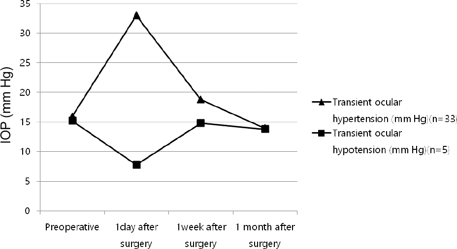
 | Figure 4.IOP change after surgery. After surgery, there were 33 cases of transient ocular hypertension and 5 cases of tran-sient ocular hypotension. At first day after surgery, the aver-age of ocular hypertension was 33.0 ± 12.42 mm Hg and average of ocular hypotension was 7.8 ± 0.83 mm Hg. The abnormal range of intraocular pressure was controlled within 1 month by conservative treatment. IOP = intraocular pressure. |
Table 1.
Causes for scleral fixation of the posterior chamber IOL
Table 2.
Patients’ characteristics
|
Age (years) |
Sex ratio (male/female) |
Follow-up period (months) |
||
|---|---|---|---|---|
| Mean ± SD | Range | Number | Mean ± SD | Range |
| 65.56 ± 13.52 | 18-89 | 46/21 | 39.47 ± 27.47 | 10-105 |
Table 3.
BCVA and refractive indexes change after surgery
| Preoperative (n = 67) | 1 week after surgery (n = 67) | 1 month after surgery (n = 67) | 3 months after surgery (n = 67) | 1 year after surgery (n = 66) | 3 years after surgery (n = 27) | 5 years after surgery (n = 15) | |
|---|---|---|---|---|---|---|---|
| Mean BCVA (log MAR) | 1.26 ± 0.94 | 0.59 ± 0.71 | 0.60 ± 0.69 | 0.58 ± 0.70 | 0.55 ± 0.70 | 0.60 ± 0.58 | 0.66 ± 0.70 |
| p-value* | <0.05 | <0.05 | <0.05 | <0.05 | <0.05 | <0.05 | |
| Mean Sph (D) | 5.80 ± 5.86 | 0.49 ± 1.70 | 0.18 ± 1.60 | 0.28 ± 1.36 | 0.26 ± 1.37 | 0.43 ± 1.64 | 0.56 ± 1.12 |
| p-value* | <0.05 | <0.05 | <0.05 | <0.05 | <0.05 | <0.05 | |
| Mean Cyl (-D) | 1.71 ± 2.95 | 2.61 ± 1.77 | 2.24 ± 1.21 | 2.18 ± 1.18 | 2.20 ± 1.12 | 1.95 ± 1.09 | 1.89 ± 1.34 |
| p-value* | <0.05 | 0.176 | 0.213 | 0.128 | 0.160 | 0.145 |
Table 4.
Postoperative complications after surgery
Table 5.
Intraocular pressure change after surgery
| Preoperative | 1 day after surgery | 1 week after surgery | 1 month after surgery | |
|---|---|---|---|---|
| Transient ocular hypertension (mm Hg, n = 33) | 15.93 ± 2.34 | 33.0 ± 12.42 | 18.81 ± 7.20 | 13.96 ± 2.98 |
| p-value* | <0.05 | 0.110 | 0.353 | |
| Transient ocular hypotension (mm Hg, n = 5) | 15.20 ± 2.38 | 7.8 ± 0.83 | 14.8 ± 10.28 | 13.8 ± 1.92 |
| p-value† | <0.05 | 0.500 | 0.416 |


 PDF
PDF ePub
ePub Citation
 Print
Print


 XML Download