

 PDF
PDF ePub
ePub Citation
 Print
Print


Abstract
Purpose
To evaluate factors affecting final visual acuity by analyzing patients referred with infectious endophthalmitis after cataract surgery.
Methods
A retrospective investigation of clinical notes of 113 patients referred with endophthalmitis following cataract surgery was conducted from January 2008 to December 2013. To evaluate factors affecting final visual acuity, initial visual acuity, onset of endophthalmitis after the cataract surgery, types of treatment, presence of hypopyon and culture results were investigated.
Results
Of the 113 patients, visual acuities at presentation were hand motions or less in 75 patients (66.3%) and final visual acuities after treatments were 0.5 or better in 73 patients (64.6%). Cases with initial visual acuity of hand motions or better achieved favorable outcomes whereas cases with gram-negative infection or endophthalmitis occurring within 2 days postoperatively showed poor prognosis. Statistically, vitrectomy versus intraocular antibiotic injection as primary means of treatment showed no differences in final visual acuity in patients with initial visual acuity of hand motion or better.
Conclusions
Visual acuities at presentation, type of cultured organism and onset of endophthalmitis after cataract surgery are significantly related to visual prognosis. Advantages of initial vitrectomy versus intraocular antibiotic injections were unclear and further investigations are necessary to clarify these issues.
Go to : 
References
1. Endophthalmitis Study Group, European Society of Cataract & Refractive Surgeons. Prophylaxis of postoperative endophthalmitis following cataract surgery: results of the ESCRS multicenter study and identification of risk factors. J Cataract Refract Surg. 2007; 33:978–88.
2. Javitt JC, Vitale S, Canner JK, et al. National outcomes of cataract extraction. Endophthalmitis following inpatient surgery. Arch Ophthalmol. 1991; 109:1085–9.
3. Kattan HM, Flynn HW Jr, Pflugfelder SC, et al. Nosocomial endophthalmitis survey. Current incidence of infection after intraocular surgery. Ophthalmology. 1991; 98:227–38.
4. Kresloff MS, Castellarin AA, Zarbin MA. Endophthalmitis. Surv Ophthalmol. 1998; 43:193–224.

5. Jeon CY, Lee TG, Na SJ. The clinical outcomes of 23-gauge trans-conjunctival sutureless vitrectomy for dndophthalmitis after cataract surgery. J Korean Ophthalmol Soc. 2011; 52:1167–72.
6. Results of the Endophthalmitis Vitrectomy Study. A randomized trial of immediate vitrectomy and of intravenous antibiotics for the treatment of postoperative bacterial endophthalmitis. Arch Ophthalmol. 1995; 113:1479–96.
7. Pijl BJ, Theelen T, Tilanus MA, et al. Acute endophthalmitis after cataract surgery: 250 consecutive cases treated at a tertiary referral center in the Netherlands. Am J Ophthalmol. 2010; 149:482–7. e1-2.
8. Altan T, Acar N, Kapran Z, et al. Acute-onset endophthalmitis after cataract surgery: success of initial therapy, visual outcomes, and related factors. Retina. 2009; 29:606–12.
9. Ding Y, Lin M, Liu H, et al. Outcomes of post-cataract surgery endophthalmitis referred to a tertiary center from local hospitals in the south of China. Infection. 2011; 39:451–60.
10. Haapala TT, Nelimarkka L, Saari JM, et al. Endophthalmitis following cataract surgery in southwest Finland from 1987 to 2000. Graefes Arch Clin Exp Ophthalmol. 2005; 243:1010–7.
11. Ohl CA, Pollack M. Infections due to Pseudomonas species and related organisms. Harrisons Principles of Internal Medicine. 2005; 16:889.
12. Bohigian GM, Olk RJ. Factors associated with a poor visual result in endophthalmitis. Am J Ophthalmol. 1986; 101:332–41.
13. Cheng JH, Chang YH, Chen CL, et al. Acute endophthalmitis after cataract surgery at a referral centre in Northern Taiwan: review of the causative organisms, antibiotic susceptibility, and clinical features. Eye (Lond). 2010; 24:1359–65.
14. Kent DG. Endophthalmitis in Auckland 1983–1991. Aust N Z J Ophthalmol. 1993; 21:227–36.
15. Somani S, Grinbaum A, Slomovic AR. Postoperative endophthalmitis: incidence, predisposing surgery, clinical course and outcome. Can J Ophthalmol. 1997; 32:303–10.
16. Wong TY, Chee SP. The epidemiology of acute endophthalmitis after cataract surgery in an Asian population. Ophthalmology. 2004; 111:699–705.
17. Kuhn F, Gini G. Ten years after… are findings of the Endophthalmitis Vitrectomy Study still relevant today? Graefes Arch Clin Exp Ophthalmol. 2005; 243:1197–9.
18. Wejde G, Montan P, Lundström M, et al. Endophthalmitis following cataract surgery in Sweden: national prospective survey 1999–2001. Acta Ophthalmol Scand. 2005; 83:7–10.
19. Kang KT, Kim KS, Kim YC. Factors affecting final visual acuity in infectious endophthalmitis following cataract surgery. J Korean Ophthalmol Soc. 2013; 54:1025–31.
20. Jung JY, Ko BY, Kim BY. Factors associated with a poor visual result in acute endophthalmitis after cataract surgery. J Korean Ophthalmol Soc. 2008; 49:1242–7.
21. Kim WJ, Kweon EY, Lee DW, Cho NC. Postoperative endophthalmitis following cataract surgery over an eight-year period. J Korean Ophthalmol Soc. 2008; 49:1771–8.
22. Pinna A, Usai D, Sechi LA, et al. An outbreak of post-cataract surgery endophthalmitis caused by Pseudomonas aeruginosa. Ophthalmology. 2009; 116:2321–6. e1-4.
23. Lalwani GA, Flynn HW Jr, Scott IU, et al. Acute-onset endophthalmitis after clear corneal cataract surgery (1996–2005). Clinical features, causative organisms, and visual acuity outcomes. Ophthalmology. 2008; 115:473–6.
24. Sandvig KU, Dannevig L. Postoperative endophthalmitis: establishment and results of a national registry. J Cataract Refract Surg. 2003; 29:1273–80.
25. Microbiologic factors and visual outcome in the endophthalmitis vitrectomy study. Am J Ophthalmol. 1996; 122:830–46.
Go to :
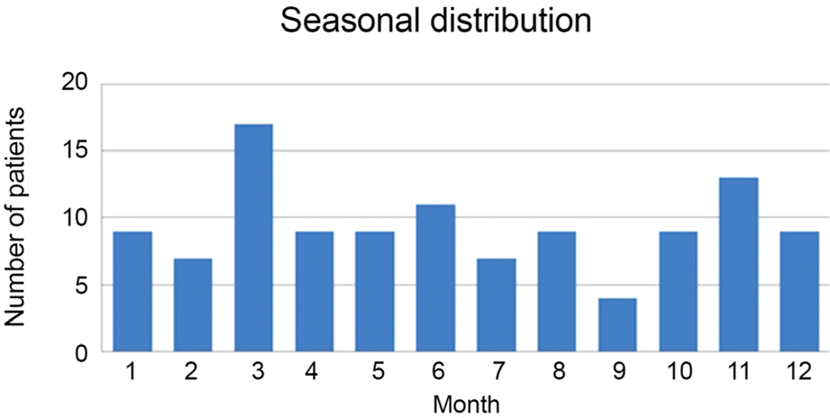 | Figure 1.Seasonal distribution of endophthalmitis after cataract surgery. The most likely month for patients to develop endophthalmitis was March. |
Table 1.
Summary of baseline patient characteristics
Table 2.
Type of treatments received before referral
| Treatments | Total count |
|---|---|
| IOAI only | 23 |
| IOAI with A/C irrigation | 2 |
| A/C irrigation | 4 |
| Primary vitrectomy | 1 |
|
|
|
| Total | 30 |
Table 3.
Microbiologic spectrum
Table 4.
Type of initial intervention according to presenting visual acuity
| Presenting vision |
Initial intervention |
Total | |
|---|---|---|---|
| PPV + IOAI | IOAI | ||
| LP | 17 | 0 | 17 |
| HM | 36 | 22 | 58 |
| Better than HM | 15 | 23 | 38 |
|
|
|||
| Total | 64 | 49 | 113 |
Table 5.
Comparison of final visual acuity after different types of treatments
| Total count |
Final VA ≥ 0.50 |
p-value | ||
|---|---|---|---|---|
| n | % | |||
| All cases | 113 | 0.184* | ||
| Primary vitrectomy | 68 | 40 | 58.8 | |
| Primary IOAI ± additional PPV | 45 | 32 | 71.1 | |
| Cases with presenting VA of HM | 58 | 0.985* | ||
| Primary vitrectomy | 36 | 23 | 63.8 | |
| Primary IOAI ± additional PPV | 22 | 14 | 63.6 | |
| Cases with presenting VA of FC | 23 | 0.178† | ||
| Primary vitrectomy | 9 | 8 | 88.9 | |
| Primary IOAI ± additional PPV | 14 | 9 | 63.6 | |
| Cases with presenting VA ≥ HM | 96 | 0.876* | ||
| Primary vitrectomy | 51 | 37 | 72.5 | |
| Primary IOAI ± additional PPV | 45 | 32 | 71.1 | |
Table 6.
Factors associated with visual outcome
| Factor |
Final visual acuity |
Significance of association (p-value) | |
|---|---|---|---|
| Bettern than 20/200 (%) | Worse than 20/200 (%) | ||
| Age (years) | 0.244* | ||
| Median (range) | 70 (44-89) | 70 (41-96) | |
| Gender | 0.057† | ||
| Male | 34 (39.1) | 15 (57.7) | |
| Female | 53 (60.9) | 11 (42.3) | |
| Involved eye | 0.981† | ||
| Right | 42 (48.3) | 12 (46.2) | |
| Left | 45 (51.7) | 14 (53.8) | |
| Latency period | 0.043† | ||
| <3 days | 34 (39.1) | 16 (61.5) | |
| ≥3 days | 53 (60.9) | 10 (38.5) | |
| Time to presentation | 0.172‡ | ||
| <7 days | 79 (90.8) | 21 (80.8) | |
| ≥7 days | 8 (9.2) | 5 (19.2) | |
| Presenting visual acuity | 0.001† | ||
| Light perception or less | 7 (8.0) | 10 (38.5) | |
| Hand motions or better | 80 (92.0) | 16 (61.5) | |
| Hypopyon | 0.565† | ||
| Yes | 49 (56.3) | 16 (61.5) | |
| No | 38 (43.7) | 10 (38.5) | |
| Causative organism | 0.030§ | ||
| Negative growth | 42 (48.3) | 8 (30.8) | |
| Gram-positive | 31 (35.6) | 8 (30.8) | |
| Gram-negative | 14 (16.1) | 10 (38.4) | |
| Immediate vitrectomy | 0.140† | ||
| Yes | 46 (52.9) | 18 (69.2) | |
| No | 41 (47.1) | 8 (30.8) | |
 XML Download