

 PDF
PDF ePub
ePub Citation
 Print
Print


Abstract
Case summary
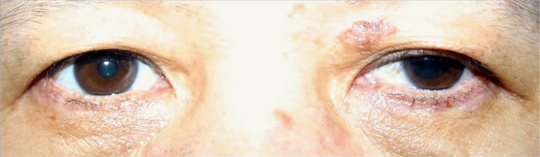
Two patients with HZO who were suffering from skin lesions, facial pain, and medicated with an antiviral agent from the dermatologic department, were diagnosed with oculomotor palsy. They presented with ptosis, dilated pupils, and ophthalmoplegia after 10 days, and 4 days after began developing skin lesions. They were diagnosed with HZO-induced oculomotor palsy and the ophthalmoplegia improved in all cases.
Conclusions
Oculomotor nerve palsy rarely occurrs in HZO patients and is usually followed by skin lesions. We recommend examinations for functions of extraocular motility, ptosis, and pupil to diagnose and treat the HZO-induced oculomotor nerve palsy even if consulted with the dermatologic department or only showing mild conjunctivitis, episcleritis, or keratitis.
REFERENCES
1). Liesegang TJ. Herpes zoster ophthalmicus natural history, risk factors, clinical presentation and morbidity. Ophthalmology. 2008; 115:(2 Suppl). S3–12.
2). Sanjay S, Chan EW, Gopal L, et al. Complete unilateral ophthalmoplegia in herpes zoster ophthalmicus. J Neuroophthalmol. 2009; 29:325–37.

3). Harthan JS, Borgman CJ. Herpes zoster ophthalmicus-induced oculomotor nerve palsy. J Optom. 2013; 6:60–5.
4). Park SH, Kim WJ, Yang WS, Kim MS. Herpes zoster ophthalmicus complicated by hyphema, glaucoma and external ophthalmoplegia. J Korean Ophthalmol Soc. 2007; 48:1573–8.
5). Edgerton AE. Ophthalmicus Herpes Zoster: report of cases and a review of the literature. Trans Am Ophthalmol Soc. 1942; 40:390–439.
6). Zaal MJ, Völker-Dieben HJ, D'Amaro J. Prognostic value of Hutchinson's sign in acute herpes zoster ophthalmicus. Graefes Arch Clin Exp Ophthalmol. 2003; 241:187–91.
7). Im M, Kim BJ, Seo YJ, et al. Complete ophthalmoplegia after herpes zoster. Clin Exp Dermatol. 2007; 32:162–4.
8). Chhabra MS, Golnik KC. Recovery of ocular motor cranial nerve palsy after herpes zoster ophthalmicus. J Neuroophthalmol. 2014; 34:20–2.
9). Savino PJ, Danesh-Meyer H. Color Atlas & Synopsis of Clinical Ophthalmology (Wills Eye Hospital): Neuro-ophthalmology. 1st ed.Vol. 3. New York: McGraw-Hill Medical Publishing Division;2003. p. 152–60. 202, 236-40.
10). Pavan-Langston D. Herpes zoster antivirals and pain management. Ophthalmology. 2008; 115:(2 Suppl). S13–20.
11). Borruat FX, Buechi ER, Piguet B, et al. Prevention of ocular complications of herpes zoster ophthalmicus by adequate treatment with acyclovir. Klin Monbl Augenheilkd. 1991; 198:358–60.
12). Jung JS, Kim DH. Factors Risk and prognosis of isolated ischemic third, fourth, or sixth cranial nerve palsies in the Korean population. J Neuroophthalmol. 2015; 35:37–40.
13). Gross G, Schöfer H, Wassilew S, et al. Herpes zoster guideline of the German Dermatology Society (DDG). J Clin Virol. 2003; 26:277–89. discussion 291-3.
14). Schoenlaub P, Grange F, Nasica X, Guillaume JC. Oculomotor nerve paralysis with complete ptosis in herpes zoster ophthalmicus: 2 cases. Ann Dermatol Venereol. 1997; 124:401–3.
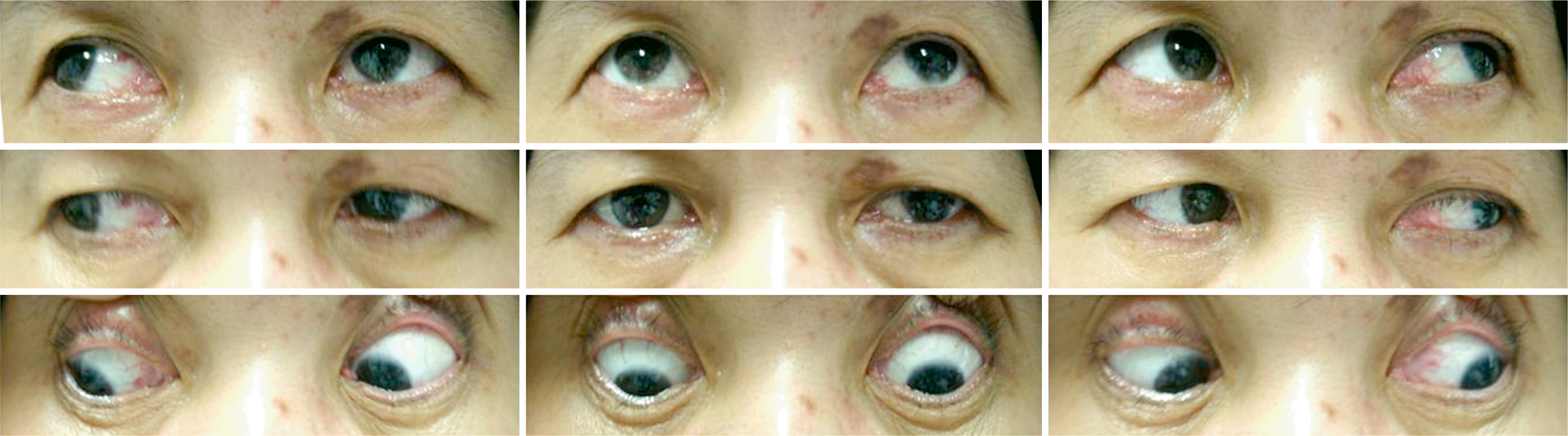
Figure 1.
Nine cardinal gaze positions of case 1. Limitation of adduction, elevation and depression were present in the right eye (the right eyelid was manually lifted due to ptosis).
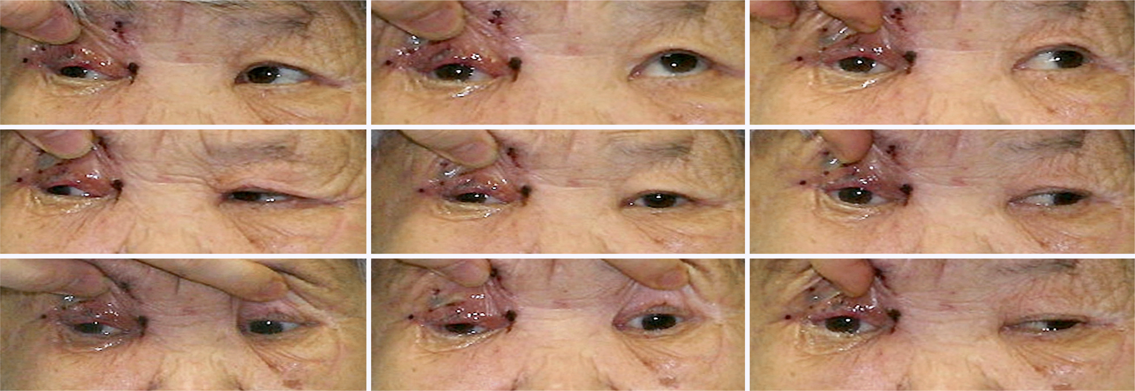
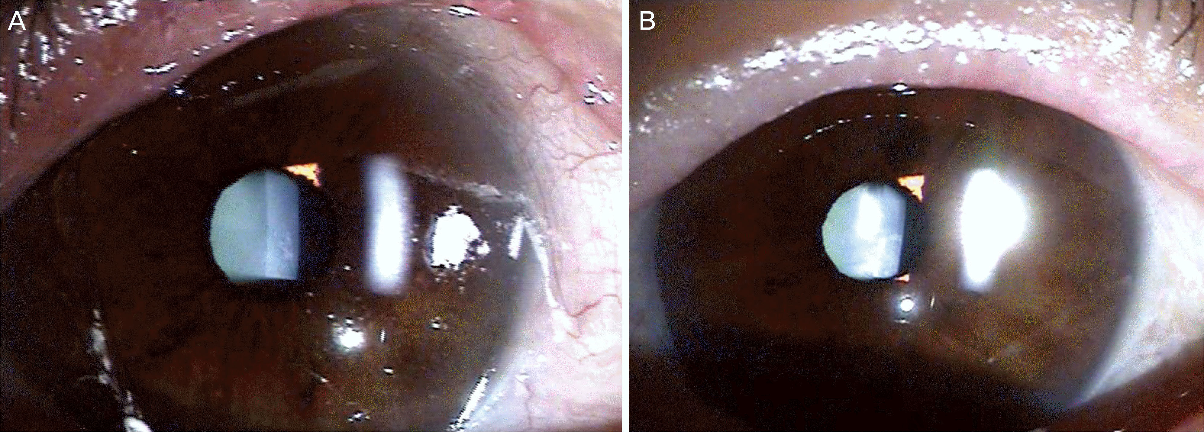
Figure 2.
Slit-lamp photographs of anisocoria. (A) Large ound pupil of the right eye. (B) Small round pupil of the left eye.

 XML Download