

 PDF
PDF ePub
ePub Citation
 Print
Print


Abstract
Case summary
A 59-year-old female with a history of epikeratophakic surgery 20 years ago complained of decreased visual acuity of both eyes for several months. She had nucleosclerotic and posterior subcapsular types of cataracts. Phacoemulsification and posterior capsule intraocular lens implantation were performed in both eyes. During surgery, corneal edema was especially prominent at the cornea with epikeratophakic lenticules in both eyes. In the left eye, severe corneal edema after one day of surgery was observed; however, after one week, corneal edema had subsided and visual acuity of both eyes had improved.
Conclusions
When it necessary that cataract surgery is performed in patients with epikeratophakic lenticules, it is important to anticipate the corneal edema intraoperatively and postoperatively. Moreover, the surgeon should consider the acute calculation of the target refraction of intraocular lens in an epikeratophakia patient.
REFERENCES
1). Kaufman HE. The correction of aphakia. XXXVI Edward Jackson Memorial Lecture. Am J Ophthalmol. 1980; 89:1–10.
2). Halliday BL. Epikeratophakia for aphakia, keratoconus, and myopia. Br J Ophthalmol. 1990; 74:67–72.

3). Kaufman HE, Werblin TP. Epikeratophakia for the treatment of keratoconus. Am J Ophthalmol. 1982; 93:342–7.
4). McDonald MB, Kaufman HE, Aquavella JV, et al. The nationwide study of epikeratophakia for aphakia in adults. Am J Ophthalmol. 1987; 103((3 Pt 2)):358–65.
5). Panda A, Gupta AK, Sharma N, et al. Anatomical and functional graft survival, 10 years after epikeratoplasty in keratoconus. Indian J Ophthalmol. 2013; 61:18–22.
6). Lee KW, Choi TH, Lee HB. The effects of epikeratoplasty & laser in situ keratomileusis(LASIK) for correction of high myopia. J Korean Ophthalmol Soc. 1998; 39:1707–15.
7). Labor PK, Ignacio T, Johnson M, Janku-Lestock L. Accommodating intraocular lens implantation in an epikeratophakia patient. J Cataract Refract Surg. 2010; 36:347–50.
8). Seitz B, Langenbucher A. Intraocular lens power calculation in eyes after corneal refractive surgery. J Refract Surg. 2000; 16:349–61.
9). Fan JC, Patel DV, McGhee CN. Long-term microstructural changes following epikeratophakia: in vivo confocal microscopy study. J Cataract Refract Surg. 2008; 34:1793–8.
10). Shin YJ, Park WC, Lee JH. Clear lens extraction and epikeratophakic lenticule removal in complicated epikeratophakic patients. J Korean Ophthalmol Soc. 2002; 43:1397–401.
11). Greenbaum A, Kaiserman I, Avni I. Long-term reversibility of epikeratophakia. Cornea. 2007; 26:1210–2.
Figure 1.
Pentacam® examination of both eyes before cataract surgery. (A) Right eye: Pentacam® shows a flattened curvature of the right cornea due to epikeratophakic lenticule. The location of epikeratophakic lenticule is slightly nasal to the center of the cornea and the keratometric value (true net power) at the center of the right cornea is 31.1 diopter. (B) Left eye: Pentacam® shows a flattened curvature of the left cornea due to epikeratophakic lenticule. The location of epikeratophakic lenticule is slightly nasal to the center of the cornea and the keratometric value (true net power) at the center of the left cornea is 36.2 diopter.
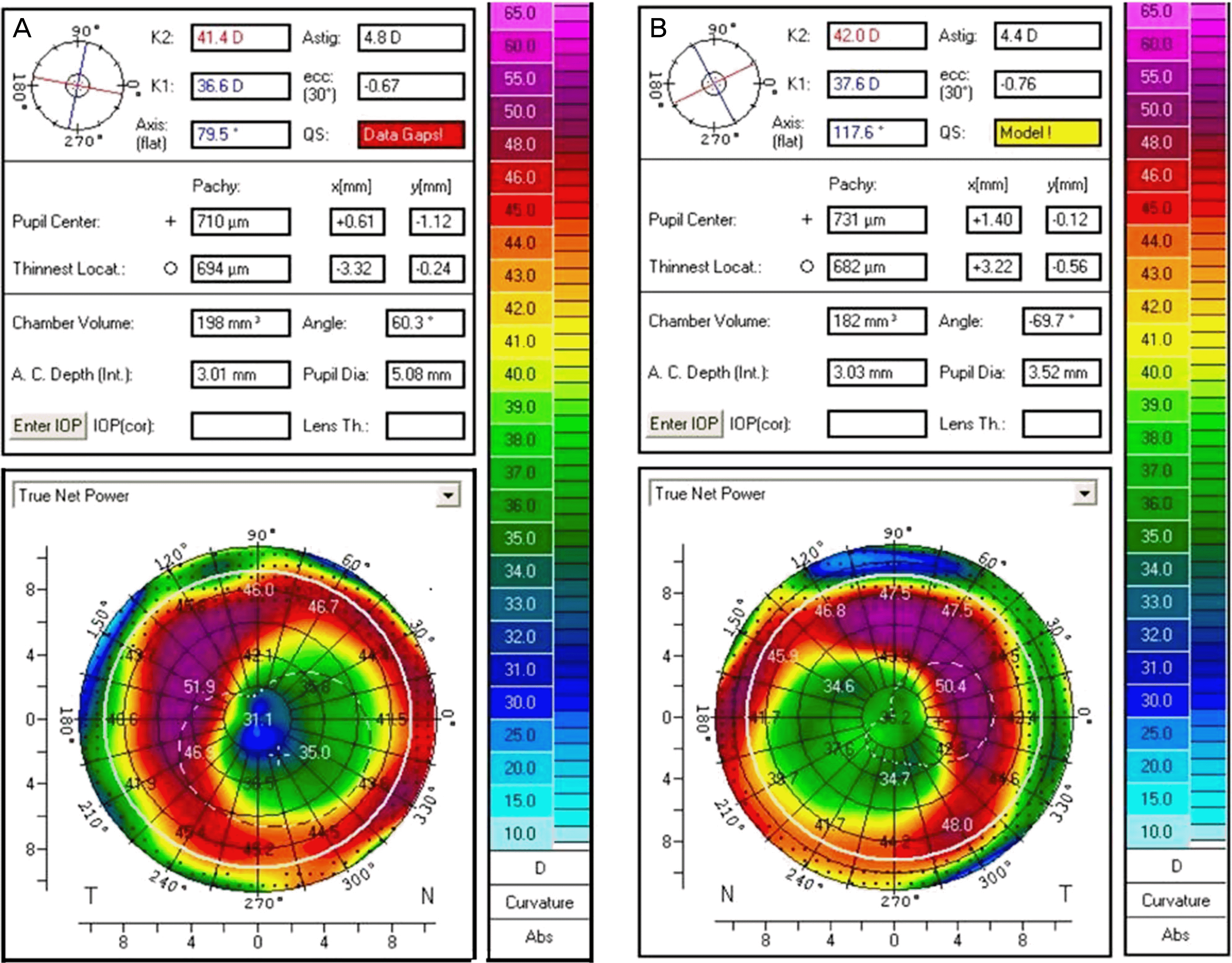
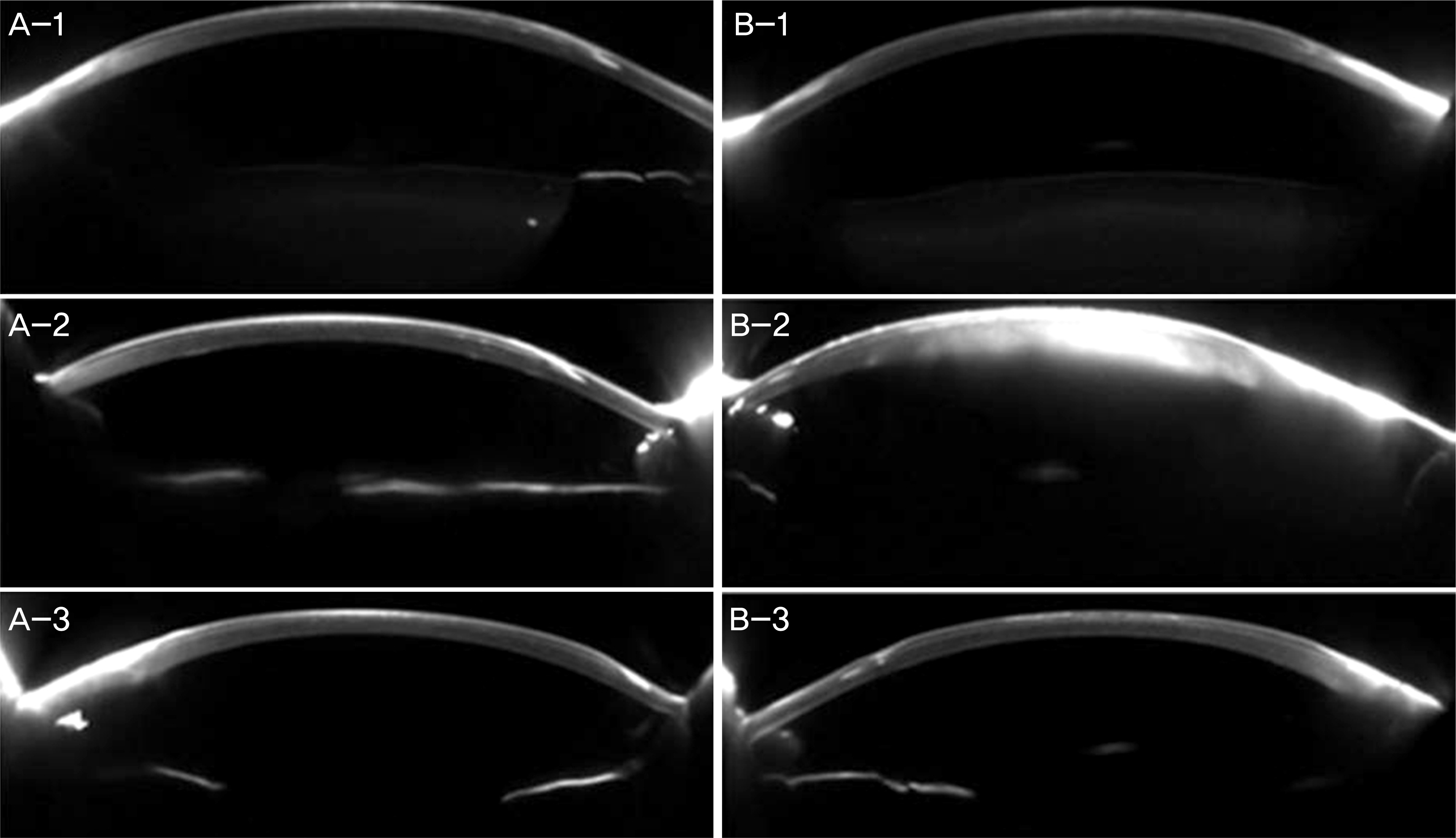
Figure 2.
Intraoperative microscopic view of the both eyes during cataract surgery. (A) Right eye. (B) Left eye. (A-1, B-1) Immediately before corneal incision: the white arrows indicate 12 o'clock position of the cornea at the intraoperative microscopic view. In each right and left eye, the epikeratophakic lenticule is located slightly nasal to the center of the cornea. (A-2, B-2) Immediately after hydrodissection: the margin of the epikeratophakic lenticule caused blurring of the image due to scattering of light, thus made it difficult to distinguish the capsulorrhexis margin before and after hydrodissection. (A-3, B-3) During phacoe-mulsificaiton: the corneal edema increased during phacoemulsification, in which the surgeon needed to decrease the illumination of the operating room to improve the microscopic view. (A-4, B-4) At the end of operation.

Figure 3.
Pentacam® Scheimpflug images of both eyes. (A) Right eye. (B) Left eye. (A-1, B-1) Preoperative images: the epikeratophakic lenticules are well attached to the corneal stroma in both eyes. (A-2, B-2) Postoperative 1 day: (A-2) In the right eye, there is mild corneal edema in both epikeratophakic lenticule and stromal bed. (B-2) In left eye, there was severe corneal edema in both epikeratophakic lenticule and stromal bed. (A-3, B-3) Postoperative 1 week: the corneal edema subsided in both eyes.
 XML Download