

 PDF
PDF ePub
ePub Citation
 Print
Print


Abstract
Purpose
The aim of this study is to analyze the influence of preoperative meibomian gland disease (MGD) on the postoperative dry eye disease after cataract surgery.
Methods
100 eyes of 85 patients who had undergone cataract surgery were enrolled. Patients were stratified into three groups by the severity of meibomian gland disease (MGD Grade I, MGD Grade II and MGD Grade III). In each group, we recorded the indexes of dry eye such as Ocular surface disease index, tear breakup time, Schirmer's test and Corneal staining at preoperatively and postoperative week 1, 2, 5, 9. We compared the indexes, preoperatively and postoperatively between three groups.
Results
In comparison of the preoperative indexes between groups, the more severe meibomian gland disease the patients have, the higher Ocular surface disease index and the shorter tear breakup time were observed (p < 0.05). In comparison the preoperative with the postoperative indexes, Ocular surface disease index was higher and tear breakup time was shorter at every postoperative moment in MGD Grade (Gr) II and Gr III. In comparison between MGD groups, MGD Gr III showed higher ocular surface disease index than MGD Gr I and Gr II at every follow up point, and shorter tear breakup time than MGD Gr I and Gr II at preoperative and postoperative week 1 and 9 (p < 0.05).
Conclusions
The severity of meibomian gland disease which exist preoperatively can affect the aggravation of dry eye disease after cataract surgery. Therefore, the treatment of meibomian gland disease before cataract surgery can be a therapeutic option to prevent aggravation of dry eye after cataract surgery.
Go to : 
REFERENCES
1). Roh YR, Lee SM, Han YK, et al. Changes in clinical manifestations of dry eye syndrome after cataract surgery and the affecting factors. J Korean Ophthalmol Soc. 2011; 52:1030–8.

2). Kohlhaas M. Corneal sensation after cataract and refractive surgery. J Cataract Refract Surg. 1998; 24:1399–409.
3). Cho YK, Kim MS. Dry eye after cataract surgery and associated intraoperative risk factors. Korean J Ophthalmol. 2009; 23:65–73.
4). Ram J, Gupta A, Brar G, et al. Outcomes of phacoemulsification in patients with dry eye. J Cataract Refract Surg. 2002; 28:1386–9.
5). Roberts CW, Elie ER. Dry eye symptoms following cataract surgery. Insight. 2007; 32:14–21. quiz 22-3.
6). Hykin PG, Bron AJ. Age-related morphological changes in lid margin and meibomian gland anatomy. Cornea. 1992; 11:334–42.
7). Mathers WD, Shields WJ, Sachdev MS, et al. Meibomian gland dysfunction in chronic blepharitis. Cornea. 1991; 10:277–85.
8). Schiffman RM, Christianson MD, Jacobsen G, et al. Reliability and validity of the Ocular Surface Disease Index. Arch Ophthalmol. 2000; 118:615–21.
9). Bron AJ, Evans VE, Smith JA. Grading of corneal and conjunctival staining in the context of other dry eye tests. Cornea. 2003; 22:640–50.
10). Cho JH, Ahn Y. Assessment of meibomian gland dysfunction and comparison of the results of BUT and Schirmer test according to meibomian gland state. J Korean Ophthalmol Soc. 2000; 41:1875–82.
11). Foulks GN. The correlation between the tear film lipid layer and dry eye disease. Survey Ophthalmol. 2007; 52:369–74.
12). Horwath-Winter J, Vidic B, Schwantzer G, Schmut O. Early changes in corneal sensation, ocular surface integrity, and tear-film function after laser-assisted subepithelial keratectomy. J Cataract Refract Surg. 2004; 30:2316–21.
13). Li XM, Hu L, Hu J, Wang W. Investigation of dry eye disease and analysis of the pathogenic factors in patients after cataract surgery. Cornea. 2007; 26((9 Suppl 1)):S16–20.
14). Ang RT, Dartt DA, Tsubota K. Dry eye after refractive surgery. Curr Opin Ophthalmol. 2001; 12:318–22.
15). Belmonte C. Eye dryness sensations after refractive surgery: impaired tear secretion or "phantom" cornea? J Refract Surg. 2007; 23:598–602.
16). Xu KP, Yagi Y, Tsubota K. Decrease in corneal sensitivity and change in tear function in dry eye. Cornea. 1996; 15:235–9.
17). Hiroko BM. Cataract Surgery in the presence of other ocular comorbidities. Steinert RF, editor. Cataract surgery: technique, complications, and management. 2nd ed.Philadelphia: WB Saunders;2004. chap. 32.
18). Walker TD. Benzalkonium toxicity. Clin Experiment Ophthalmol. 2004; 32:657.
19). Bron AJ, Tiffany JM. The contribution of meibomian disease to dry eye. Ocul Surf. 2004; 2:149–65.
20). Foulks GN. Blepharitis: lid margin disease and the ocular surface. Holland EJ, Mannis MJ, editors. Ocular Surface Disease: Medical and Surgical Management. 1st ed.New York: Springer;2002. chap. 3.
21). Foulks GN, Bron AJ. Meibomian gland dysfunction: a clinical scheme for description, diagnosis, classification, and grading. Ocul Surf. 2003; 1:107–26.
22). Goto E, Endo K, Suzuki A, et al. Tear evaporation dynamics in normal subjects and subjects with obstructive meibomian gland dysfunction. Invest Ophthalmol Vis Sci. 2003; 44:533–9.
23). Perry HD, Donnenfeld ED. Dry eye diagnosis and management in 2004. Curr Opin Ophthalmol. 2004; 15:299–304.
24). Smith J, Nichols KK, Baldwin EK. Current patterns in the use of diagnostic tests in dry eye evaluation. Cornea. 2008; 27:656–62.
25). Nichols KK, Mitchell GL, Zadnik K. The repeatability of clinical measurements of dry eye. Cornea. 2004; 23:272–85.
26). Nichols KK, Nichols JJ, Mitchell GL. The lack of association between signs and symptoms in patients with dry eye disease. Cornea. 2004; 23:762–70.
27). Nichols KK. Patient-reported symptoms in dry dye disease. Ocul Surf. 2006; 4:137–45.
Go to :
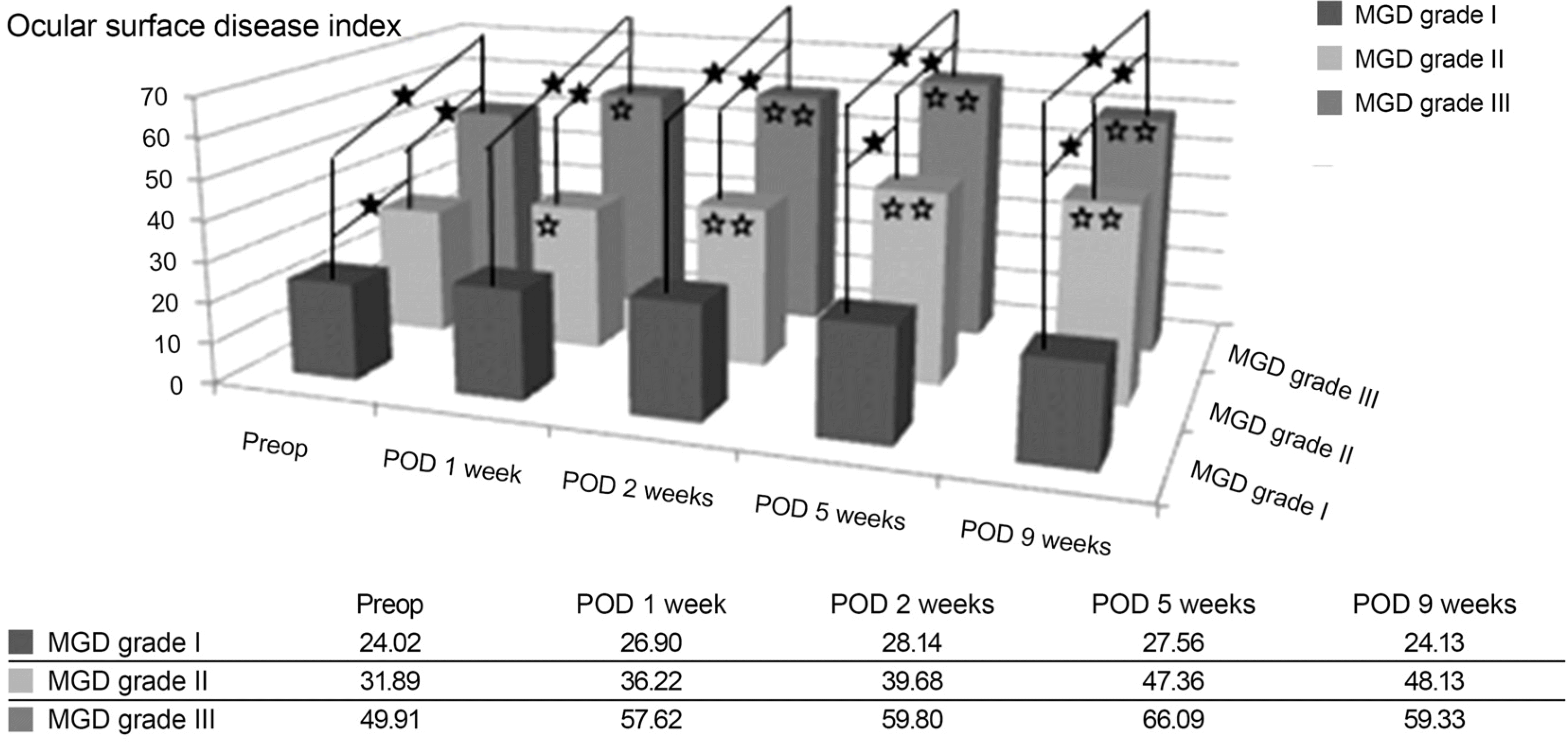 | Figure 1.Changes of Ocular surface disease index stratified by Meibomian gland disease severity (Wilcoxon signed rank test, one white star symbol [p < 0.05] and two white star symbols [p < 0.01] inside the bar means the statistical p-value compared with pre-operative Ocular surface disease index; Kruskal Wallis test & Mann Whitney test, one black star symbol [p < 0.05] means the statistical p-value compared between the Ocular surface disease index of two groups stratified by MGD severity at the same point). Preop = preoperative; POD = postoperative day; MGD = meibomian gland disease. |
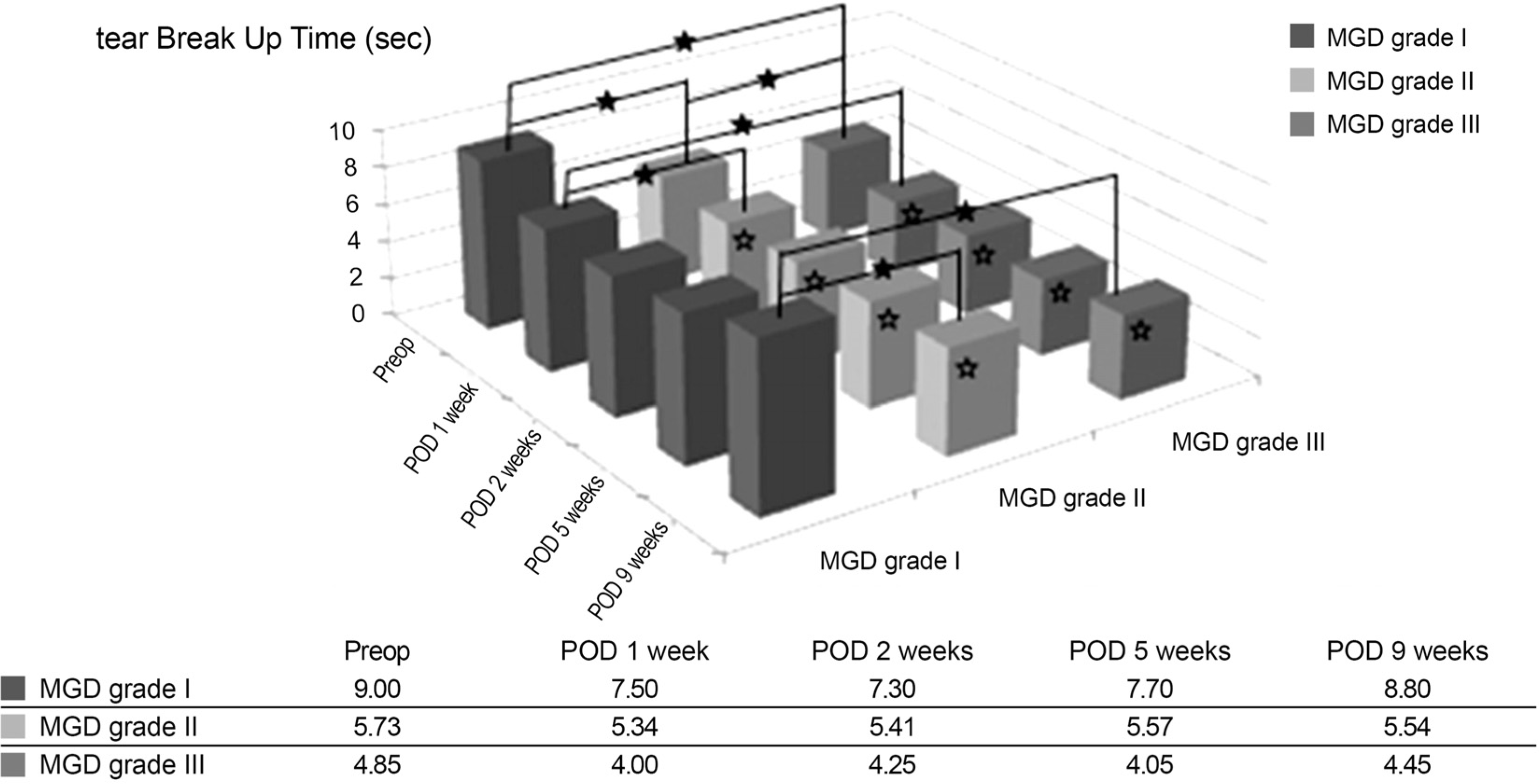 | Figure 2.Changes of tear breakup time stratified by Meibomian gland disease severity (Wilcoxon signed rank test, one white star symbol [p < 0.05] inside the bar means the statistical p-value compared with preoperative tear Break Up Time; Kruskal Wallis test & Mann Whitney test, one black star symbol [p < 0.05] means the statistical p-value compared between the tear Breakup time of two groups stratified by MGD severity at the same point). Preop = preoperative; POD = postoperative day; MGD = meibomian gland disease. |
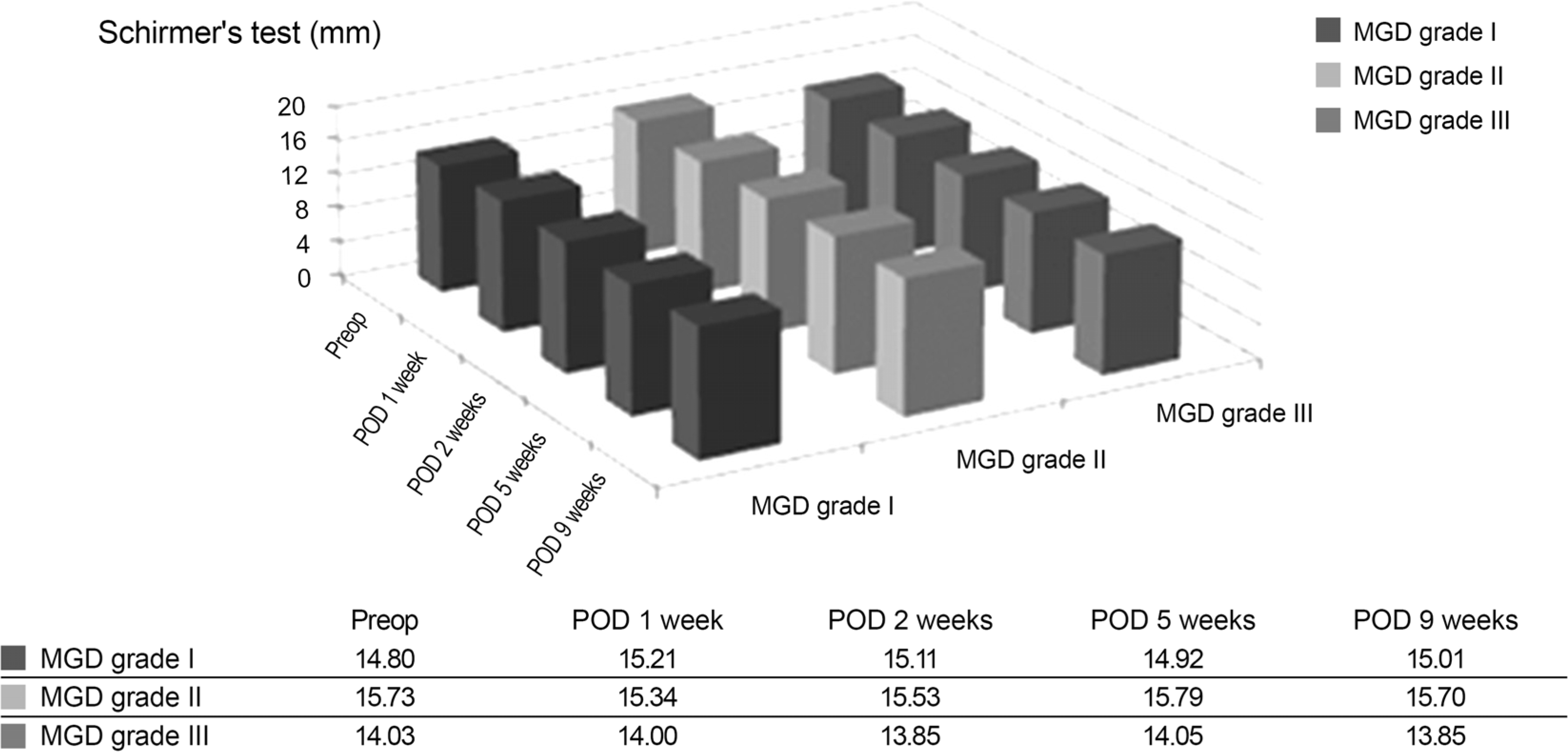 | Figure 3.Changes of Schirmer's test stratified by Meibomian gland disease severity (Wilcoxon signed rank test, compared with pre-operative Schirmer's test score; Kruskal Wallis test & Mann Whitney test, compared between the Schirmer's test score of two groups stratified by MGD severity at the same point). Preop = preoperative; POD = postoperative day; MGD = meibomian gland disease. |
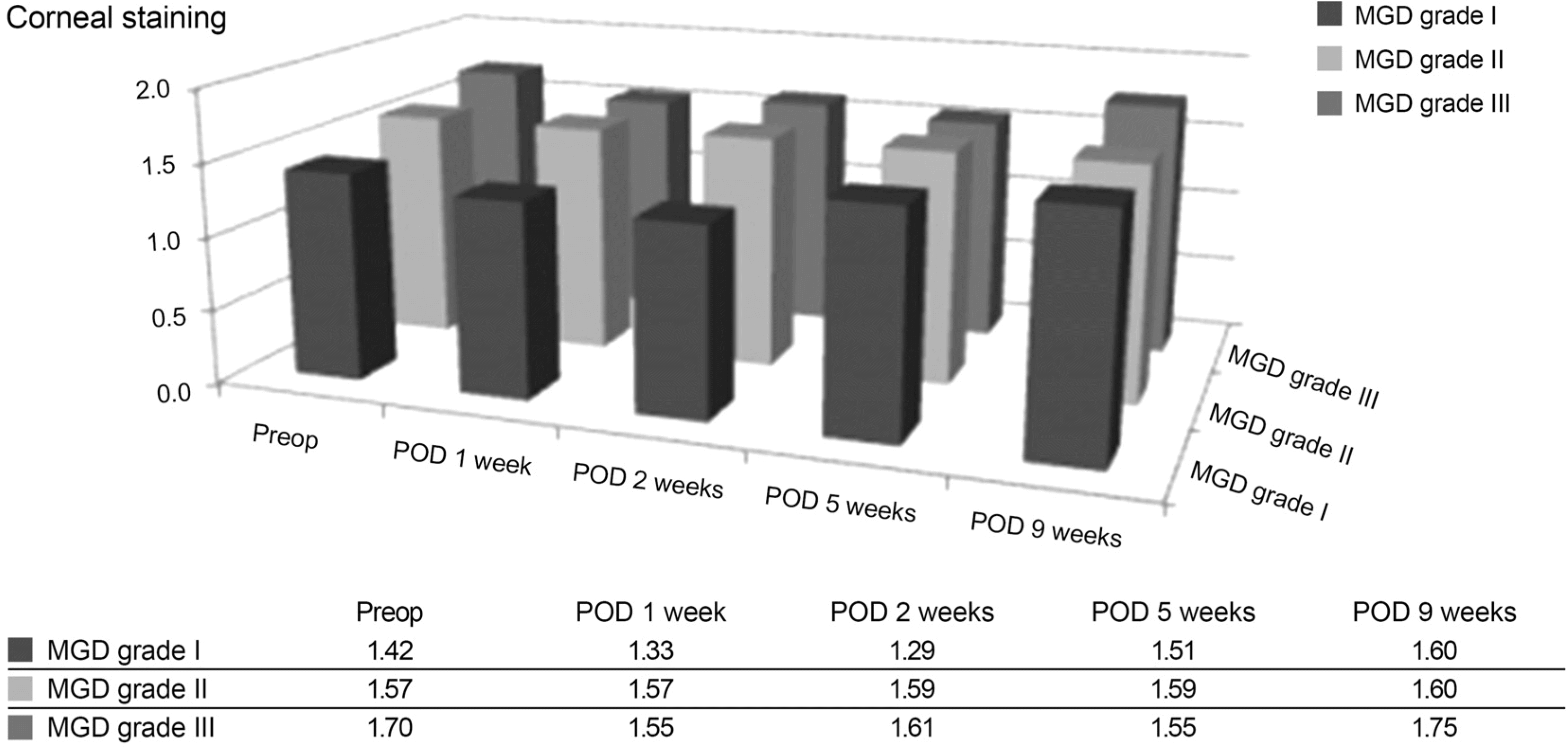 | Figure 4.Changes of corneal staining stratified by Meibomian gland disease severity (Wilcoxon signed rank test, compared with preoperative Corneal staining score; Kruskal Wallis test & Mann Whitney test, compared between the Corneal staining score of two groups stratified by MGD severity at the same point). Preop = preoperative; POD = postoperative day; MGD = meibomian gland disease. |
Table 1.
Demographics of study group
Table 2.
Preoperative characteristics of groups stratified by Meibomian gland disease severity
| No. | Age (years) | OSDI | tBUT (sec) | Schirmer's test (mm) | Corneal staining | |
|---|---|---|---|---|---|---|
| MGD Gr I | 11 | 58.40 ± 13.43 | 24.02 ± 9.43 | 9.00 ± 1.89 | 14.80 ± 8.70 | 1.40 ± 0.70 |
| MGD Gr II | 68 | 68.29 ± 10.13 | 31.89 ± 18.61 | 5.72 ± 1.74 | 15.73 ± 6.97 | 1.57 ± 0.67 |
| MGD Gr III | 21 | 66.90 ± 12.61 | 49.94 ± 14.19 | 4.85 ± 0.88 | 14.02 ± 6.62 | 1.70 ± 0.57 |
| p-value* | >0.05 | <0.01 | <0.01 | >0.05 | >0.05 |
Table 3.
Changes of MGD, OSDI, tBUT, Schirmer's test, Corneal staining
| Preop | Postop 1 week | Postop 2 weeks | Postop 5 weeks | Postop 9 weeks | |
|---|---|---|---|---|---|
| MGD | 2.10 ± 0.54 | 2.19 ± 0.58 | 2.34 ± 0.54 | 2.24 ± 0.59 | 2.18 ± 0.59 |
| p-value* | >0.05 | <0.01 | 0.01 | 0.06 | |
| OSDI | 34.71 ± 18.76 | 39.37 ± 20.51 | 42.30 ± 21.01 | 48.70 ± 25.27 | 47.85 ± 23.48 |
| p-value* | <0.01 | <0.01 | <0.01 | <0.01 | |
| tBUT (sec) | 5.88 ± 1.95 | 5.18 ± 1.88 | 5.33 ± 1.97 | 5.53 ± 1.83 | 5.51 ± 2.04 |
| p-value* | <0.01 | <0.01 | <0.01 | <0.01 | |
| Schirmer's test (mm) | 15.30 ± 7.05 | 15.06 ± 7.32 | 15.15 ± 7.24 | 15.31 ± 7.21 | 15.26 ± 7.06 |
| p-value* | >0.05 | >0.05 | >0.05 | >0.05 | |
| Corneal staining | 1.58 ± 0.65 | 1.54 ± 0.74 | 1.55 ± 0.74 | 1.57 ± 0.74 | 1.62 ± 0.75 |
| p-value* | >0.05 | >0.05 | >0.05 | >0.05 |
 XML Download