Abstract
Purpose
In this study, a case of toxic encephalopathy and optic neuropathy due to methyl bromide poisoning is reported.
Case summary
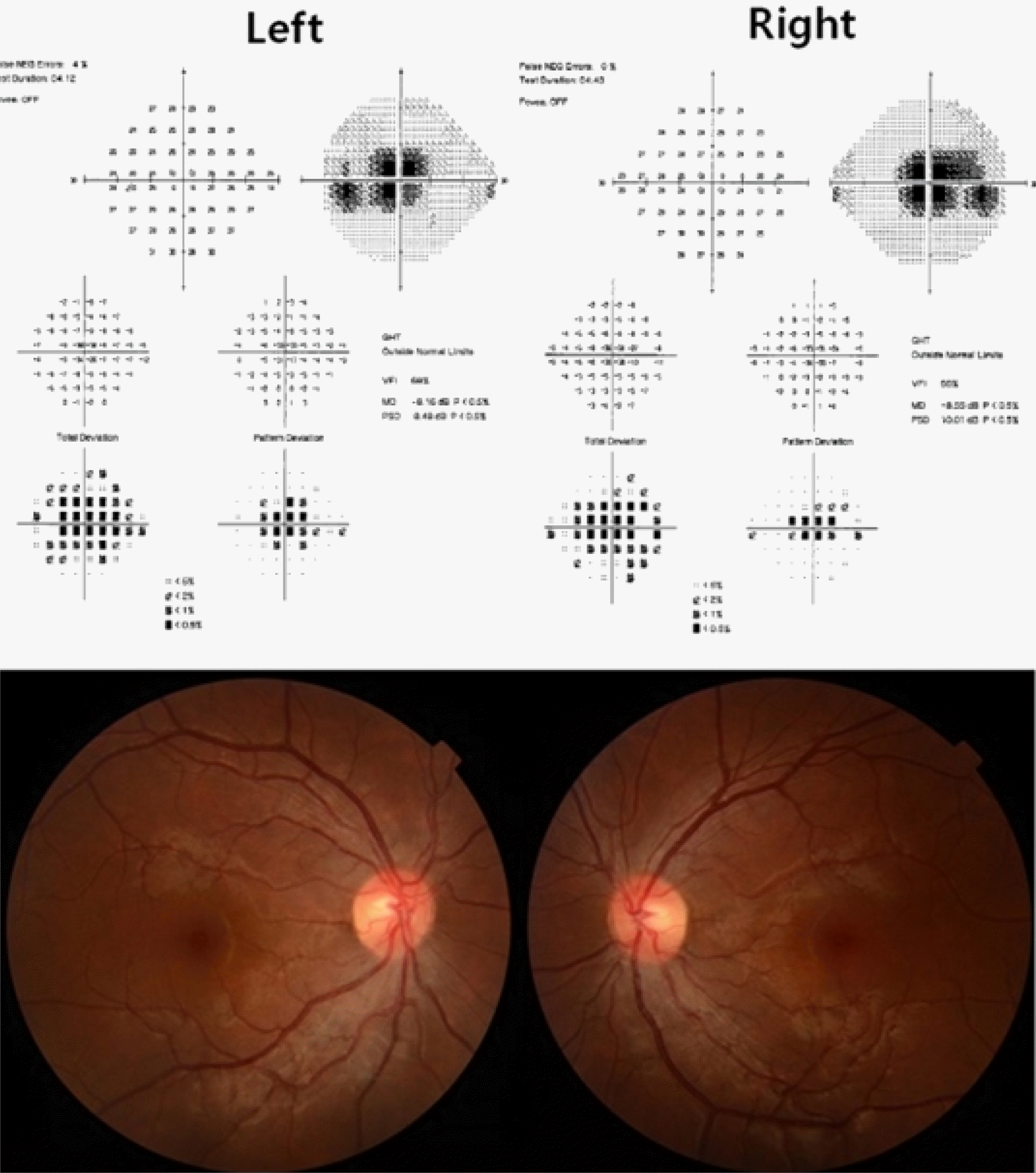
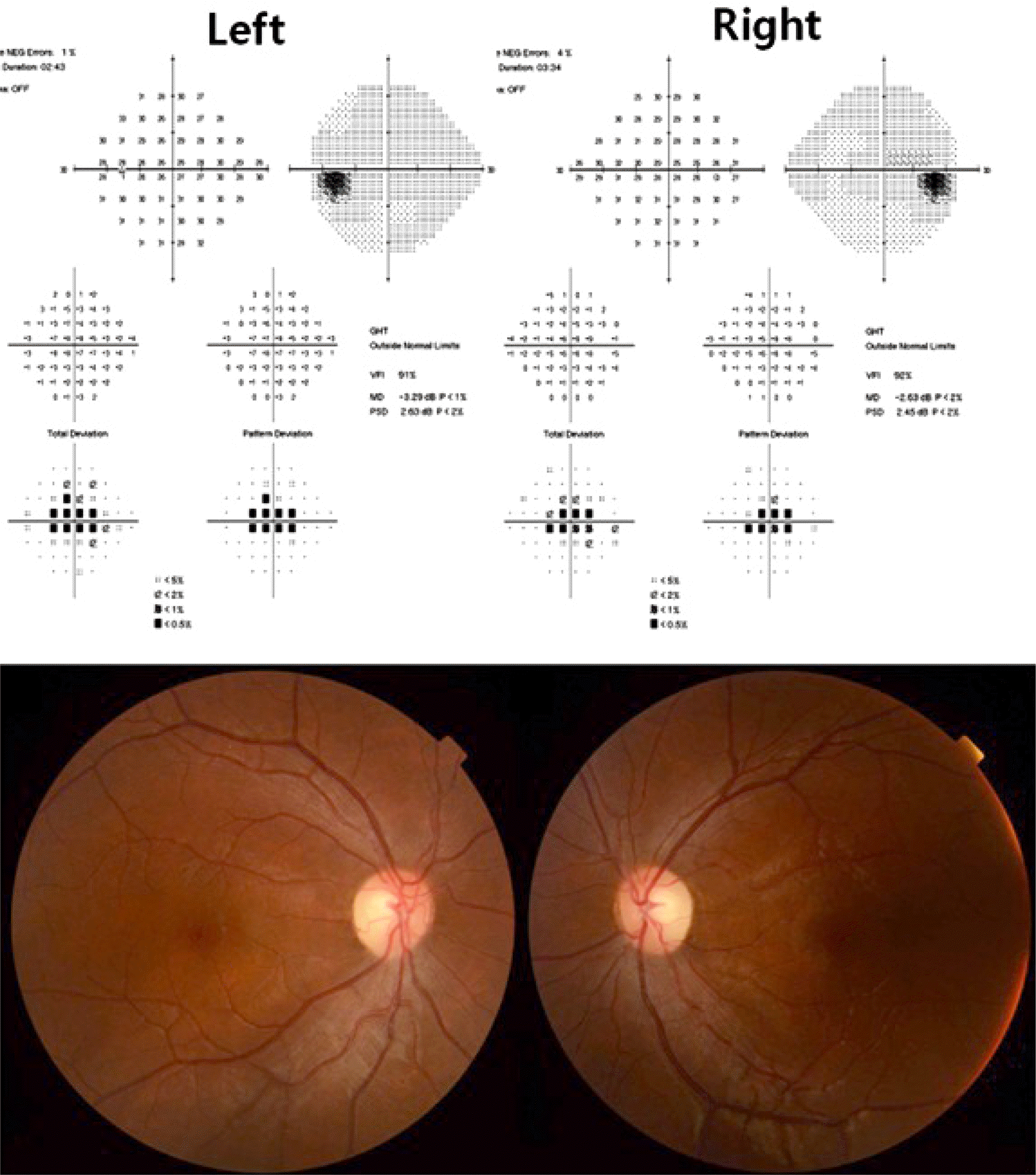
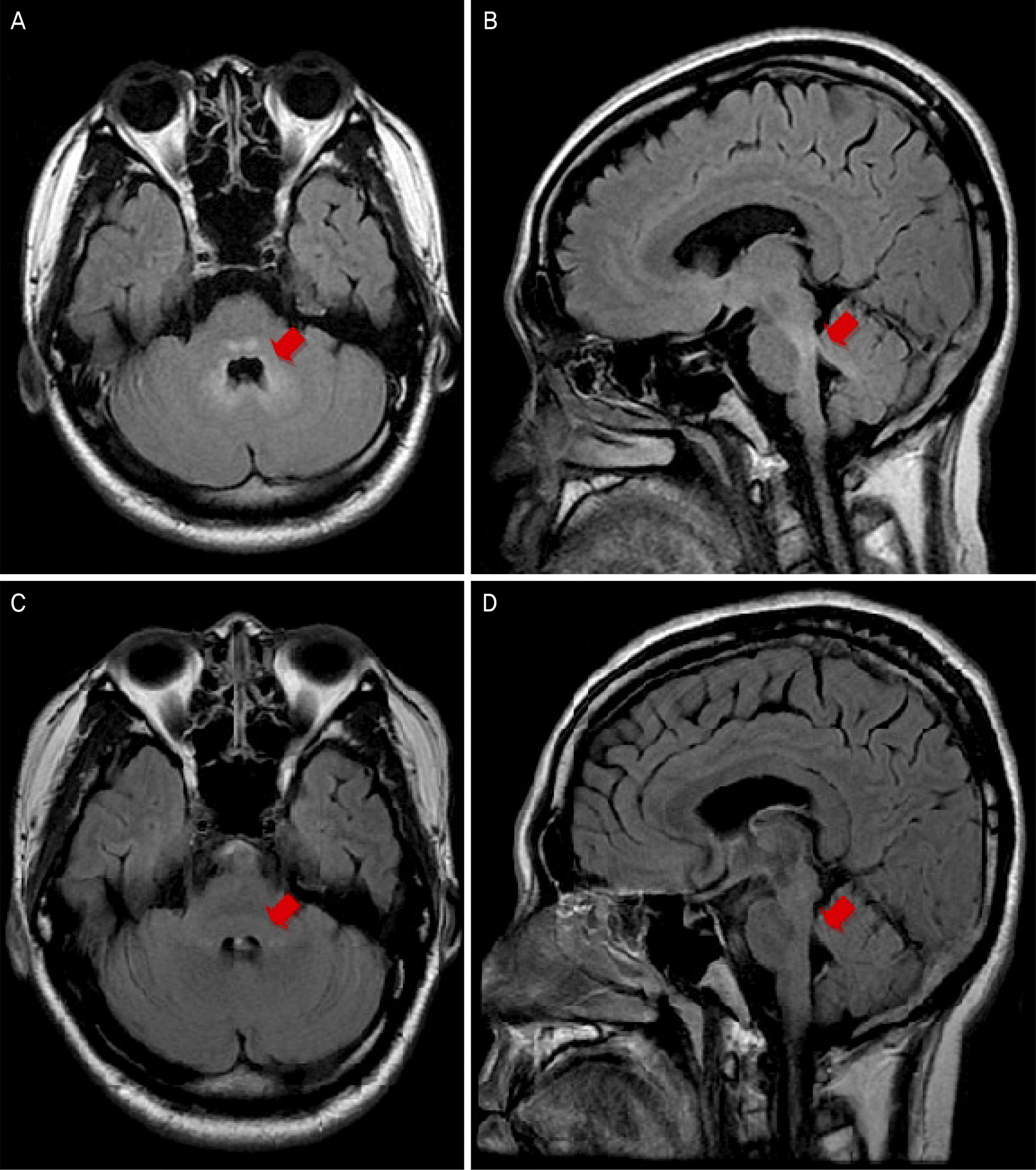
A 31-year-old male presented with dysarthria, gait disturbance and bilateral visual impairment. He was treated with intravenous methylprednisolone for bilateral optic neuritis 1 year prior. He previously worked in a fumigation warehouse and was exposed to methyl bromide in the past 3 years. His corrected visual acuity was 20/30 in both eyes. The patient had reduced color vision and enlarged central scotoma in both eyes. His mentality was alert but exhibited slow response, ataxia and dysarthria. Brain magnetic resonance imaging (MRI) revealed high signals in the brainstem, cerebellum and midbrain. His serum and urine methyl bromide concentrations were significantly elevated. The patient was treated with intravenous methylprednisolone 1.0 g/day for 5 days. MRI showed resolution of the multiple brain lesions observed previously. Ten days after steroid therapy, his visual acuity was 20/20 in both eyes and his neurologic manifestations were completely recovered at 2 months after treatment.
Go to : 
References
1. Bulathsinghala AT, Shaw IC. The toxic chemistry of methyl bromide. Hum Exp Toxicol. 2014; 33:81–91.

2. De Souza A, Narvencar KP, Sindhoora KV. The neurological abdominals of methyl bromide intoxication. J Neurol Sci. 2013; 335:36–41.
3. De Haro L, Gastaut JL, Jouglard J, Renacco E. Central and abdominal neurotoxic effects of chronic methyl bromide intoxication. J Toxicol Clin Toxicol. 1997; 35:29–34.
4. Park TH, Kim JI, Son JE, et al. Two cases of neuropathy by methyl bromide intoxication during fumigation. Korean J Occup Environ Med. 2000; 12:547–53.

5. Geyer HL, Schaumburg HH, Herskovitz S. Methyl bromide abdominal causes reversible symmetric brainstem and cerebellar MRI lesions. Neurology. 2005; 64:1279–81.
6. Chavez CT, Hepler RS, Straatsma BR. Methyl bromide optic atrophy. Am J Ophthalmol. 1985; 99:715–9.

7. Choi KD, Shin JH, Kim DS, et al. A case of chronic methyl abdominal poisoning associated with cerebellar ataxia, polyneuropathy and optic neuropathy. J Korean Neurol Assoc. 2002; 20:307–10.
Go to : 
 | Figure 1.Visual field and fundus photography at initial attack. The patient presented with the central scotoma and mild optic disc swelling in both eyes at the initial attack. |
 | Figure 2.Visual field and fundus photography at second attack. The patient presented with mild central scotoma and the optic disc was temporally pallor at the second attack. |
 | Figure 3.Magnetic resonance images. Fluid-attenuated inversion recovery images for a patient showing high signal changes in peri-aqueductal gray matter of midbrain (arrow) (A), the pons, the medulla, and cerebellum (arrow) (B). Ten days after treatment, the brain lesions previously observed was rapidly disappeared (arrows) (C, D). |
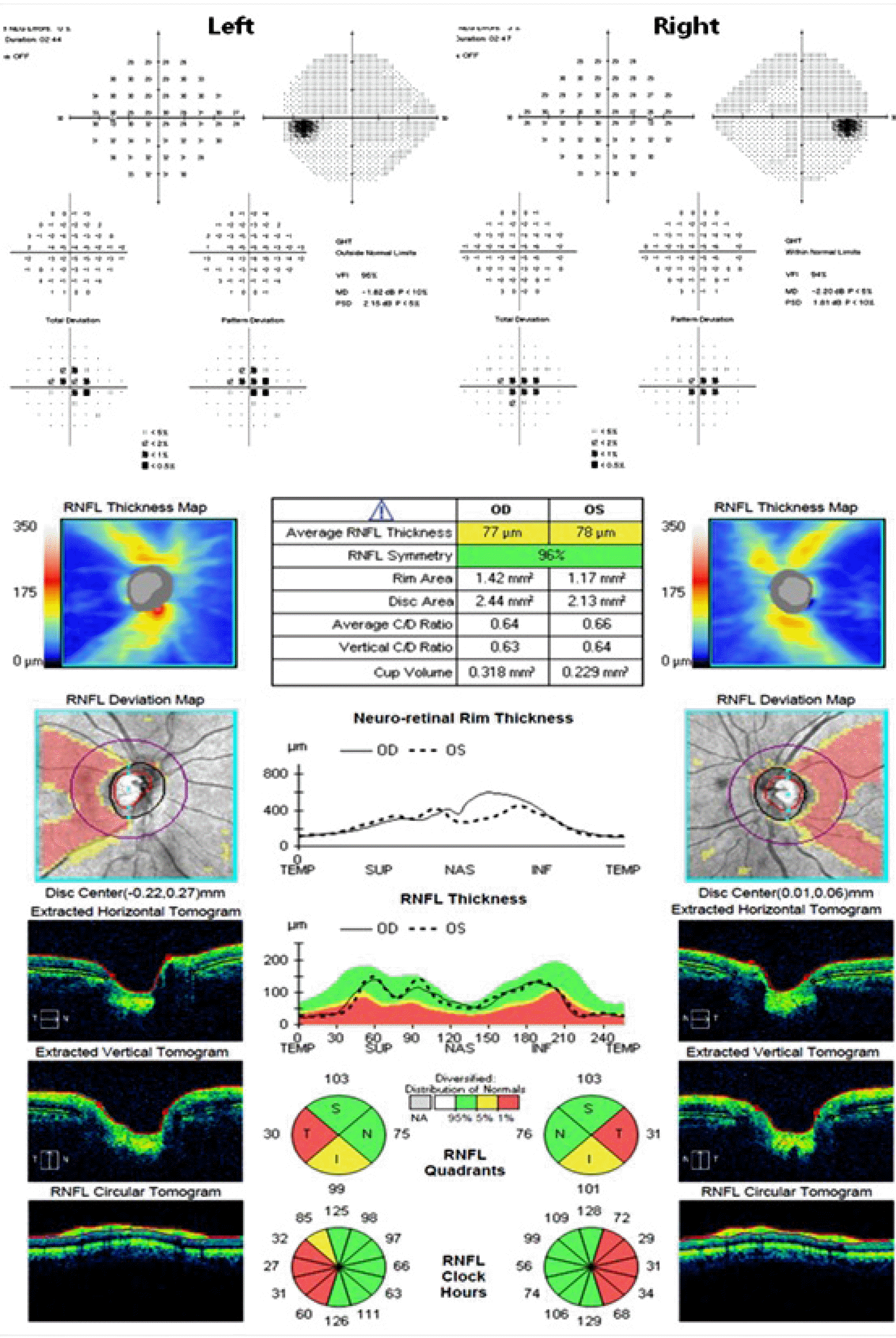
 | Figure 4.Visual field and disc optical coherence tomography at one and one-half years after treatment. One and one-half years after treatment, the patient was presented with mild central scotoma and decreased retinal nerve fiber thickness of temporal area in optic coherence tomography. RNFL = retinal nerve fiber layer; OD = oculus dexter; OS = oculus sinister; C/D = cup/disc; TEMP = temporal; SUP = superior; NAS = nasal; INF = inferior; S = superior; N = nasal; I = inferior; T = temporal. |


 PDF
PDF ePub
ePub Citation
 Print
Print


 XML Download