

 PDF
PDF ePub
ePub Citation
 Print
Print


Abstract
Purpose
In the present study, the prognosis of ocular injury caused by a wasp sting was evaluated in two cases: Case 1 was treated by anterior chamber irrigation and Case 2 was simultaneously treated by anterior chamber irrigation and vitrectomy.
Case summary
Both patients had unilateral damage and complained of severe eye pain and blurred vision. Severe corneal edema, conjunctival injection, marked anterior chamber inflammatory reaction and the wasp sting through the cornea at the anterior chamber were observed in both cases. In Case 1, anterior chamber irrigation was performed, however, corneal edema was not recovered. Six months after the wasp sting, phthisis was observed. In Case 2, anterior chamber irrigation and vitrectomy were simultaneously performed, corneal edema decreased and epithelial healing occurred. Four months after the wasp sting, the eyeball was stable, but there was no wave on the electroretinogram.
Go to : 
References
1. Vetter RS, Visscher PK, Camazine S. Mass envenomations by honey bees and wasps. West J Med. 1999; 170:223–7.
2. Park JK, Chang KC. Corneal endothelial changes induced by abdominal bee sting injury. J Korean Ophthalmol Soc. 2010; 51:435–9.
3. Habermann E. Bee and wasp venoms. Science. 1972; 177:314–22.

4. Gilboa M, Gdal-On M, Zonis S. Bee and wasp stings of the eye. Retained intralenticular wasp sting: a case report. Br J Ophthalmol. 1977; 61:662–4.
5. Arcieri ES, França ET, Oliveria HB, et al. Ocular lesions arising abdominal stings by hymenopteran insects. Cornea. 2002; 21:328–30.
6. Teoh SC, Lee JJ, Fam HB. Corneal honeybee sting. Can J Ophthalmol. 2005; 40:469–71.
7. Chen CJ, Richardson CD. Bee sting-induced ocular changes. Ann Ophthalmol. 1986; 18:285–6.
8. Choe GY, Chi KU. Wasp sting on the eyeball. J Korean Ophthalmol Soc. 1968; 9:39–42.
9. Li Z, Oh HJ, Ji Y, Yoon KC. Wasp sting of the cornea: a case treated with amniotic membrane transplantation. Graefes Arch Clin Exp Ophthalmol. 2013; 251:1039–40.
10. Lai P, Yang J, Cui H, Xie H. Prognosis of corneal wasp sting: case report and review of the literature. Cutan Ocul Toxicol. 2011; 30:325–7.
11. Kim JM, Kang SJ, Kim MK, et al. Corneal wasp sting abdominal by optic neuropathy and retinopathy. Jpn J Ophthalmol. 2011; 55:165–7.
12. Lin PH, Wang NK, Hwang YS, et al. Bee Sting of the cornea and conjunctiva: management and outcomes. Cornea. 2011; 30:392–4.
13. King TP, Spangfort MD. Structure and biology of stinging insect venom allergens. Int Arch Allergy Immunol. 2000; 123:99–106.
14. Smolin G, Wong I. Bee sting of the cornea: case report. Ann Ophthalmol. 1982; 14:342–3.
15. George P, Pawar B, Calton N, Mathew P. Wasp sting: an unusual abdominal outcome. Saudi J Kidney Dis Transpl. 2008; 19:969–72.
16. Haspel G, Libersat F. Wasp venom blocks central cholinergic syn-apses to induce transient paralysis in cockroach prey. J Neurobiol. 2003; 54:628–37.
17. Nakatani Y, Nishimura A, Sugiyama K. Successful treatment of corneal wasp sting-induced panuveitis with vitrectomy. J Ophthalmic Inflamm Infect. 2013; 3:18.
18. Yildirim N, Erol N, Basmak H. Bee sting of the cornea: a case report. Cornea. 1998; 17:333–4.
19. Al-Towerki AE. Corneal honeybee sting. Cornea. 2003; 22:672–4.
20. Razmjoo H, Abtahi MA, Roomizadeh P, et al. Management of abdominal bee sting. Clin Ophthalmol. 2011; 5:1697–700.
21. Kitagawa K, Hayasaka S, Setogawa T. Wasp sting-induced retinal damage. Ann Ophthalmol. 1993; 25:157–8.
Go to :
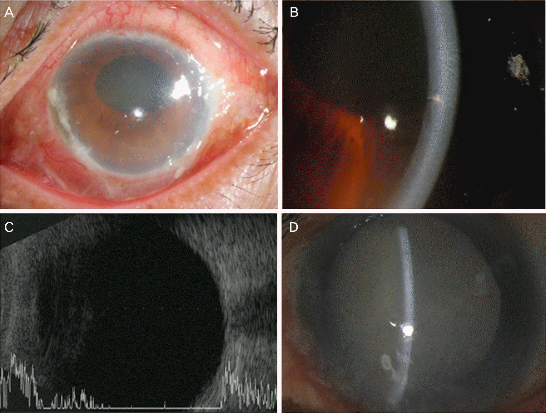 | Figure 1.Slit-lamp biomicroscopic photograph and vertical ultrasonography scan images of case 1. (A) Mucopurulent keratoconjunctivitis with chemosis and conjunctival injection was detected. (B) Severe anterior chamber reaction and a retained wasp stinger at the 6 o'clock position. (C) Ultrasonography scan image showed no significant abnormality. (D) Cornea edema and anterior chamber collapse with cataract was detected 1 month after injury. |
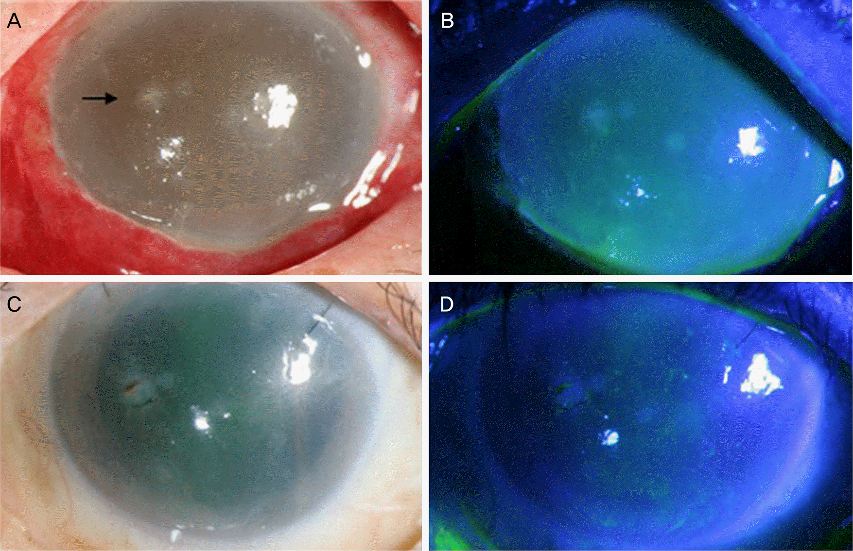 | Figure 2.Slit-lamp biomicroscopic findings of case 2. (A, B) Initial presentation showed severe conjunctival chemosis, conjunctival injection, and total epithelial defect with severe corneal edema. Penentrating site of the wasp sting at 9 o'clock at the mid-periphery of the cornea (arrow). (C, D) Decreased corneal edema, corneal re-epithelization, and improvement of anterior chamber inflammation 1 month after initial visit. |
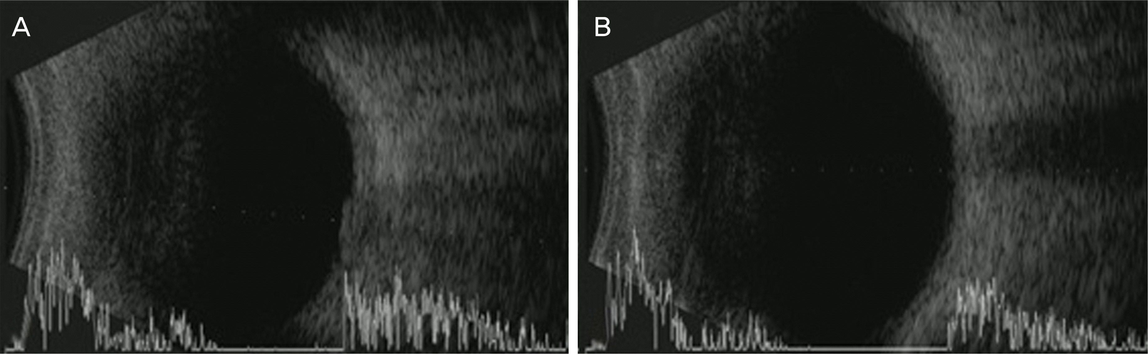 | Figure 3.Vertical ultrasonography scan images of case 2. (A) Ultrasonography scan image showed choroidal edema at initial presentation. (B) Improved choroidal edema 1 month after injury. |
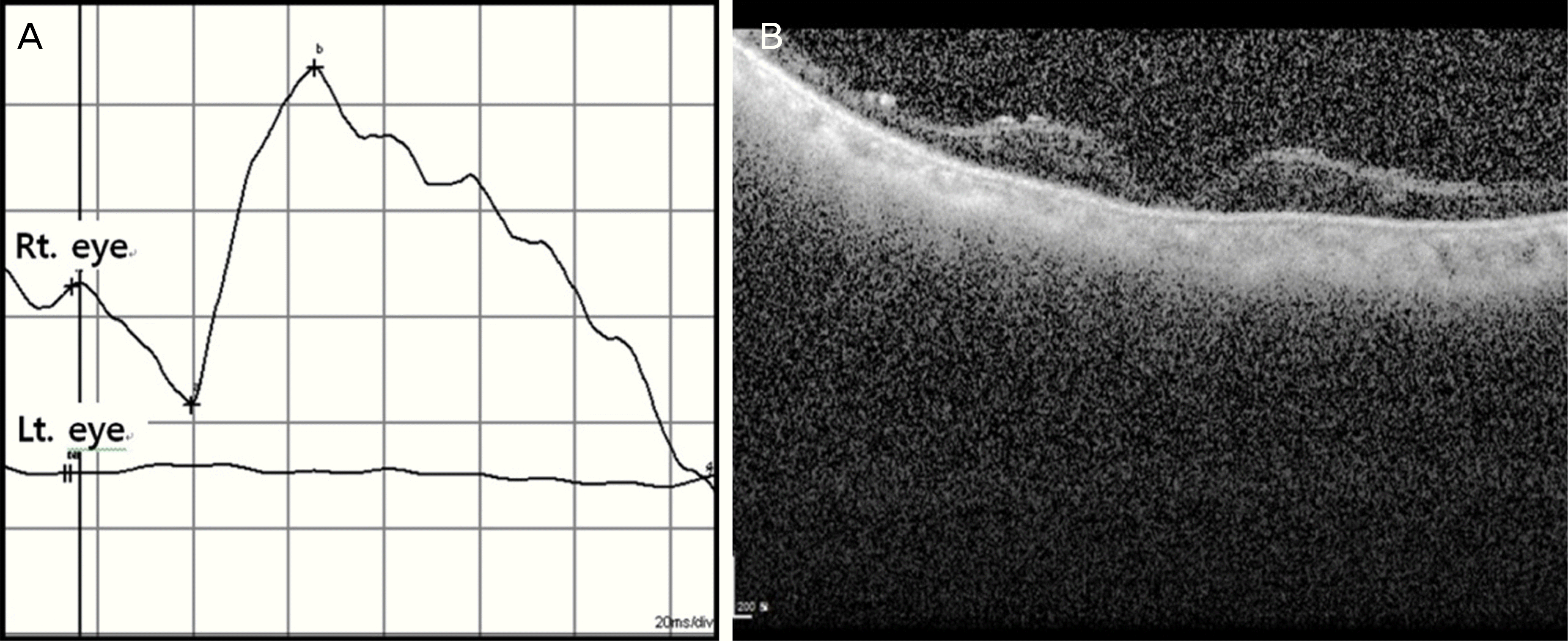 | Figure 4.Examination from 4 months after injury in case 2. (A) The electroretinogram was normal on the right eye, but absent on the left eye. (B) Optical coherence tomography image from 4 months after injury demonstrating vitreous haze and atropic retina. Rt. = right; Lt. = left. |
Table 1.
Systemic review of corneal Bee and Wasp sting
| Species | Authors | Initial findings* | 1st visit | Initial VA | Surgical Treatment | Follow-up VA |
|---|---|---|---|---|---|---|
| Bee | ||||||
| Chen et al7 (1986) | Hyphema | 3 hr | – | Lentectomy | 20/30 | |
| Lens dislocation | Anterior vitrectomy | |||||
| Yildirim et al18 (1998) | Iris atropy | <1 hr | 7/10 | A/C irrigation | 10/10 | |
| Al-Towerki19 (2003) | Mild A/C reaction | 3 days | 2/200 | None | 20/60 | |
| Teoh et al6 (2005) | Mild A/C reaction | 12 hrs | Hand motion | Unsuccessful stinger remova | al Counting fingers | |
| RAPD (−)/RAPD (+)† | ||||||
| Razmjoo et al20 (2011) | – | 20 hrs | 160/200 | None | 180/200 | |
| Wasp | ||||||
| Kitagawa et al21 (1993) | Increased IOP | 1 hr | Hand motion | None | Light perception | |
| Severe media opacity in A/C | ||||||
| Lai et al10 (2011) | Severe media opacity in A/C | 7 days | Light perception | None | Light perception | |
| RAPD(+) | ||||||
| Kim et al11 (2011) | Increased IOP | 4 days | Hand motion | A/C irrigation | Phthisis | |
| Severe media opacity in A/C | AMT | |||||
| Nakatani et al17 (2013) | Severe media opacity in A/C | 1 hr | Hand motion | A/C irrigation & vitrectomy | 7/10 | |
| Vitreous opacity |
 XML Download