

 PDF
PDF ePub
ePub Citation
 Print
Print


Abstract
Purpose
In the present study, a case of recurrent nasolacrimal duct obstruction as ectrodactyly-ectodermal dysplasia-cleft syndrome is reported.
Case summary
An 18-year-old male complained of epiphora in both eyes. By the age of 1, he was diagnosed with nasolacrimal duct obstruction and received left side dacryocystotomy, both sides silicone tube insertion and, right side endoscopic dacryocystorhinostomy. The general findings showed microdontia and, bilateral ectrodactyly. An irrigation test showed ‘regurgitation without pus' and Jones test showed ‘negative’ in both sides. Complete obstruction was observed on dacryocystography and the patient underwent endoscopic conjunctivodacryocystorhinostomy with Jones tube at right side and endoscopic dacryocystorhinostomy at left side. The chromosome test showed normal findings.
Conclusions
Nasolacrimal duct obstruction in ectrodactyly-ectodermal dysplasia-cleft syndrome is usually caused by dysplasia of the nasolacrimal duct and accompanied by dysplasia of lacrimal punctum and canaliculus. Providing proper care for nasolacrimal duct obstruction in ectrodactyly-ectodermal dysplasia-cleft syndrome is important. Furthermore, the high failure rate should be considered.
Go to : 
References
1. Cockayne EA. Cleft palate, hare lip, dacryocystitis, and cleft hand and feet. Biometrika. 1936; 28:60–3.

2. Walker JC, Clodius L. The syndrome of cleft lip, cleft palate and lobster-claw deformities of the hands and feet. Plast Reconstr Surg. 1963; 32:627–36.
3. Rüdiger RA, Haase W, Passage E. Association of ectrodactyly, abdominal dysplasia, and cleft lip–palate. Am J Dis Child. 1970; 120:160–3.
4. Koul M, Dwivedi R, Upadhyay V. Ectrodactyly-ectodermal abdominal clefting syndrome (EEC syndrome). J Oral Biol Craniofac Res. 2014; 4:135–9.
5. Ray AK, Marazita ML, Pathak R, et al. TP63 mutation and clefting modifier genes in an EEC syndrome family. Clin Genet. 2004; 66:217–22.
6. Brunner HG, Hamel BC, Van Bokhoven H. The p63 gene in EEC and other syndromes. J Med Genet. 2002; 39:377–81.
7. Rinne T, Brunner HG, van Bokhoven H. p63-associated disorders. Cell Cycle. 2007; 6:262–8. Epub 2007 Feb 3.
8. Qumsiyeh MB. EEC syndrome (ectrodactyly, ectodermal abdominal and cleft lip/palate) is on 7p11.2-q21.3. Clin Genet. 1992; 42:101.
9. Maas SM, de Jong TP, Buss P, Hennekam RC. EEC syndrome and genitourinary anomalies: an update. Am J Med Genet. 1996; 63:472–8.
10. O'Quinn JR, Hennekam RC, Jorde LB, Bamshad M. Syndromic ectrodactyly with severe limb, ectodermal, urogenital, and palatal defects maps to chromosome 19. Am J Hum Genet. 1998; 62:130–5.
11. van Bokhoven H, Hamel BC, Bamshad M, et al. p63 gene abdominal in EEC syndrome, limb-mammary syndrome, and isolated split hand-foot malformation suggest a genotype-phenotype correlation. Am J Hum Genet. 2001; 69:481–92.
12. Buss PW, Hughes HE, Clarke A. Twenty-four cases of the EEC syndrome: clinical presentation and management. J Med Genet. 1995; 32:716–23.
13. Felipe AF, Abazari A, Hammersmith KM, et al. Corneal changes in ectrodactyly-ectodermal dysplasia-cleft lip and palate syndrome: case series and literature review. Int Ophthalmol. 2012; 32:475–80.
14. Elmann S, Hanson SA, Bunce CN, Shinder R. Ectrodactyly abdominal dysplasia clefting (EEC) syndrome: a rare cause of abdominal lacrimal anomalies. Ophthal Plast Reconstr Surg. 2015; 31:e35–7.
15. Tien AM, Tien DR. Bilateral congenital lacrimal sac fistulae in a patient with ectrodactyly-ectodermal dysplasia-clefting syndrome. J AAPOS. 2006; 10:577–8.
16. Van Straten C, Butow KW. Gene p63: in ectrodactyly-ectodermal dysplasia clefting, ankyloblepharon-ectodermal dysplasia, Rapp-Hodgkin syndrome. Ann Maxillofac Surg. 2013; 3:58–61.
17. McNab AA, Potts MJ, Welham RA. The EEC syndrome and its abdominal manifestations. Br J Ophthalmol. 1989; 73:261–4.
Go to :
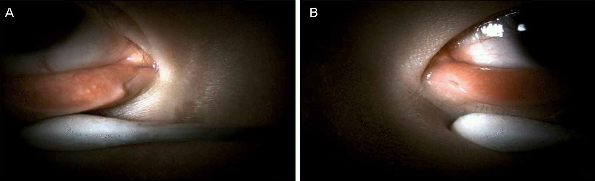 | Figure 1.Slit photographs of the patient's punctum. (A) right lower punctum shows cheese wiring shape. (B) left small lower punctum. |
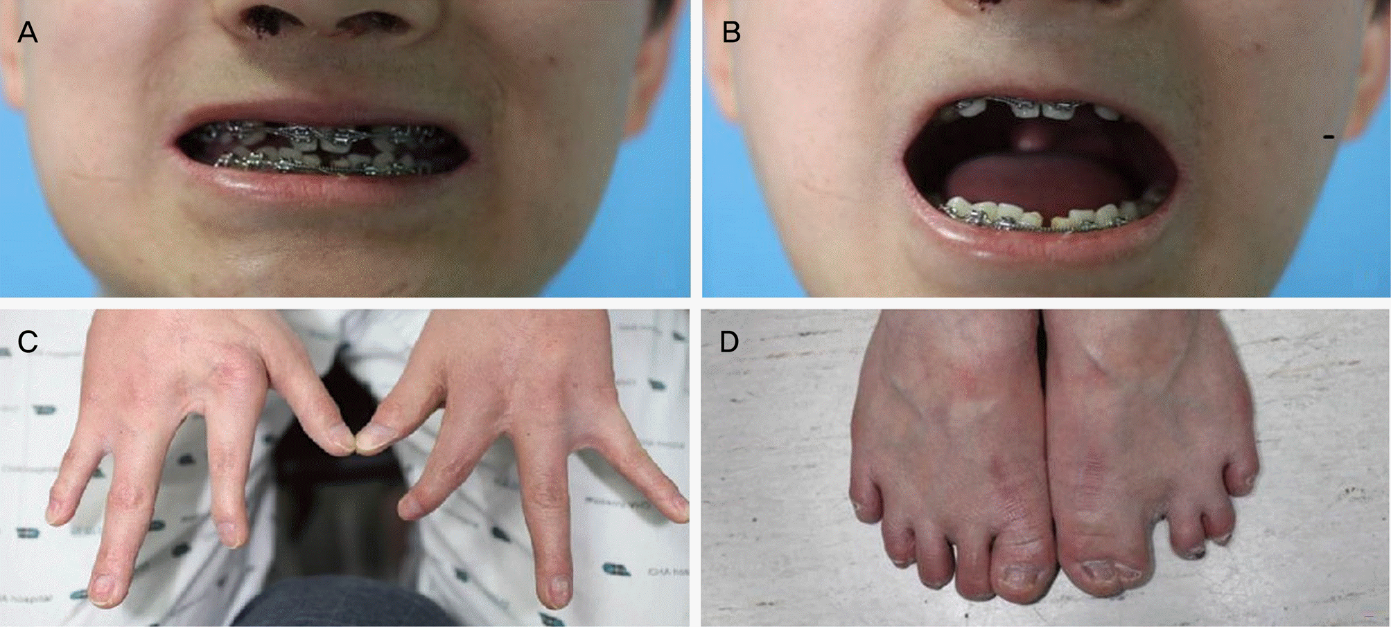 | Figure 2.Clinical photographs of the patient. (A, B) Teeth defect. (C, D) Fingers and toes. Odontodysplasia, ectrodactylia of finger and syndactylia of toe were observed. |
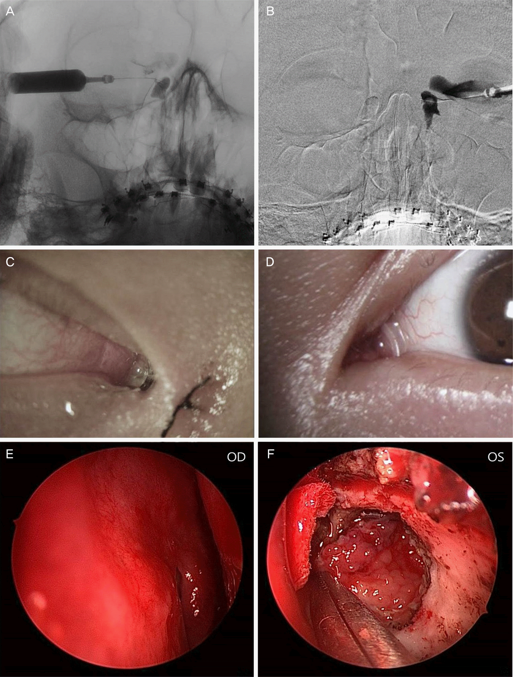 | Figure 3.The clinical findings of the patients. (A, B) dacryocystographic finding of the patient shows complete obstruction of nasolacrimal duct (right, left). (C, D) Slit photographs of the patient after operation. Jones tube at right side and silicone tube at left side were observed. (E) previous endoscopic dacryocystorhinostomy (DCR) opening was noted at the right side. (F) Neo opening of DCR was created. Severe inflammatory and follicular change at lacrimal sac was observed. OD = oculus dexter; OS = oculus sinister. |
 XML Download