

 PDF
PDF ePub
ePub Citation
 Print
Print


Abstract
Purpose
In this study, the changes in ocular surface parameters and tear meniscus after strabismus surgery when treated with or without artificial tears were evaluated using optical coherence tomography (OCT).
Methods
The present study included 30 patients who received bilateral lateral rectus recession surgery for exotropia. The patients instilled artificial tear eye drops only in the left eye. Before and 2, 4, 8, and 12 weeks after surgery, tear film break-up time (BUT), Schirmer's test, corneal staining test, tear meniscus height and area were measured using OCT and compared in both eyes. Before and 8 weeks after surgery, conjunctival compression cytology test was performed.
Results
The mean patient age was 8.7 years. After 8 weeks, BUT and corneal staining scores were 12.3 ± 1.5 seconds and 1.3 ± 0.4 in patients treated with artificial tears and 9.5 ± 1.0 seconds and 2.0 ± 0.7 in patients not treated with artificial tears (both p < 0.000). Four weeks after surgery, tear meniscus height and area using OCT were 290.2 ± 42.3 μm and 566.7 ± 48.2 pixels in patients treated with artificial tears and 246 ± 45.5 μ m and 504.0 ± 29.7 pixels in patients not treated with artificial tears (p = 0.045 and p = 0.019, respectively). Goblet cell count was significantly different between the eyes 8 weeks after surgery (p = 0.033).
Conclusions
Instability of tear meniscus can be detected after strabismus surgery using BUT, Schirmer's test, corneal staining test, tear meniscus height and area, and OCT. After surgery, artificial tears help treat dry eye symptoms by corneo-conjunctival wound healing mechanism and increasing tear meniscus stability.
Go to : 
References
1. Lemp MA, Foulks GN. The definition and classification of dry eye disease: report of the Definition and Classification Subcommittee of the International Dry Eye WorkShop (2007). Ocul Surf. 2007; 5:75–92.
2. Mantelli F, Massaro-Giordano M, Macchi I, et al. The cellular mechanisms of dry eye: from pathogenesis to treatment. J Cell Physiol. 2013; 228:2253–6.

3. Sullivan BD, Whitmer D, Nichols KK, et al. An objective approach to dry eye disease severity. Invest Ophthalmol Vis Sci. 2010; 51:6125–30.
4. Cher I. Fluids of the ocular surface: concepts, functions and physics. Clin Exp Ophthalmol. 2012; 40:634–43.
5. Li XM, Hu L, Hu J, Wang W. Investigation of dry eye disease and analysis of the pathogenic factors in patients after cataract surgery. Cornea. 2007; 26(Suppl 1):S16–20.
6. Nichols KK, Foulks GN, Bron AJ, et al. The International Workshop on meibomian gland dysfunction: executive summary. Invest Ophthalmol Vis Sci. 2011; 52:1922–9.
7. Konomi K, Chen LL, Tarko RS, et al. Preoperative characteristics and a potential mechanism of chronic dry eye after LASIK. Invest Ophthalmol Vis Sci. 2008; 49:168–74.
8. Srinivasan S, Chan C, Jones L. Apparent time-dependent abdominal in inferior tear meniscus height in human subjects with mild dry eye symptoms. Clin Exp Optom. 2007; 90:345–50.
9. Qiu X, Gong L, Sun X, Jin H. Age-related variations of human tear meniscus and diagnosis of dry eye with Fourier-domain anterior segment optical coherence tomography. Cornea. 2011; 30:543–9.
10. Ibrahim OM, Dogru M, Takano Y, et al. Application of visante abdominalal coherence tomography tear meniscus height measurement in the diagnosis of dry eye disease. Ophthalmology. 2010; 117:1923–9.
11. Jeon S, Park SH, Choi JS, Shin SY. Ocular surface changes after lateral rectus muscle recession. Ophthalmic Surg Lasers Imaging. 2011; 42:428–33.
12. Bron AJ, Evans VE, Smith JA. Grading of corneal and conjunctival staining in the context of other dry eye tests. Cornea. 2003; 22:640–50.
13. Anshu , Munshi MM, Sathe V, Ganar A. Conjunctival impression cytology in contact lens wearers. Cytopathology. 2001; 12:314–20.
14. Nelson JD. Impression cytology. Cornea. 1988; 7:71–81.
15. Apt L, Cullen BF. Newborns do secrete tears. JAMA. 1964; 189:951–3.
16. Axelrod FB. Familial dysautonomia. Muscle Nerve. 2004; 29:352–63.
17. Makari G, Hoffman WH, Carroll JE, et al. Autonomic dysfunction and adrenocortical unresponsiveness to ACTH. J Child Neurol. 1988; 3:174–6.
18. Arya SK, Chaudhuri Z, Jain R, et al. Congenital alacrima in Pierre Robin sequence. Cornea. 2004; 23:632–4.
19. Mohammadpour M, Javadi MA. Keratitis associated with multiple endocrine deficiency. Cornea. 2006; 25:112–4.
20. Thouret MC, Sirvent N, Triolo V, et al. Primary Gougerot-Sjögren syndrome in a 13-year-old girl. Arch Pediatr. 2002; 9:142–6.
21. Leite SC, de Castro RS, Alves M, et al. Risk factors and abdominal of ocular complications, and efficacy of autologous serum tears after haematopoietic progenitor cell transplantation. Bone Marrow Transplant. 2006; 38:223–7.
22. Dogru M, Okada N, Asano-Kato N, et al. Atopic ocular surface abdominal: implications on tear function and ocular surface mucins. Cornea. 2005; 24(8 Suppl):S18–23.
23. Akinci A, Cakar N, Uncu N, et al. Keratoconjunctivitis sicca in abdominal rheumatoid arthritis. Cornea. 2007; 26:941–4.
24. Sommer A. Xerophthalmia and vitamin A status. Prog Retin Eye Res. 1998; 17:9–31.
25. Nichols KK, Mitchell GL, Zadnik K. The repeatability of clinical measurements of dry eye. Cornea. 2004; 23:272–85.
26. Yokoi N, Komuro A. Non-invasive methods of assessing the tear film. Exp Eye Res. 2004; 78:399–407.
27. Mainstone JC, Bruce AS, Golding TR. Tear meniscus abdominal in the diagnosis of dry eye. Curr Eye Res. 1996; 15:653–61.
28. Oguz H, Yokoi N, Kinoshita S. The height and radius of the tear meniscus and methods for examining these parameters. Corena. 2000; 19:497–500.
29. Wang J, Palakuru JR, Aquavella JV. Correlations among upper and lower tear menisci, noninvasive tear break-up time, and the Schirmer test. Am J Ophthalmol. 2008; 145:795–800.
30. Kinoshita S, Kiorpes TC, Friend J, Thoft RA. Goblet cell density in ocular surface disease. A better indicator than tear mucin. Arch Ophthalmol. 1983; 101:1284–7.
31. Benelli U, Nardi M, Posarelli C, Albert TG. Tear osmolarity abdominal using the TearLab Osmolarity System in the assessment of dry eye treatment effectiveness. Cont Lens Anterior Eye. 2010; 33:61–7.
32. Lozano JS, Chay EY, Healey J, et al. Activation of the epidermal growth factor receptor by hydrogels in artificial tears. Exp Eye Res. 2008; 86:500–5.
33. Schmidl D, Schmetterer L, Witkowska KJ, et al. Tear film abdominal after treatment with artificial tears in patients with moderate dry eye disease. Cornea. 2015; 34:421–6.
34. Chang YH, Yoon JS, Chang JH, et al. Changes in corneal and abdominal sensitivitiy, tear film stability, and tear secretion after strabismus surgery. J Pediatr Ophthalmol Strabismus. 2006; 43:95–9.
Go to :
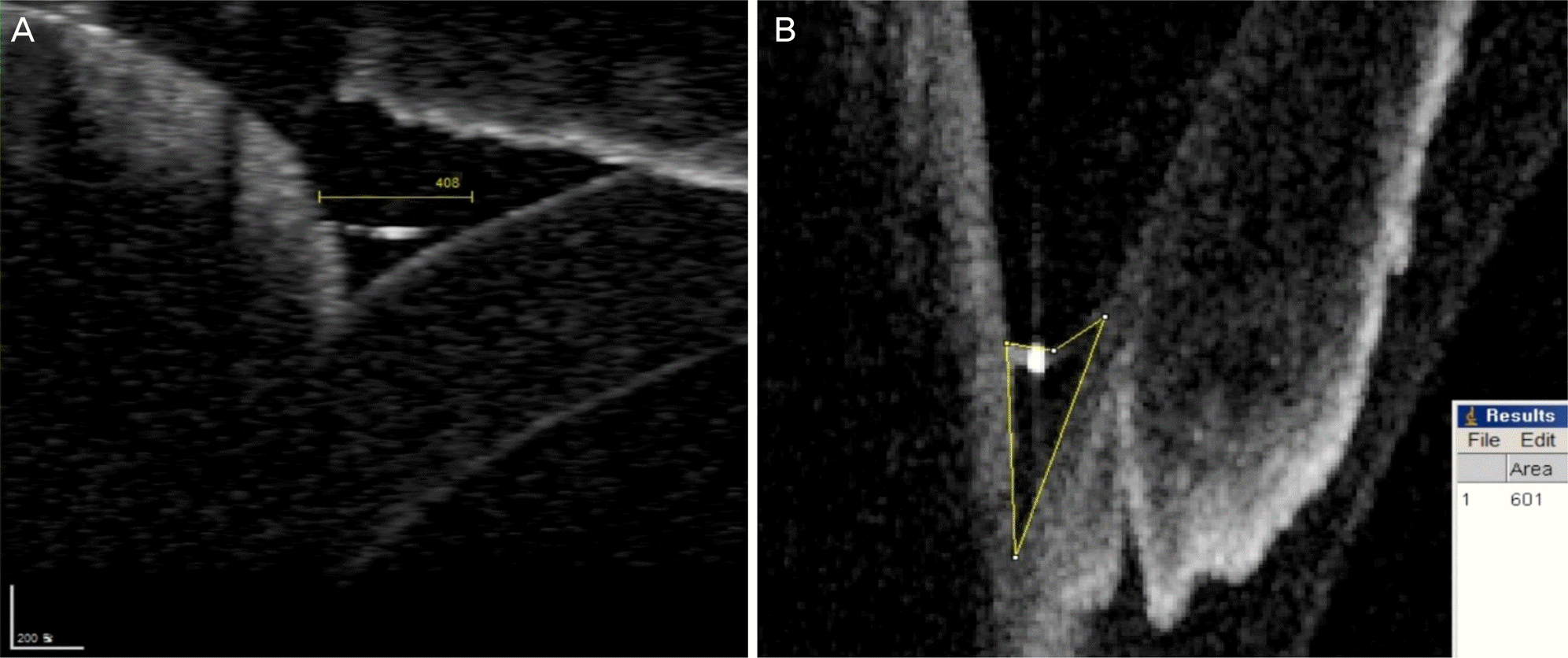 | Figure 1.Tear meniscus measurement. (A) Optical coherence tomography image of the lower tear meniscus showing the tear me-niscus height. (B) Optical coherence tomography image of the lower tear meniscus exported to ImageJ software for measuring me-niscus area. |
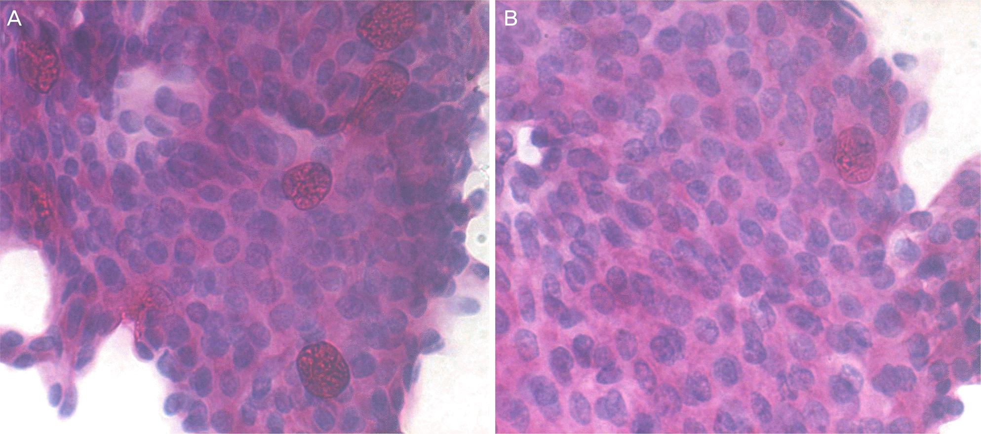 | Figure 2.Impression cytology (Periodic acid-Schiff, ×400). (A) 8 weeks after surgery, specimen from a patient treated artificial tear shows normal nuclear/cytoplasmic ratio and goblet cells. (B) 8 weeks after surgery, specimen from a control group patient shows normal nuclear/cytoplasmic ratio and depletion of goblet cells. |
Table 1.
Changes of corneal surface parameters
| Group 1 | Group 2 | p-value† | |
|---|---|---|---|
| TBUT (sec) | |||
| Baseline | 14.2 ± 1.9 | 14.0 ± 1.8 | 0.683 |
| 2 weeks | 8.2 ± 2.0* | 6.8 ± 1.3* | 0.030 |
| 4 weeks | 9.9 ± 1.6* | 8.1 ± 1.2* | 0.000 |
| 8 weeks | 12.3 ± 1.5* | 9.5 ± 1.0* | 0.000 |
| 12 weeks | 14.0 ± 1.3 | 13.6 ± 1.8 | 0.417 |
| Schirmer test (mm) | |||
| Baseline | 20.9 ± 2.2 | 21.5 ± 2.7 | 0.329 |
| 2 weeks | 19.0 ± 3.0* | 20.4 ± 2.8 | 0.061 |
| 4 weeks | 19.5 ± 2.4 | 19.1 ± 2.5* | 0.534 |
| 8 weeks | 21.2 ± 2.4 | 22.3 ± 2.6 | 0.089 |
| 12 weeks | 19.7 ± 2.0 | 20.2 ± 2.5 | 0.441 |
| Corneal staining score | |||
| Baseline | 0.1 ± 0.3 | 0.1 ± 0.3 | 0.694 |
| 2 weeks | 5.1 ± 1.7* | 6.0 ± 1.4* | 0.024 |
| 4 weeks | 2.2 ± 0.8* | 4.1 ± 0.7* | 0.000 |
| 8 weeks | 1.3 ± 0.4* | 2.0 ± 0.7* | 0.000 |
| 12 weeks | 0.2 ± 0.4 | 0.2 ± 0.4 | 0.759 |
Table 2.
Changes of tear meniscus height and area
| Group 1 | Group 2 | p-value† | |
|---|---|---|---|
| Height (μ m) | |||
| Baseline | 294.7 ± 35.4 | 286 ± 42.5 | 0.476 |
| 2 weeks | 288.3 ± 52.5 | 236 ± 38.7* | 0.037 |
| 4 weeks | 290.2 ± 42.3 | 246 ± 45.5* | 0.045 |
| 8 weeks | 296.5 ± 47.2 | 279 ± 31.9 | 0.144 |
| 12 weeks | 292.1 ± 57.3 | 295 ± 44.3 | 0.743 |
| Area (pixels) | |||
| Baseline | 601.6 ± 52.1 | 587.4 ± 65.3 | 0.374 |
| 2 weeks | 553.1 ± 57.6* | 503.7 ± 40.8* | 0.021 |
| 4 weeks | 566.7 ± 48.2 | 504.0 ± 29.7* | 0.019 |
| 8 weeks | 588.4 ± 60.5 | 578.9 ± 56.6 | 0.624 |
| 12 weeks | 579.3 ± 51.9 | 584.2 ± 48.7 | 0.771 |
 XML Download