

 PDF
PDF ePub
ePub Citation
 Print
Print


Abstract
Case summary
A 45-year-old women presented with decreased visual acuity in her left eye without eye movement pain. Her best corrected visual acuity (BCVA) in that eye was 0.3. She showed a relative afferent pupillary defect, abnormal color vision test, and inferior visual field defect in her left eye. The optic disc showed slight blurring superiorly and pallor temporally. Fluorescein angiography showed choroidal filling defect, and ischemic optic neuropathy was suspected. The carotid artery sonography showed normal results. The BCVA of the left eye was 0.5 after 7 months. The visual field test and color vision test were improved after 7 months. The optic disc was pale. The magnetic resonance imaging was performed because of persistent headache, and that showed a suprasellar mass that was removed by surgical resection and diagnosed as meningioma on biopsy.
References
1. McDermott MW, Wilson CB. Meningiomas. Youmans JR, editor. Neurological Surgery. 4th ed.Philadelphia: WB Saunders;1996. p. 2782–825.
2. Young HY, Min A. A case of bilateral visual loss due to an olfactory groove meningioma. J Korean Ophthalmol Soc. 2012; 53:906–9.

3. Lee EK, Kim SJ, Park SH, et al. Clinical features and management outcome of optic nerve sheath meningioma in Korea. J Korean Ophthalmol Soc. 2011; 52:74–85.
4. Park JH, Seol ER, Choi HY, Lee JW. A case of suprasellar arach-noid cyst with compressive optic neuropathy. J Korean Ophthalmol Soc. 2012; 53:1532–9.
5. Beri M, Klugman MR, Kohler JA, Hayreh SS. Anterior ischemic optic neuropathy. VII. Incidence of bilaterality and various abdominal factors. Ophthalmology. 1987; 94:1020–8.
6. Kim DH, Hwang JM. Risk factors for Korean patients with anterior ischemic optic neuropathy. J Korean Ophthalmol Soc. 2007; 48:1527–31.
7. Bergmann M, Brück W, Neubauer U, Probst-Cousin S. Diagnostic pitfall: optic neuritis mimicking optic nerve glioma. Neuropathology. 2009; 29:450–3.
8. Kim SB, Kyung SE. Fluorescein angiographic findings of non-arteritic anterior ischemic optic neuropathy and optic neuritis. J Korean Ophthalmol Soc. 2012; 53:1143–9.
9. Janáky M, Fülöp Z, Pálffy A, et al. Non-arteritic ischemic optic neuropathy (NAION) in patients under 50 years of age. Acta Ophthalmol Scand. 2005; 83:499–503.
10. Jung JW, Jin HC, Kim KS, Kim YC. A case of compressive optic neuropathy due to breast cancer metastasis. J Korean Ophthalmol Soc. 2010; 51:1161–5.
11. Kim I, Kim M, Chung Y. A case of compressive optic neuropathy caused by sphenoid sinus mucocele. J Korean Ophthalmol Soc. 1989; 30:1025–9.
12. Danesh-Meyer HV, Papchenko T, Savino PJ, et al. In vivo retinal nerve fiber layer thickness measured by optical coherence abdominal predicts visual recovery after surgery for parachiasmal tumors. Invest Ophthalmol Vis Sci. 2008; 49:1879–85.
13. Schick U, Hassler W. Surgical management of tuberculum sellae meningiomas: involvement of the optic canal and visual outcome. J Neurol Neurosurg Psychiatry. 2005; 76:977–83.
14. Schlezinger NS, Alpers BJ, Weiss BP. Suprasellar meningiomas associated with scotomatous field defects. Arch Ophthal. 1946; 35:624–42.
15. Lee JH, Jeun SS, Evans J, Kosmorsky G. Surgical management of clinoidal meningiomas. Neurosurgery. 2001; 48:1012–9. discussion 1019–21.
16. Monney AJ, McConnell AA. Visual scotomata with intracranial abdominal affecting the optic nerve. J Neurol Neurosurg Psychiatry. 1949; 12:205–18.
17. Zevgaridis D, Medele RJ, Müller A, et al. Meningiomas of the sell-ar region presenting with visual impairment: impact of various prognostic factors on surgical outcome in 62 patients. Acta Neurochir (Wien). 2001; 143:471–6.
18. Hayreh SS, Joos KM, Podhajsky PA, Long CR. Systemic diseases associated with nonarteritic anterior ischemic optic neuropathy. Am J Ophthalmol. 1994; 118:766–80.
19. Hayreh SS, Zimmerman MB, Podhajsky P, Alward WL. Nocturnal arterial hypotension and its role in optic nerve head and ocular abdominal disorders. Am J Ophthalmol. 1994; 117:603–24.
Figure 1.
Humphrey visual field test at the first visit. (A) The test showed inferior arcuate defect with central scotoma in left eye. (B) No defect in right eye.
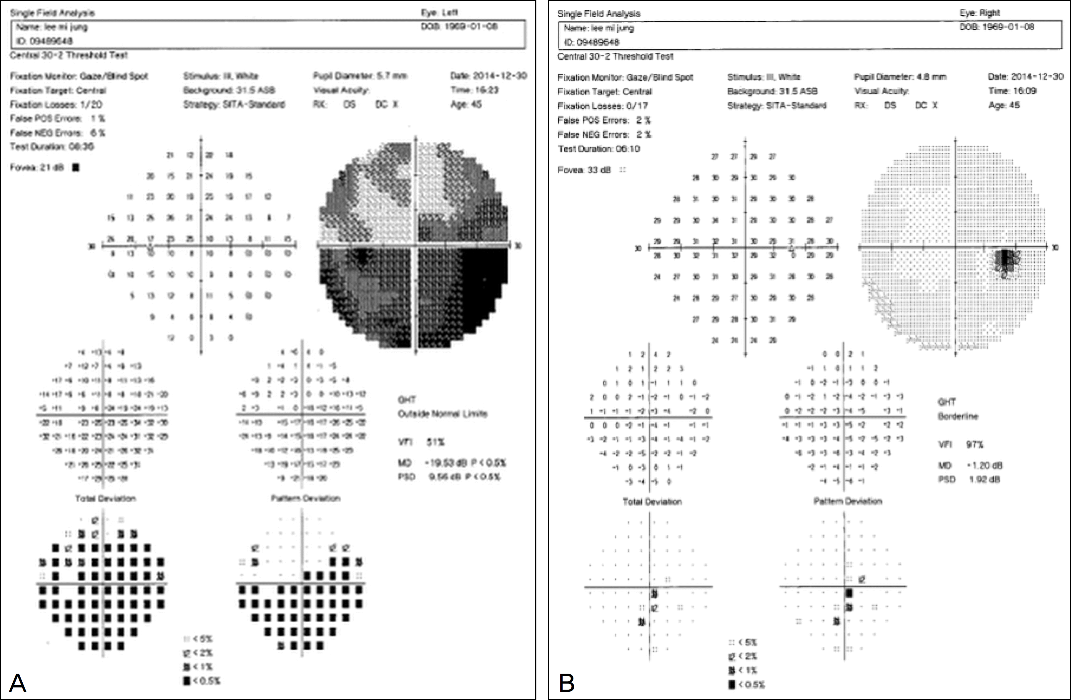
Figure 2.
The left optic disc at the first visit. The slight blurred superiorly and pallor temporally optic disc was suspected to be ischemic optic neuropathy.
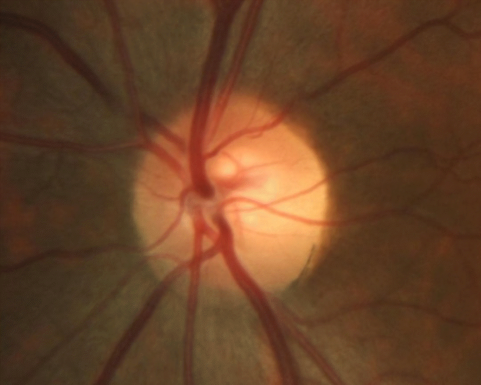
Figure 3.
The optical coherence tomography at the first visit. The test showed decreased retinal nerve fiber layer (RNFL) thickness temporally of left eye corresponding to optic disc pallor. ONH = optic nerve head; OU = oculus unitas; OD = oculus dexter; OS = oculus sinister; C/D = cup/disc; TEMP = temporal; SUP = superior; NAS = nasal; INF = inferior; S = superior; N = nasal; I = inferior; T = temporal.
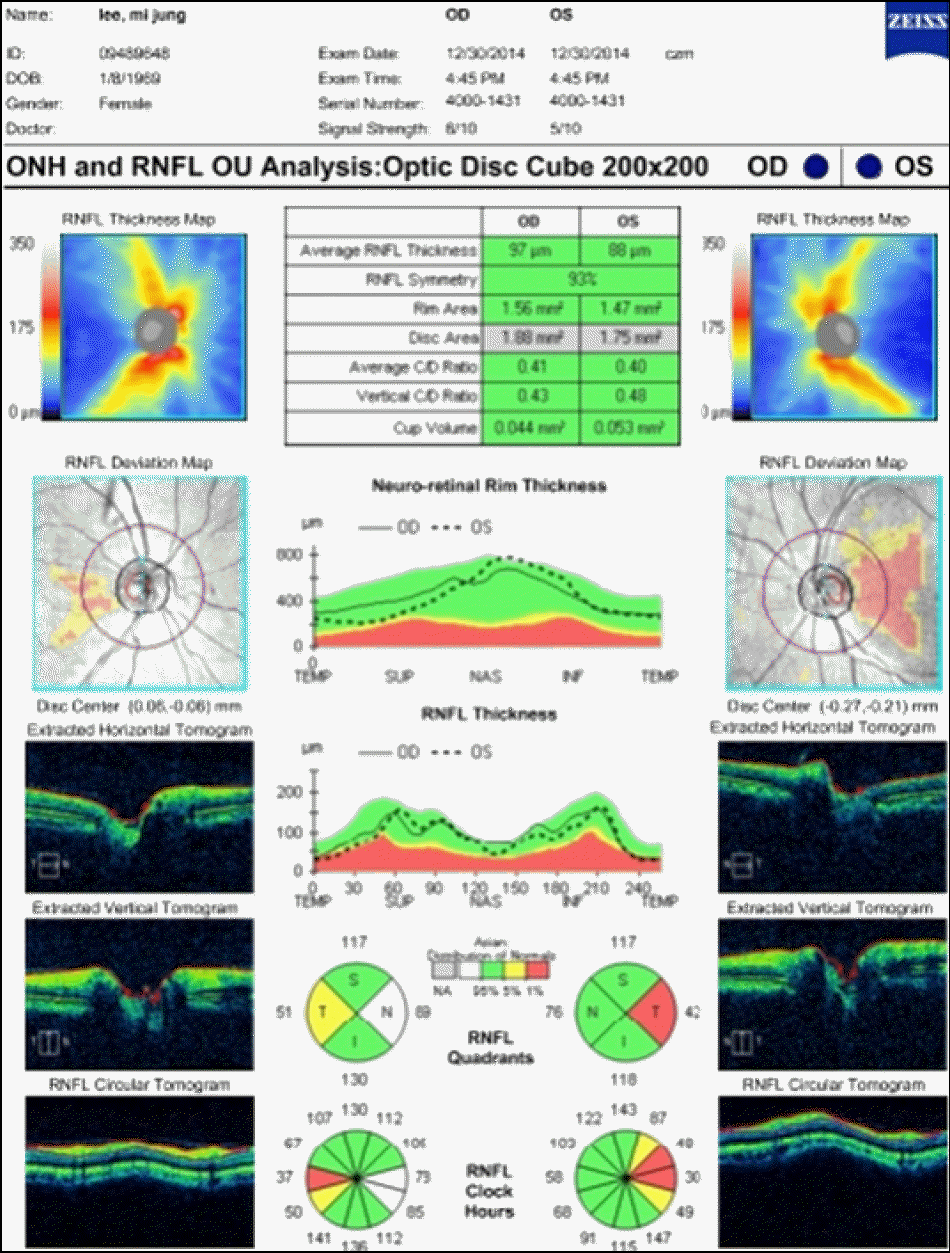
Figure 4.
Fluorescein angiograph of left eye. The test showed delayed peripapillary choroidal filling at 12 seconds that was suspected to be ischemic optic neuropathy at the first time.
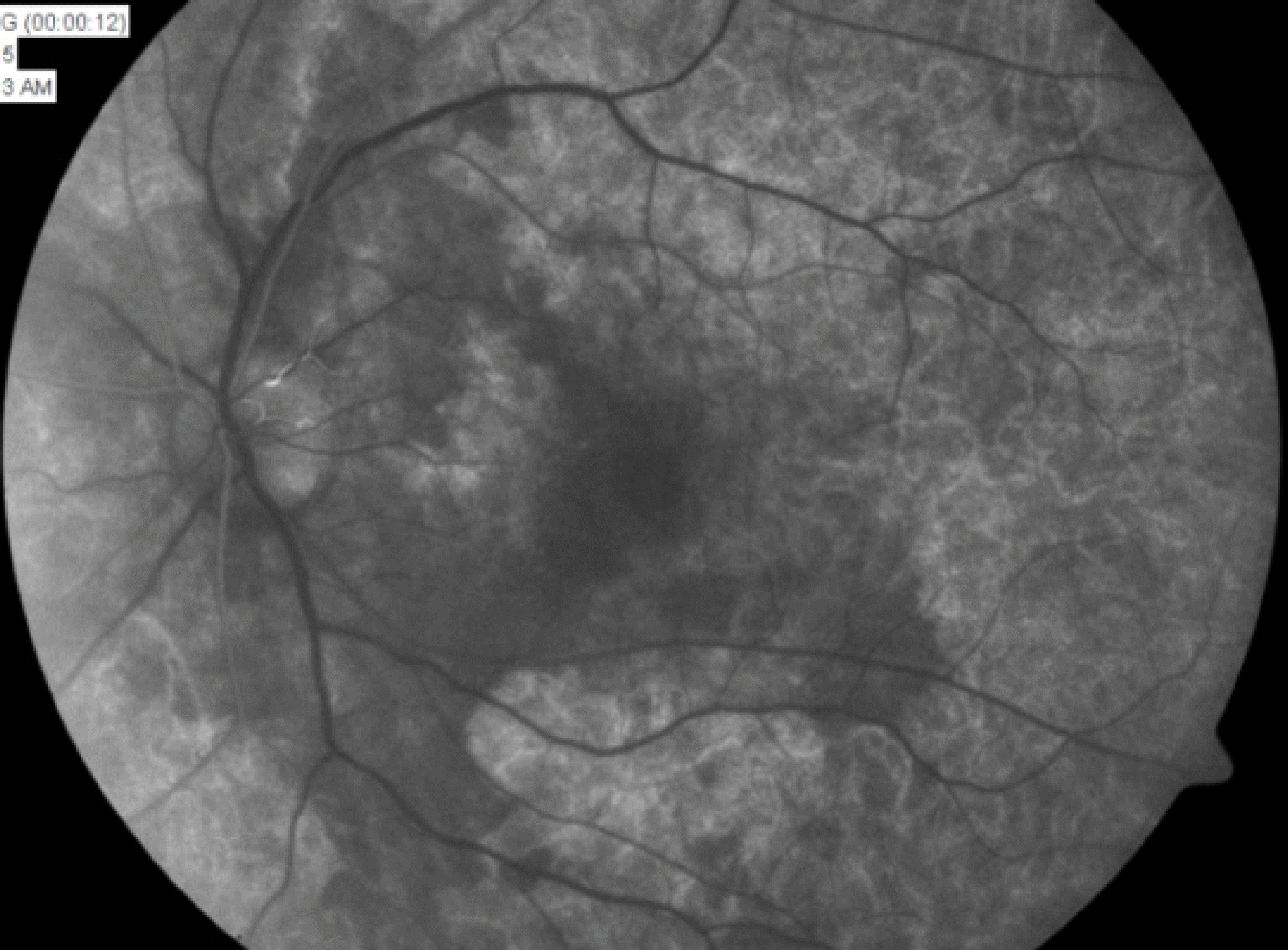
Figure 5.
Humphrey visual field test in left eye at 7 months. The test showed improvement compared to that of at the first visit after steroid therapy.
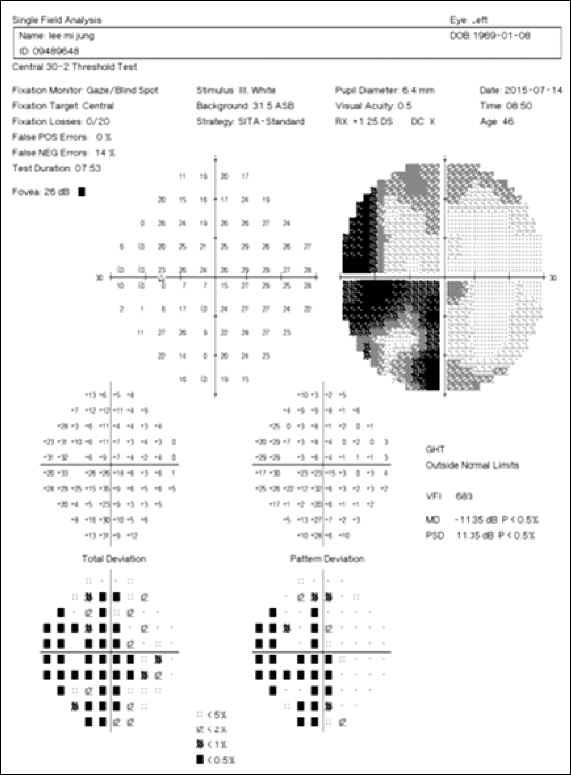
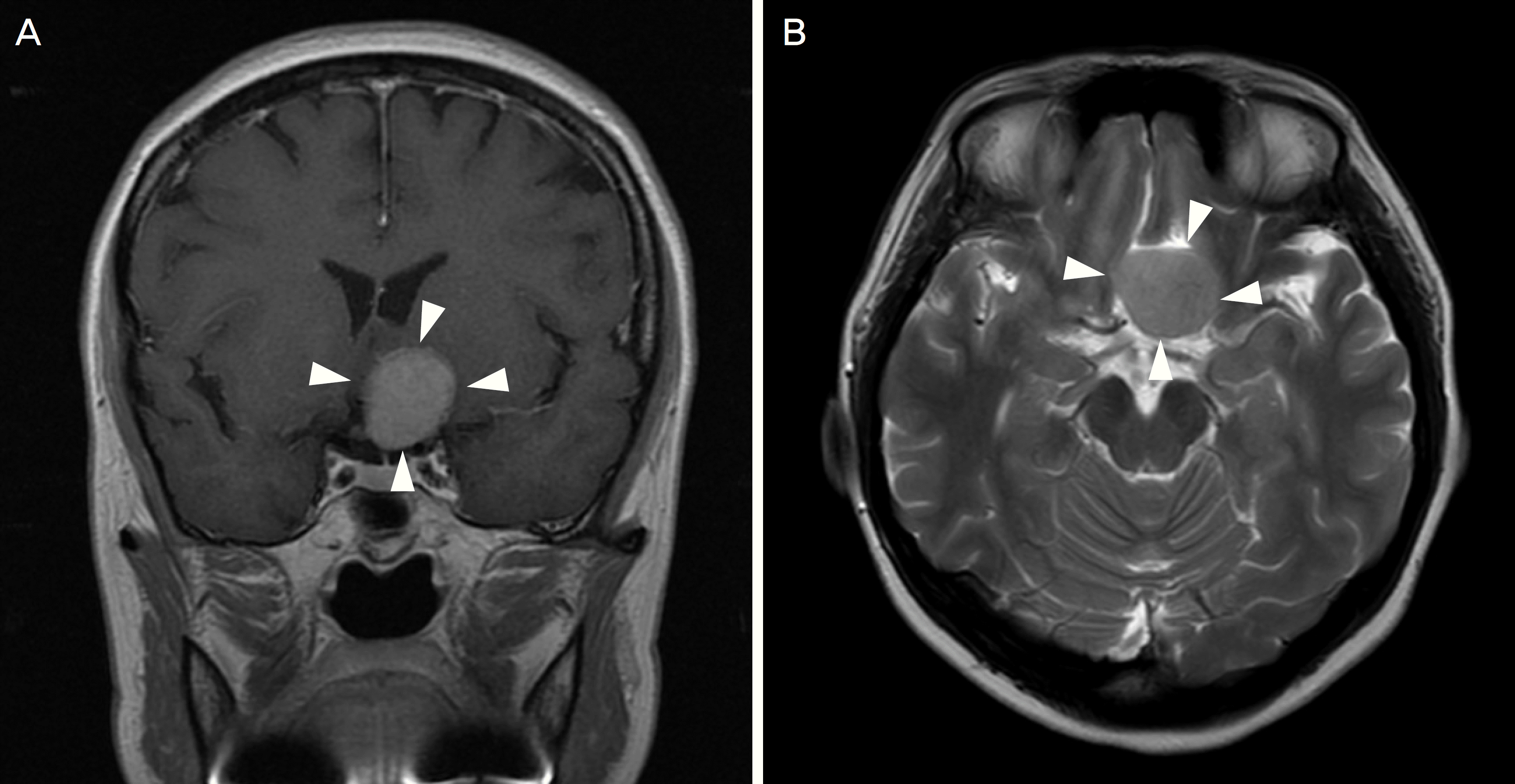
Figure 6.
Brain magnetic resonance image. (A) 2.7 × 2.3 cm sized well circumscribed mass (white arrowheads) of left suprasellar area with direct contact to optic chiasm in axial T1-enhanced images. (B) Low signal intensity mass intensified higher than gray matter in transverse T2-weighted images (white arrowheads).
 XML Download