Abstract
Purpose
Systemic lupus erythematosus (SLE) is a chronic autoimmune disorder with widespread manifestations that rarely include the eye. We present a case of SLE-associated choroidoretinopathy and secondary angle closure attack in both eyes.
Case summary
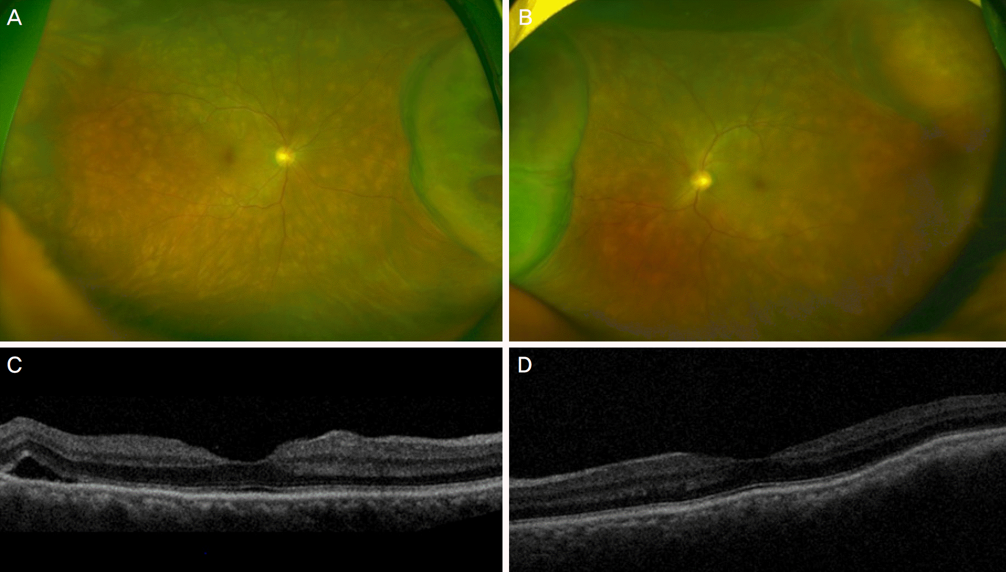
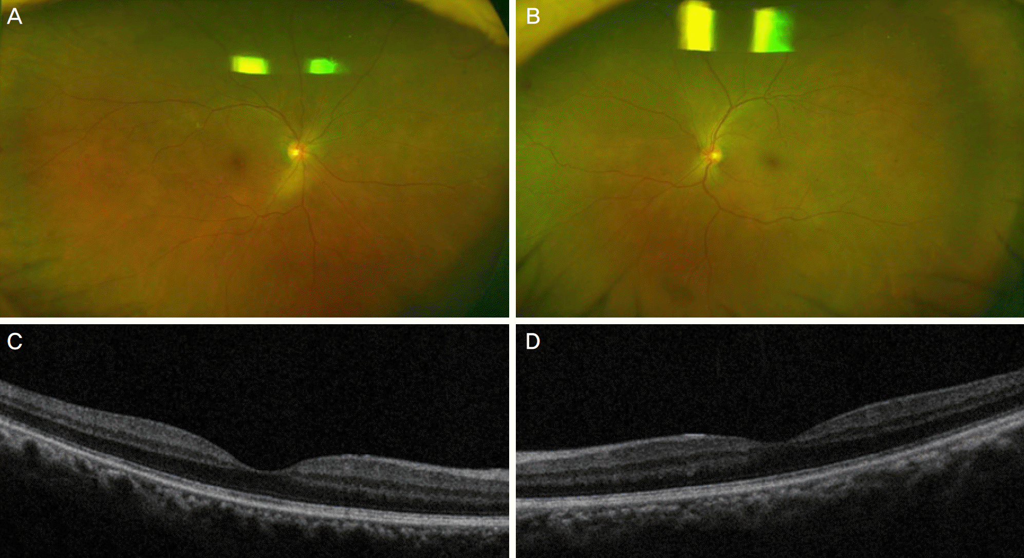
A 58-year-old male was admitted into the urologic department complaining of right scrotal swelling, and then consulted with the ophthalmology department regarding both ocular pain and eye injection. The patient was diagnosed with acute angle closure attack using a slit lamp test and tonometry secondary to choroidoretinitis with choroidal detachment at fundus examination in both eyes. The rheumatologist performed systemic evaluation, including serologic tests, and then diagnosed the patient with SLE. After systemic steroid therapy, intraocular pressure was decreased and choroidal detachment disappeared with improvements of choroidoretinitis in both eyes.
Go to : 
References
1. Sivaraj RR, Durrani OM, Denniston AK, et al. Ocular abdominal of systemic lupus erythematosus. Rheumatology (Oxford). 2007; 46:1757–62.
2. Hochberg MC. Updating the American college of rheumatology revised criteria for the classification of systemic lupus erythematosus. Arthritis Rheum. 1997; 40:1725.

3. Yoon CK, Park JH, Yu HG. Retinopathy associated with systemic lupus erythematosus. J Korean Ophthalmol Soc. 2009; 50:1215–20.

4. Edouard S, Douat J, Sailler L, et al. Bilateral choroidopathy in abdominalic lupus erythematosus. Lupus. 2011; 20:1209–10.
5. Han YS, Yang CM, Lee SH, et al. Secondary angle closure abdominal by lupus choroidopathy as an initial presentation of systemic lupus erythematosus: a case report. BMC ophthalmol. 2015; 15:148.

6. Lavina AM, Agarwal A, Hunyor A, Gass JD. Lupus choroidopathy and choroidal effusions. Retina. 2002; 22:643–7.

7. Wisotsky BJ, Magat-Gordon CB, Puklin JE. Angle-closure abdominal as an initial presentation of systemic lupus erythematosus. Ophthalmology. 1998; 105:1170–2.
8. Gäckle HC, Lang GE, Freissler KA, Lang GK. Central serous chorioretinopathy. Clinical, fluorescein angiography and abdominal aspects. Ophthalmologe. 1998; 95:529–33.
9. Chaine G, Haouat M, Menard-Molcard C, et al. Central serous abdominal and systemic steroid therapy. J Fr Ophtalmol. 2001; 24:139–46.
10. Baglio V, Gharbiya M, Balacco-Gabrieli C, et al. Choroidopathy in patients with systemic lupus erythematosus with or without nephropathy. J Nephrol. 2011; 24:522–9.

11. Bengtsson AA, Rönnblom L. Systemic lupus erythematosus: still a challenge for physicians. J Intern Med. 2016; Jun 16:doi:. DOI: 10.1111/joim.12529. [Epub ahead of print].

12. Kamdar NV, Erko A, Ehrlich JS, et al. Choroidopathy and kidney disease: a case report and review of the literature. Cases J. 2009; 2:7425.

13. Kuehn MW, Oellinger R, Kustin G, Merkel KH. Primary testicular manifestation of systemic lupus erythematosus. Eur Urol. 1989; 16:72–3.

14. Elagouz M, Stanescu-Segall D, Jackson TL. Uveal effusion syndrome. Surv Ophthalmol. 2010; 55:134–45.

15. Ikeda N, Ikeda T, Nomura C, Mimura O. Ciliochoroidal effusion syndrome associated with posterior scleritis. Jpn J Ophthalmol. 2007; 51:49–52.

16. Palejwala NV, Walia HS, Yeh S. Ocular manifestations of systemic lupus erythematosus: a review of the literature. Autoimmune Dis. 2012; 2012:290898.

17. Shimura M, Tatehana Y, Yasuda K, et al. Choroiditis in systemic abdominal erythematosus: systemic steroid therapy and focal laser treatment. Jpn J Ophthalmol. 2003; 47:312–5.
Go to : 
 | Figure 1.Initial ultrawide fundus photography of the right eye, left eye and optical coherence tomography (OCT) of the right eye and left eye. Diffuse choroiditis foci with choroidal detachment in both eyes were found in fundus examination(A, B). Irregular choroidal folding with subretinal fluid was found in OCT (C, D). |


 PDF
PDF ePub
ePub Citation
 Print
Print


 XML Download