Abstract
Purpose
To report a case of Descemet's membrane detachment and corneal edema caused by an iatrogenic corneal perforation created while performing a local anesthetic (lidocaine) injection into the eyelid for a hordeolum incision and a drainage procedure. The detachment resolved after 14% C3 F8 gas and air injections into the anterior chamber.
Case summary
An 8-year-old female visited our clinic after the onset of severe pain and decreased visual acuity while receiving a local anesthetic injection into the upper eyelid in preparation for a hordeolum incision and drainage procedure. Corneal optical coherence tomography (OCT) showed Descemet's membrane detachment. Three days after the first visit, the corneal epithelium had entirely healed. However, Descemet's membrane detachment persisted even after three weeks of follow-up. A corneal OCT was repeated after three weeks and showed a partial Descemet's membrane rupture. A more aggressive treatment method was deemed necessary, and gas and air injections into the anterior chamber were performed. After 48 hours, aside from some Descemet's membrane rolling at the site of rupture, overall reattachment of Descemet's membrane was noted. After three months of follow-up, the patient showed a stable corneal state and normalized vision.
Conclusions
Descemet's membrane detachment and rupture resulting from an iatrogenic corneal perforation during an injection of lidocaine to the eyelid led to decreased visual acuity from corneal edema. As a more aggressive treatment method, 14 % C3 F8 gas and air injections into the anterior chamber were performed and resulted in near complete reattachment of Descemet's membrane's and normalization of the patient's visual acuity.
References
1. Iradier MT, Moreno E, Aranguez C, et al. Late spontaneous abdominal of a massive detachment of Descemet's membrane after phacoemulsification. J Cataract Refract Surg. 2002; 28:1071–3.
2. Minkovitz JB, Schrenk LC, Pepose JS. Spontaneous resolution of an extensive detachment of Descemet's membrane following phacoemulsification. Arch Ophthalmol. 1994; 112:551–2.

3. Ghosh S, Mukhopadhyay S, Mukhopadhyay S, et al. Inadvertent intracorneal injection of local anesthetic during lid surgery. Cornea. 2010; 29:701–2.

4. Schellini SA, Creppe MC, Grego'rio EA, Padovani CR. Lidocaine effects on corneal endothelial cell ultrastructure. Vet Ophthalmol. 2007; 10:239–44.

5. Rosenwasser GO. Complications of topical ocular anesthetics. Int Ophthalmol Clin. 1989; 29:153–8.

6. Britton B, Hervey R, Kasten K, et al. Intraocular irritation evaluation of benzalkonium chloride in rabbits. Ophthalmic Surg. 1976; 7:46–55.

7. Najjar DM, Rapuano CJ, Cohen EJ. Descemet membrane abdominal with hemorrhage after alkali burn to the cornea. Am J Ophthalmol. 2004; 137:185–7.
8. Liu DT, Lai JS, Lam DS. Descemet membrane detachment after abdominal argon-neodymium: YAG laser peripheral iridotomy. Am J Ophthalmol. 2002; 134:621–2.
9. Lee SE, Cho KJ, Cho WH, et al. Spontaneous reattachment of Descmet's membrane detachment at postoperative two months, which occurred during cataract surgery. J Korean Ophthalmol Soc. 2013; 54:351–6.
10. Koh JW, Woon WJ, Na KS. A case of total Desmet's Nembrane abdominal treated by non-expansible SF6 gas infusion. J Korean Ophthalmol Soc. 2002; 43:2598–602.
11. Hoover DL, Giangiacomo J, Benson RL. Descemet's membrane detachment by sodium hyaluronate. Arch Ophthalmol. 1985; 103:805–8.

12. Assia EI, Levkovich-Verbin H, Blumenthal M. Management of Descemet's membrane detachment. J Cataract Refract Surg. 1995; 21:714–7.

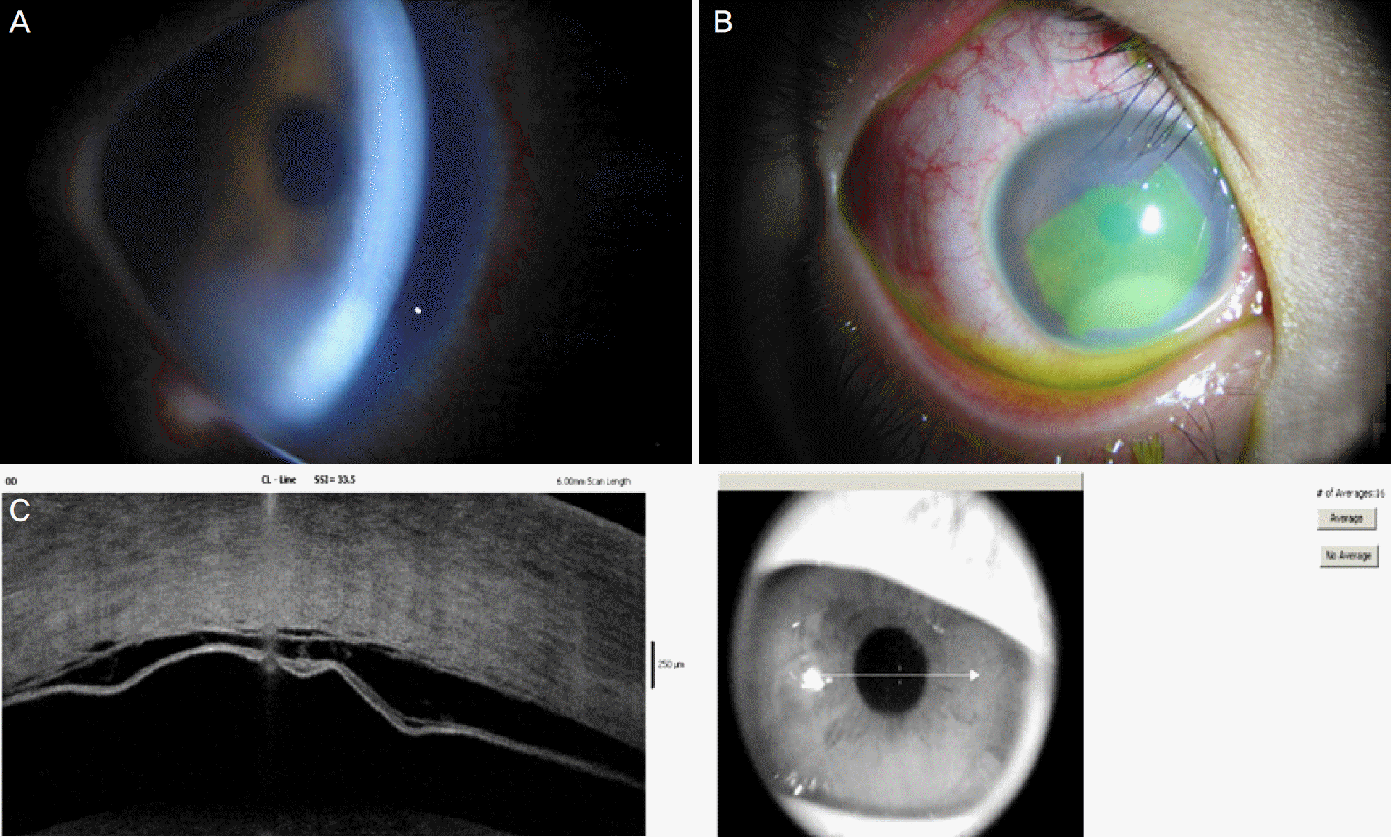
Figure 1.
The images were taken on the injury day. On slit lamp examination, corneal edema (A) could be visualized with large corneal abrasion (B). Anterior segment optical coherence tomography across the plane marked on the anterior segment photo revealing Descemet's membrane detachment (C).

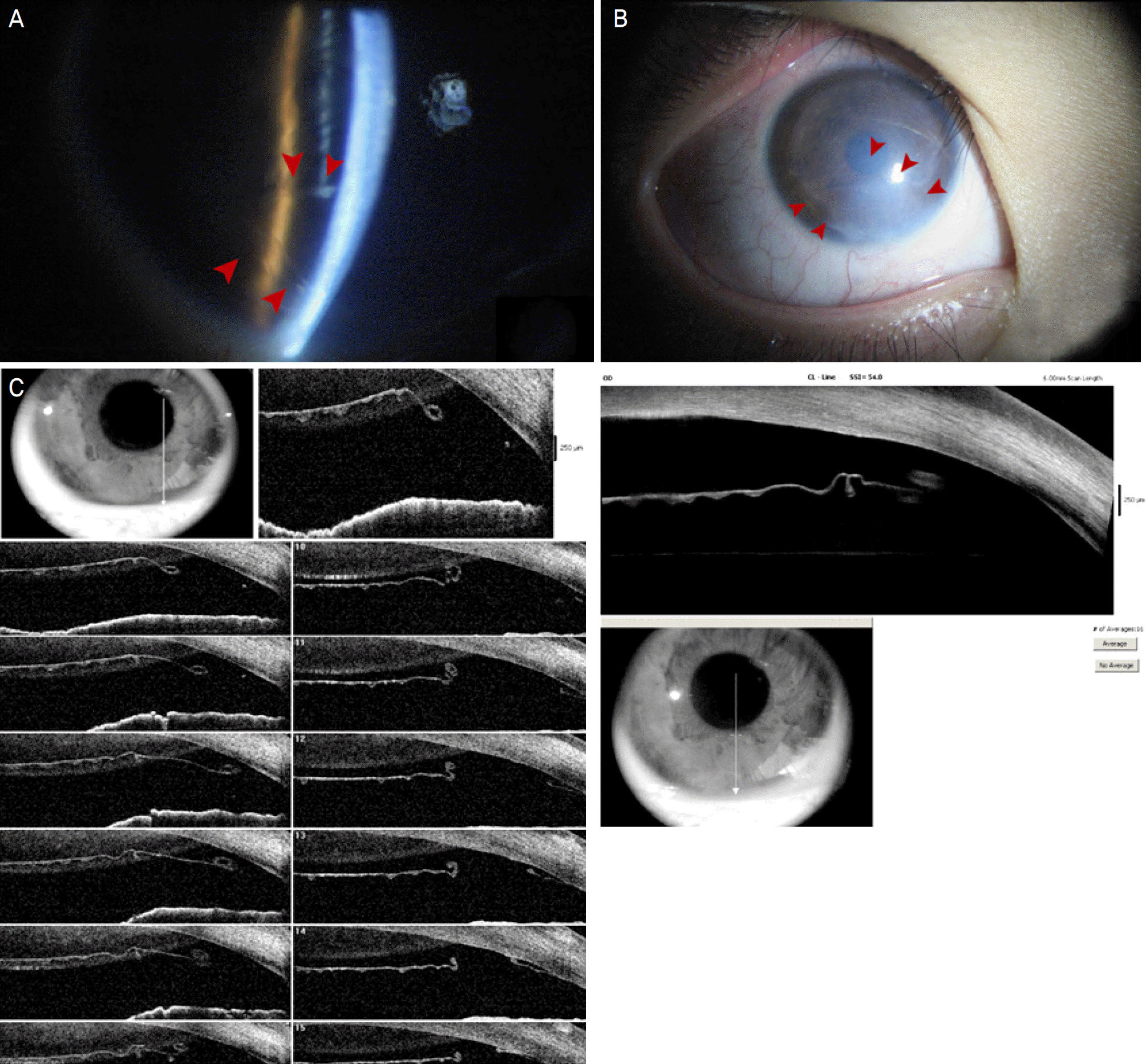
Figure 2.
The images were taken on the third week after injury. Arrows mark newly detected Descemet's membrane rupture at the primary puncture site by slit lamp examination (A, B). Anterior segment optical coherence tomography across the plane remarked on the anterior segment image showing Desmet's membrane rupture (C).

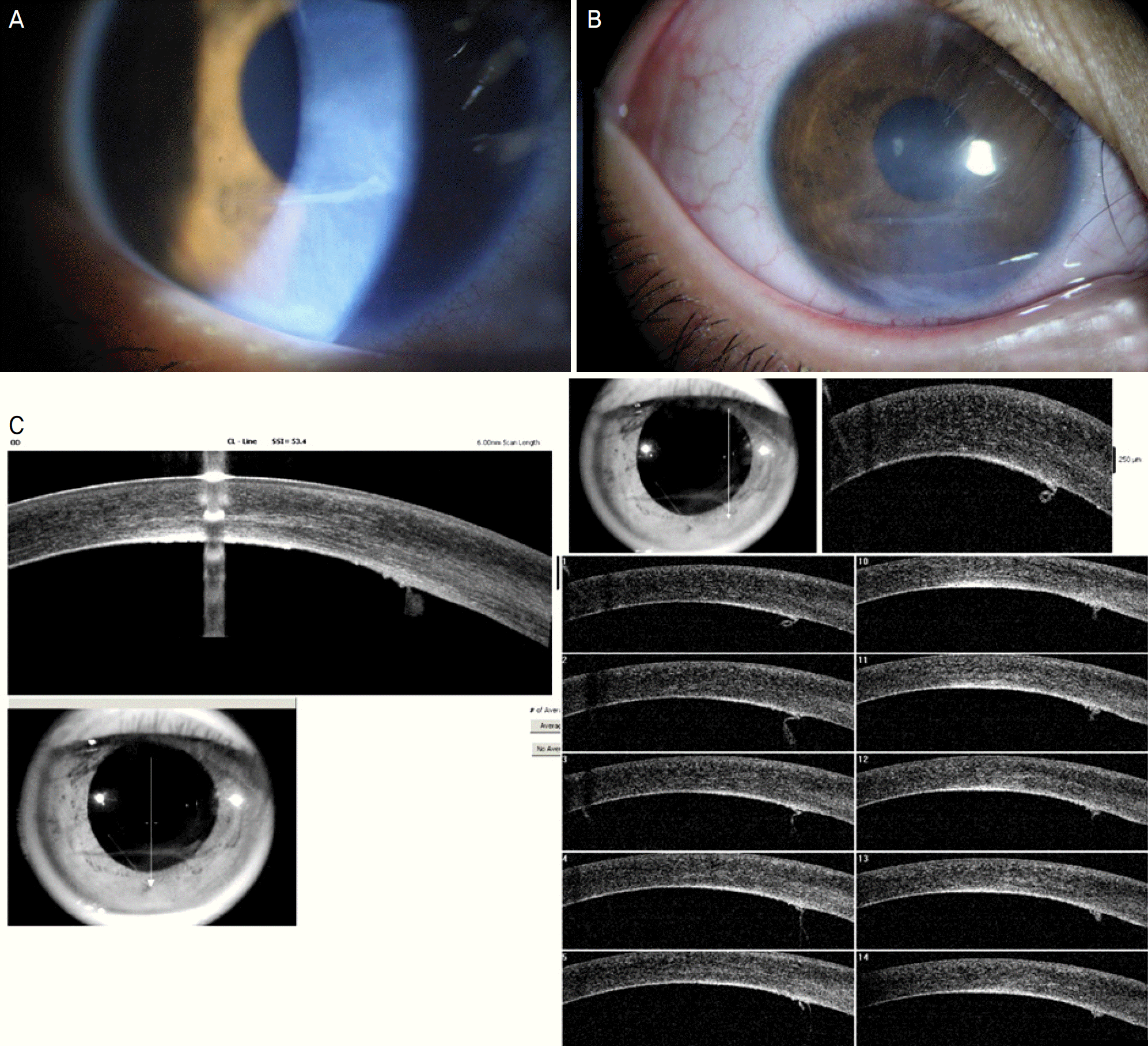
Figure 3.
The images were taken at 1 month after the procedure. Corneal edema was resolved and Descemet's membrane was attached in the area comprising the visual axis (A, B). Remaining strand-like Descemet's membrane folds can be seen by anterior segment optical coherence tomography at the injury site (C).


 PDF
PDF ePub
ePub Citation
 Print
Print


 XML Download