

 PDF
PDF ePub
ePub Citation
 Print
Print


Abstract
Purpose
We report the first case of extra-axial anaplastic meningioma with direct orbital extension for differential diagnosis of orbital tumors.
Case summary
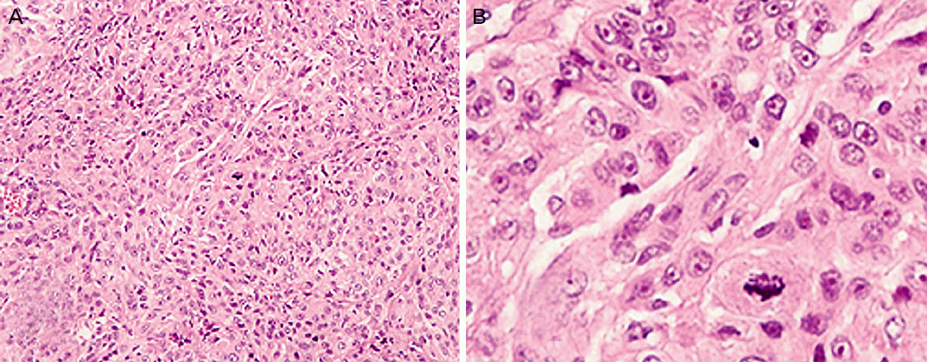
An 83-year-old woman presented with a protruding left eye and a palpable mass on the forehead. A brain computed tomography scan revealed a huge sarcomatous mass that had directly invaded the surrounding tissues. We removed the tumor by craniotomy and found that it involved the extradural and intradural spaces, brain parenchyma, subcutaneous tissue, left temporalis, orbital roof, and the other frontal lobe across the midline. A cranioplasty was performed, and the orbital roof was reconstructed with artificial bone and bone cement. The final histological diagnosis was anaplastic meningioma. The left eyeball was restored to its normal position 1 month after the surgery. Also, visual acuity and eye movement of the left eye were preserved.
References
1. Mahnood A, Caccamo DV, Tomecek F, Malik GM. Atypical and malignant meningiomas: a clinicopathological review. Neurosurgery. 1993; 33:955–63.
2. Karikari IO, Syed NA, Cummings TJ. Secretory meningiomas of the orbit. Orbit. 2009; 28:408–11.

3. Perry A, Stafford SL, Scheithauer BW, et al. Menigioma grading: an analysis of histologic parameters. Am J Surg Pathol. 1997; 21:1455–65.
4. Gündüz K, Kurt RA, Erden E. Ectopic orbital meningioma: report of two cases and literature review. Surv Ophthalmol. 2014; 59:643–8.
5. Modha A, Gutin PH. Diagnosis and treatment of atypical and abdominal meningiomas: a review. Neurosurgery. 2005; 57:538–50.
6. Al-Mefty O, Topsakal C, Pravdenkova S, et al. Radiation-induced meningiomas: clinical, pathological, cytokinetic, and cytogenetic characteristic. J Neurosurg. 2004; 100:1002–13.
Figure 1.
Anterior photographs. 9-cardinal segment photographs. (A) The protruding mass in the left forehead with orbital dystopia. The left globe was displaced inferiorly. Up gaze eye movement disorder is also shown. (B) Proptosis of the left eye.
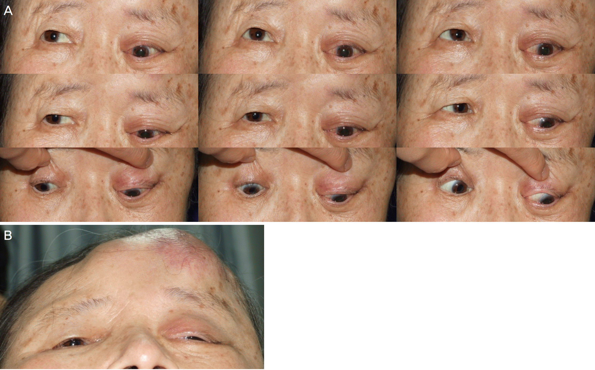
Figure 2.
Initial coronal brain images recorded 1 year ago. (A) Computed tomography. (B) Magnetic resonance imaging. These are showing a heterogeneously enhanced extra-axial mass in the left frontoparietal bone. Bony destruction and intracranial extension are also shown.
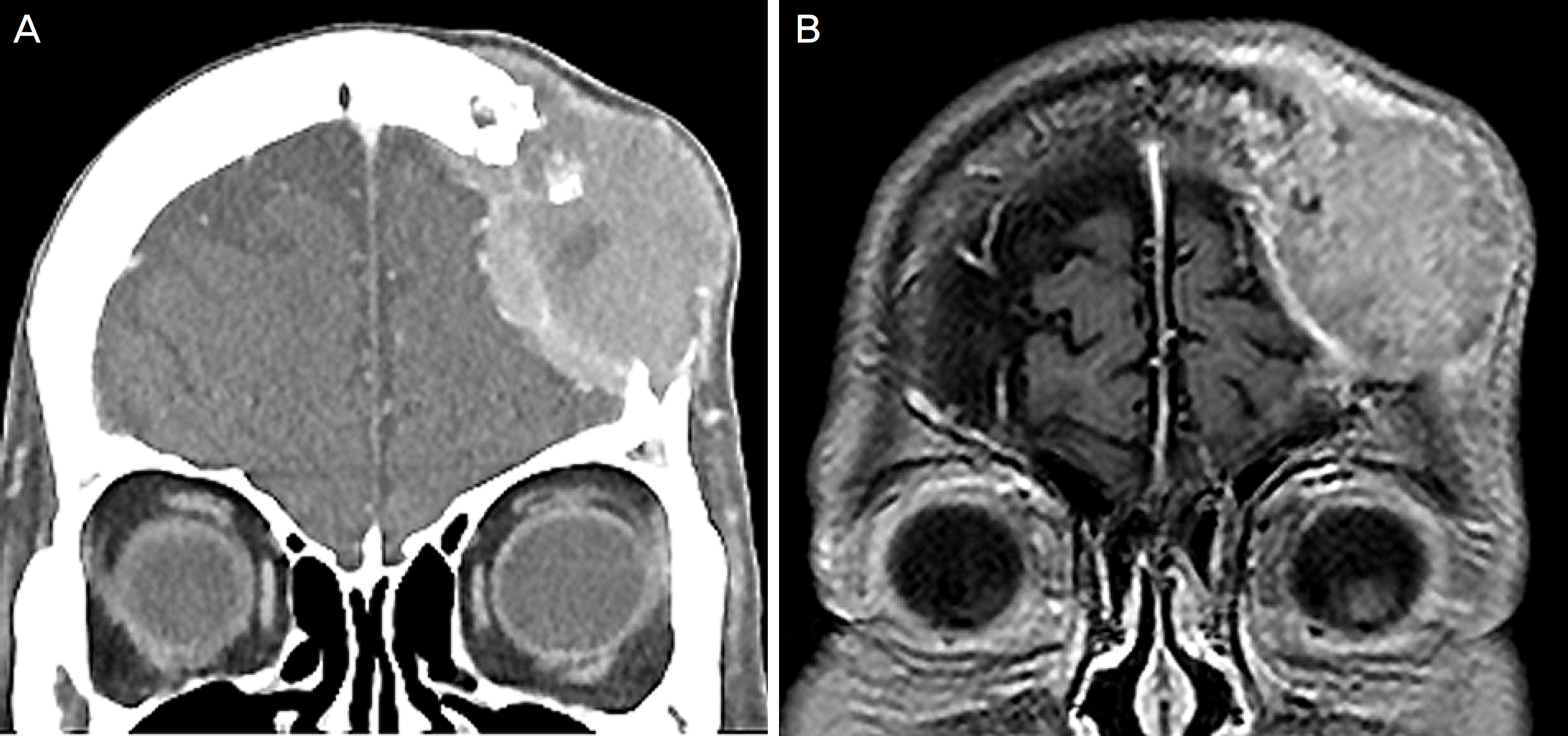
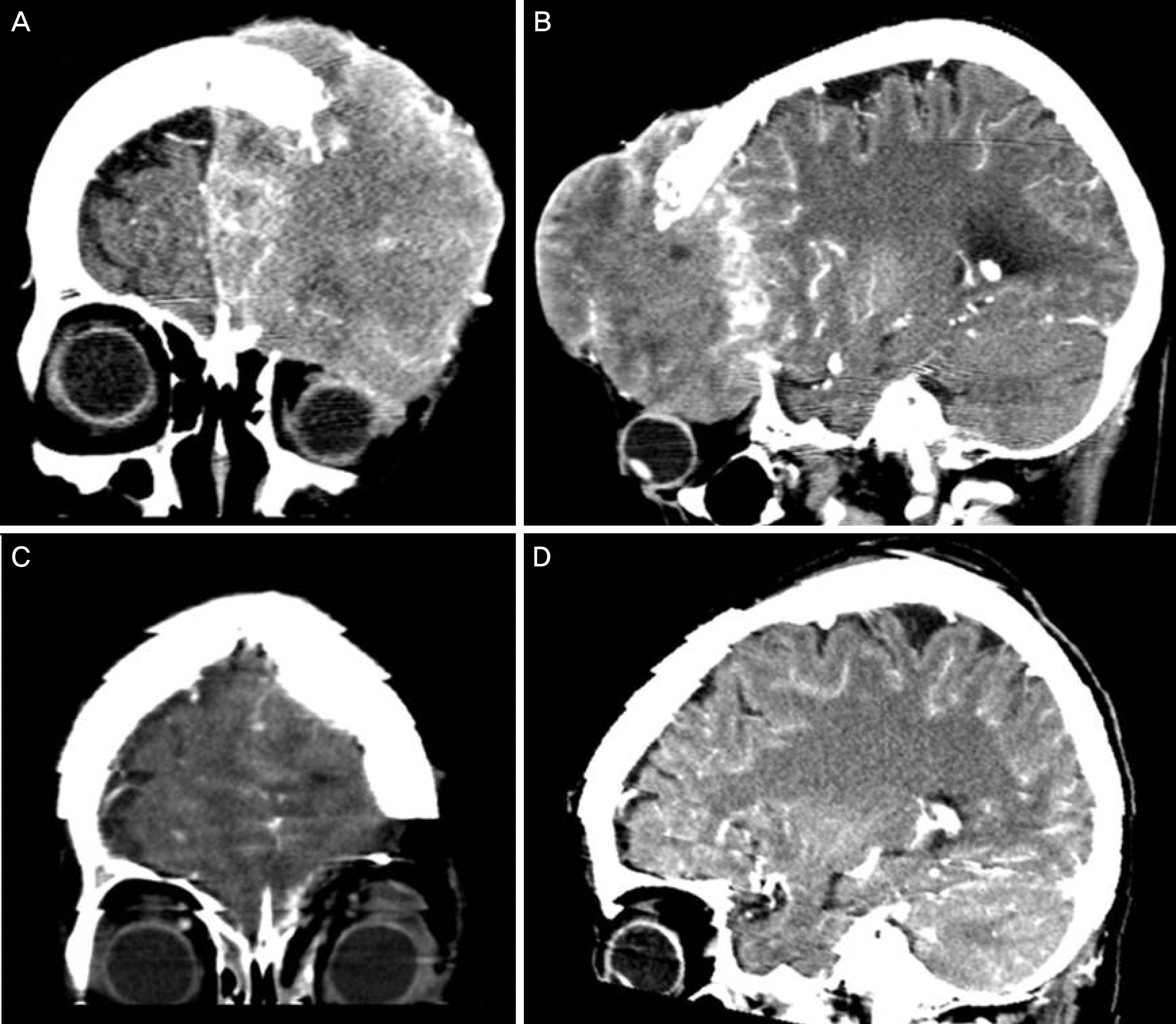
Figure 3.
Pre-operative brain computed tomography. (A, B) 10.9 × 9.9 × 7.0 cm heterogeneously enhanced extra-axial scalp mass with central necrosis on the left forehead. The left eyeball is deviated inferiorly and the left frontoparietal bone, left supero-lateral/medial bony orbit, left frontal bone, and left temporalis muscle were directly invaded. (C, D) Post-operative brain computed tomography. The inferior deviation of left eyeball was recovered.
 XML Download