

 PDF
PDF ePub
ePub Citation
 Print
Print


Abstract
Purpose
Dermatofibrosarcoma in the orbit is a rare malignant neoplasm. We report an extremely rare case of primary dermatofibrosarcoma in the orbit.
Case summary
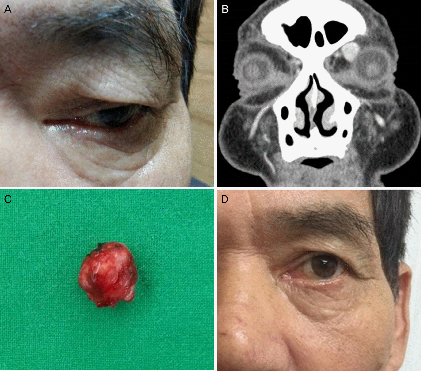
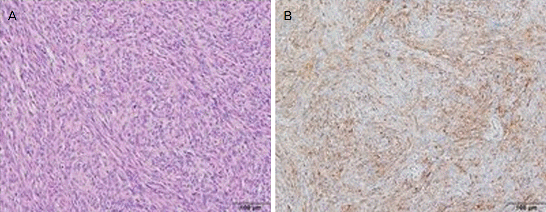
A 66-year-old male presented with a slowly progressing periorbital mass on his left upper eyelid which developed 3 weeks earlier. On physical examinations, a palpable firm mass under the skin was observed at the superomedial aspect of the left upper eyelid. However, there was no surface nodule or demarcated line on the eyelid. An approximately 1.2 × 1 × 1 cm sized well defined and clearly demancated mass was observed on orbital computed tomography. Excisional biopsy was performed under local anesthesia and pathological examination revealed dermatofibrosarcoma. There was no metabolic evidence of regional or distant metastasis based on positron emission tomography-computed tomography. Nine months after surgical excision there was no evidence of local recurrence.
References
1. Saiag P, Grob JJ, Lebbe C, et al. Diagnosis and treatment of abdominal protuberans. European consensus-based abdominal guideline. Eur J Cancer. 2015; 51:2604–8.
2. Erickson BP, Henry C, Alabiad CR. Recurrent dermatofibrosarcoma protuberans masquerading as a lacrimal sac neoplasm: a case report and review. Ophthal Plast Reconstr Surg. 2015; 31:e135–8.
3. Kim MS, Han TY, Lee JH, Son SJ. A case of congenital abdominal protuberans. Korean J Dermatol. 2010; 48:624–6.
4. Schittkowski MP, Wrede A. Dermatofibrosarcoma protuberans with primary orbital manifestation. Orbit. 2013; 32:117–9.

5. Nakra T, Cook T, Douglas RS, Goldberg RA. Dermatofibrosarcoma protuberans metastatic to the orbit. Arch Ophthalmol. 2004; 122:1240–1.
6. Ko HC, Jang BS, Kim MB, et al. A case of dermatofibrosarcoma protuberans on the face with various cutaneous lesions. Koreran J Dermatol. 2006; 44:1122–5.
7. Park JY, Jang YH, Kim YC. Subcutaneous dermatofibrosarcoma protuberans on the breast. Korean J Dermatol. 2011; 49:1025–27.
8. Li J, Ge X, Ma JM, Li M. Dermatofibrosarcoma protuberans. Ophthalmology. 2012; 119:197.e1–3.
9. Collazo Lorduy A, Obispo B, Villar K, et al. Dermatofibrosarcoma protuberans with lung metastasis requiring pneumonectomy. Rare Tumors. 2015; 7:5981.
Figure 1.
Clinical photos and Orbital computed tomography. Photograph of (A) 66-year-old man with a palpable firm mass under the skin at the superomedial aspect of the left upper eyelid. (B) Orbital computed tomography showed 1.2 × 1 × 1 cm sized well demarcated enhancing mass. (C) The orbital mass immediate after surgical excision. (D) The 9 months after operation showed no evidence of recurrence.
 XML Download