Abstract
Purpose
To investigate the clinical manifestations, management, ophthalmologic complications, and prognosis of traumatic optic neuropathy.
Methods
A retrospective survey of 55 patients who visited Chosun Hospital from April 2009 to February 2016 was performed. The sex, age, causes, fracture characteristics, neurologic injury, and combined craniofacial bone fractures of patients who were diagnosed with traumatic optic neuropathy were statistically analyzed. Also, we investigated the rate of visual impairment in the patients with intracranial hemorrhaging and craniofacial fracture on radiologic examination and development of sensory strabismus.
Results
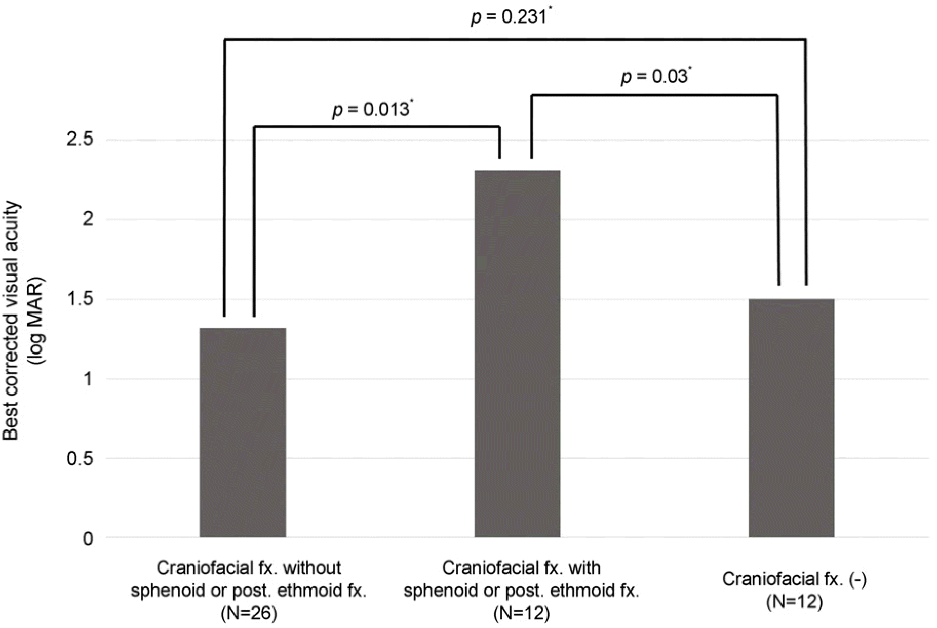
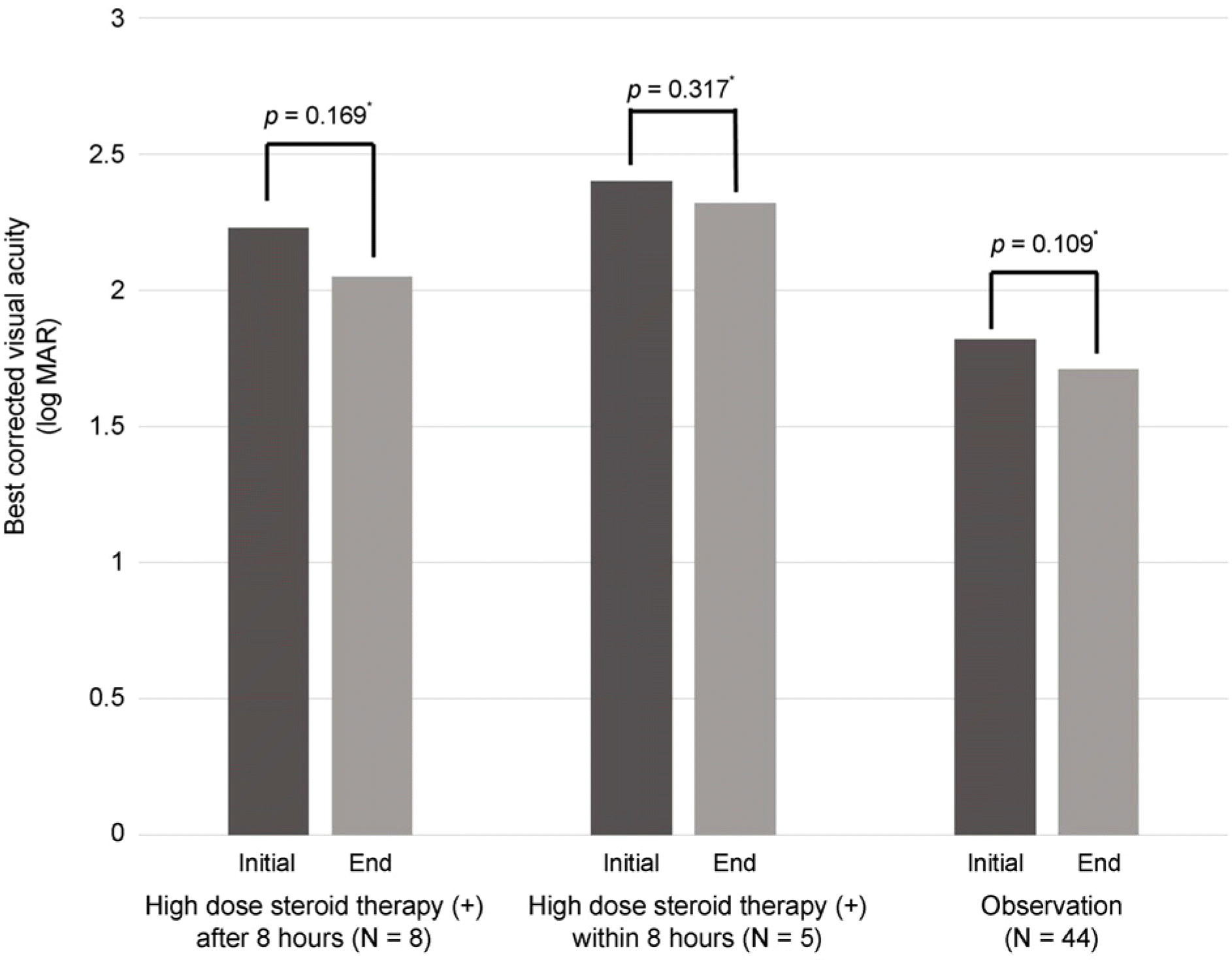
Traffic accidents were the most common cause of traumatic optic neuropathy. Among the patients, more than 60% showed severe visual impairment of less than 0.1 that lasted until the final observation. Altitudinal visual defects were the most common visual field defect and presented as marginal atrophy and central scotoma. While intracranial hemorrhaging was showed in 52.4% of the patients, craniofacial fracture was observed in 90.5% of the patients. The initial visual acuity was decreased when the patient presented with orbital fracture located in the retrobulbar area. Intravenous high-dose steroid injection did not affect visual prognosis. Sensory strabismus occurred more commonly under conditions of poor initial vision (p = 0.007) or young age (p < 0.001).
Go to : 
References
1. Burch FE. Ocular evidence of head trauma. Wisc Med J. 1942; 41:1092–7.
3. Seo JS, Jeong SK, Cho JS. Clinical evaluation of the traumatic abdominal neuropathy. J Korean Ophthalmol Soc. 1995; 36:1790–7.
4. Park JW, Jeong SK, Park YG. Clinical evaluation of the traumatic optic neuropathy. J Korean Ophthalmol Soc. 1999; 40:3497–505.
6. Sabates NR, Gonce MA, Farris BK. Neuro-ophthalmological abdominal in closed head trauma. J Clin Neuroophthalmol. 1991; 11:273–7.
7. Thurman DJ, Jeppson L, Burnett CL, et al. Surveillance of abdominal brain injuries in Utah. West J Med. 1996; 165:192–6.
8. Pirouzmand F. Epidemiological trends of traumatic optic nerve abdominal in the largest Canadian adult trauma center. J Craniofac Surg. 2012; 23:516–20.
9. Edmund J, Godtfredsen E. Unilateral optic atrophy following head injury. Acta Ophthalmol (Copenh). 1963; 41:693–7.

10. Levin LA, Beck RW, Joseph MP, et al. The treatment of traumatic optic neuropathy: the international optic nerve trauma study. Ophthalmology. 1999; 106:1268–77.
12. Chou PI, Sadun AA, Chen YC, et al. Clinical experiences in the management of traumatic optic neuropathy. Neuro-ophthalmology. 1996; 16:325–36.

13. Suchoff IB, Kapoor N, Ciuffreda KJ, et al. The frequency of abdominal, types, and characteristics of visual field defects in acquired brain injury: a retrospective analysis. Optometry. 2008; 79:259–65.
14. Warner N, Eggenberger E. Traumatic optic neuropathy: a review of the current literature. Curr Opin Ophthalmol. 2010; 21:459–62.

16. Dratviman-Storobinsky O, Hasanreisoglu M, Offen D, et al. Progressive damage along the optic nerve following induction of crush injury or rodent anterior ischemic optic neuropathy in trans-genic mice. Mol Vis. 2008; 14:2171–9.
17. Agrawal R, Shah M, Mireskandari K, et al. Controversies in ocular trauma classification and management: review. Int Ophthalmol. 2013; 33:435–45.

18. Ong HS, Qatarneh D, Ford RL, et al. Classification of orbital abdominal using the AO/ASIF system in a population surveillance abdominal of traumatic optic neuropathy. Orbit. 2014; 33:256–62.
19. Al-Qurainy IA, Stassen LF, Dutton GN, et al. The characteristics of midfacial fractures and the association with ocular injury: a abdominal study. Br J Oral Maxillofac Surg. 1991; 29:291–301.
20. Steinsapir KD, Goldberg RA. Traumatic optic neuropathy: a abdominal update. Compr Ophthalmol Update. 2005; 6:11–21.
21. Jeong JS, Kim DH. Associated injuries and prognosis in traumatic isolated 3rd, 4th, and 6th cranial nerve palsies. J Korean Ophthalmol Soc. 2014; 55:596–601.

22. Young W, Bracken MB. The Second National Acute Spinal Cord Injury Study. J Neurotrauma. 1992; 9(Suppl 1):S397–405.
23. Edwards P, Arango M, Balica L, et al. Final results of MRC CRASH, a randomized placebo-controlled trial of intravenous corticosteroid in adults with head injury-outcomes as 6 months. Lancet. 2005; 365:1957–9.
24. Braughler JM, Hall ED, Means ED, et al. Evaluation of an abdominal methylprednisolone sodium succinate dosing regimen in experimental spinal cord injury. J Neurosurg. 1987; 67:102–5.
25. Rajiniganth MG, Gupta AK, Gupta A, Bapuraj JR. Traumatic optic neuropathy: visual outcome following combined therapy protocol. Arch Otolaryngol Head Neck Surg. 2003; 129:1203–6.
26. Ropposch T, Steger B, Meço C, et al. The effect of steroids in abdominal with optic nerve decompression surgery in traumatic optic neuropathy. Laryngoscope. 2013; 123:1082–6.
27. Wang BH, Robertson BC, Girotto JA, et al. Traumatic optic abdominal: a review of 61 patients. Plast Reconstr Surg. 2001; 107:1655–64.
28. Carta A, Ferrigno L, Slavo M, et al. Visual prognosis after indirect traumatic optic neuropathy. J Neurol Neurosurg Psychiatry. 2003; 74:246–8.

29. Havertape SA, Cruz OA, Chu FC. Sensory strabismus–eso or exo? J Pediatr Ophthalmol Strabismus. 2001; 38:327–30. quiz 354–5.

30. Sidikaro Y, Von Noorden GK. Observation in sensory heterotropia. J Pediatr Ophthalmol Starabismus. 1982; 19:12–9.
31. Kim KS, Park SC. The clinical consideration of sensory strabismus. J Korean Ophthalmol Soc. 2005; 46:316–22.
Go to : 
 | Figure 1.Initial visual acuity difference between the groups of patients classified by involvement of posterior wall fracture in craniofacial fracture. fx. = fracture; Post. = posterior. * Mann whitney U-test. |
 | Figure 2.Initial and final visual acuity in group of patients classified by steroid treatment. * Wilcoxson signed rank test. |
Table 1.
Visual acuity and color vision of traumatic optic neuropathy patients
| First visit | Final visit | |
|---|---|---|
| Visual acuity | ||
| >0.5 | 4 (7.0%) | 5 (8.7%) |
| 0.1–0.5 | 18 (31.5%) | 17 (29.8%) |
| <0.1 | 35 (61.4%) | 35 (61.4%) |
| Ishihara test | 2.4 ± 1.5 | 1.9 ± 1.1 |
Table 2.
Visual field defect distribution in traumatic optic neuropathy patients
Table 3.
Accompanying injury of traumatic optic neuropath patients
Table 4.
Univariate analysis with visual prognostic factor for traumatic optic neuropathy
| Independent variable | No. of cases | No. of recovery of visual acuity (N, %) | RR* | CI (95%) |
|---|---|---|---|---|
| Initial visual acuity below finger count | 29 | 27 (93.1) | 3.77 | 1.65–6.78 |
| Sphenoid or Posterior ethmoid Fx. | 12 | 10 (83.3) | 2.12 | 1.26–3.74 |
| Optic canal Fx. | 4 | 3 (75.0) | 1.98 | 1.43–2.79 |
| Loss of consciousness | 35 | 32 (91.4) | 4.18 | 1.52–2.47 |
| Nerve palsy associated | 10 | 5 (50) | 0.87 | 0.84–3.51 |
Table 5.
Influencing factors, angle of deviation, and type of sensory strabismus after traumatic optic neuropathy
| Sensory strabismus (+)(N = 8) | Sensory strabismus (−)(N = 12) | p-value* | |
|---|---|---|---|
| Age (years) | 14.1 ± 4.4 | 43.0 ± 15.8 | p < 0.001 |
| BCVA (log MAR) | 2.8 ± 0.4 | 1.6 ± 1.1 | p = 0.007 |
| Angle of deviation | 26.3 ± 14.4 (exotropia) |


 PDF
PDF ePub
ePub Citation
 Print
Print


 XML Download