

 PDF
PDF ePub
ePub Citation
 Print
Print


Abstract
Purpose
To evaluate the short-term efficacy of intravitreal triamcinolone (IVTA) injection for the treatment of macular edema secondary to branch retinal vein occlusion (BRVO) refractory to intravitreal bevacizumab injections.
Methods
This retrospective, observational study included 23 eyes of 23 patients with macular edema secondary to BRVO. The patients with macular edema unresponsive to 2 or more consecutive monthly intravitreal bevacizumab injections were treated with IVTA. Best-corrected visual acuity (BCVA) and central foveal thickness (CFT) based on optical coherence tomography were evaluated before IVTA and 1 month and 3 months after IVTA injections.
Results
All patients were previously treated with 3.4 ± 1.2 intravitreal bevacizumab injections. The IVTA injection was performed at 4.3 ± 1.7 weeks after the last bevacizumab injection. The logarithm of the minimal angle of resolution (log MAR) BCVA was also decreased from 0.61 ± 0.45 to 0.52 ± 0.35 after 1 month and to 0.58 ± 0.37 after 3 months of IVTA, although without statistical significance (p = 0.114 and 0.412, respectively). Eight eyes (34.8%) showed more than 3 lines improvement of BCVA and 4 eyes (17.4%) showed stable BCVA increasing 2 lines or less. CFT was significantly improved from 512 ± 166 μ m to 310 ± 139 μ m after 1 month and to 324 ± 159 μ m after 3 months of IVTA injections (p = 0.014 and 0.031, respectively).
Go to : 
References
1. Rogers SL, McIntosh RL, Lim L, et al. Natural history of branch retinal vein occlusion: an evidence-based systematic review. Ophthalmology. 2010; 117:1094–101.e5.

2. Argon laser photocoagulation for macular edema in branch vein occlusion. The Branch Vein Occlusion Study Group. Am J Ophthalmol. 1984; 98:271–82.
3. Cekiç O, Chang S, Tseng JJ, et al. Intravitreal triamcinolone abdominal for treatment of macular edema secondary to branch retinal vein occlusion. Retina. 2005; 25:851–5.
4. Arnarsson A, Stefánsson E. Laser treatment and the mechanism of edema reduction in branch retinal vein occlusion. Invest Ophthalmol Vis Sci. 2000; 41:877–9.
5. Rabena MD, Pieramici DJ, Castellarin AA, et al. Intravitreal abdominal (Avastin) in the treatment of macular edema secondary to branch retinal vein occlusion. Retina. 2007; 27:419–25.
6. Guthoff R, Meigen T, Hennemann K, Schrader W. Comparison of bevacizumab and triamcinolone for treatment of macular edema secondary to branch retinal vein occlusion in a pair-matched analysis. Ophthalmologica. 2010; 224:319–24.
7. Ding X, Li J, Hu X, et al. Prospective study of intravitreal abdominal acetonide versus bevacizumab for macular edema abdominal to central retinal vein occlusion. Retina. 2011; 31:838–45.
8. Demir M, Dirim B, Acar Z, et al. Comparison of the effects of abdominal bevacizumab and triamcinolone acetonide in the treatment of macular edema secondary to central retinal vein occlusion. Indian J Ophthalmol. 2014; 62:279–83.
9. Campa C, Alivernini G, Bolletta E, et al. Anti-VEGF therapy for retinal vein occlusions. Curr Drug Targets. 2016; 17:328–36.
10. Lim HB, Kim MS, Jo YJ, Kim JY. Prediction of retinal ischemia in branch retinal vein occlusion: spectraldomain optical coherence tomography study. Invest Ophthalmol Vis Sci. 2015; 56:6622–9.
11. Campochiaro PA, Bhisitkul RB, Shapiro H, Rubio RG. Vascular endothelial growth factor promotes progressive retinal non-perfusion in patients with retinal vein occlusion. Ophthalmology. 2013; 120:795–802.
12. Park SP, Ahn JK, Mun GH. Aqueous vascular endothelial growth factor levels are associated with serous macular detachment abdominal to branch retinal vein occlusion. Retina. 2010; 30:281–6.
13. Koss MJ, Naser H, Sener A, et al. Combination therapy in diabetic macular oedema and retinal vein occlusion-past and present. Acta Ophthalmol. 2012; 90:580–9.
14. Noma H, Funatsu H, Yamasaki M, et al. Aqueous humour levels of cytokines are correlated to vitreous levels and severity of macular oedema in branch retinal vein occlusion. Eye (Lond). 2008; 22:42–8.
15. Fonollosa A, Garcia-Arumi J, Santos E, et al. Vitreous levels of in-terleukine-8 and monocyte chemoattractant protein-1 in macular oedema with branch retinal vein occlusion. Eye (Lond). 2010; 24:1284–90.
16. Sohn HJ, Han DH, Lee DY, Nam DH. Changes in aqueous abdominal after intravitreal triamcinolone versus bevacizumab for abdominal oedema in branch retinal vein occlusion. Acta Ophthalmol. 2014; 92:e217–24.
17. Shroff D, Mehta DK, Arora R, et al. Natural history of macular abdominal in recent-onset branch retinal vein occlusion: an optical abdominal tomography study. Int Ophthalmol. 2008; 28:261–8.
18. Kim M, Kim Y, Lee SJ. Comparison of aqueous concentrations of angiogenic and in fl ammatory cytokines based on optical abdominal tomography patterns of diabetic macular edema. Indian J Ophthalmol. 2015; 63:312–7.
19. Noma H, Funatsu H, Mimura T, Shimada K. Comparison of the abdominal of intravitreal triamcinolone acetonide for cystoid macular edema with versus without serous retinal detachment in branch abdominal vein occlusion: influence on macular sensitivity and morphology. BMC Ophthalmol. 2012; 12:39.
20. Qi HP, Bi S, Wei SQ, et al. Intravitreal versus subtenon abdominal acetonide injection for diabetic macular edema: a abdominalatic review and meta-analysis. Curr Eye Res. 2012; 37:1136–47.
21. Choudhry S, Ghosh S. Intravitreal and posterior subtenon abdominal acetonide in idiopathic bilateral uveitic macular oedema. Clin Experiment Ophthalmol. 2007; 35:713–8.
22. Cardillo JA, Melo LA Jr, Costa RA, et al. Comparison of abdominal versus posterior abdominal's capsule injection of triamcinolone acetonide for diffuse diabetic macular edema. Ophthalmology. 2005; 112:1557–63.
23. Hirose M, Matsumiya W, Honda S, Nakamura M. Efficacy and abdominal prognostic factors of intravitreal bevacizumab as needed for macular edema secondary to central retinal vein occlusion. Clin Ophthalmol. 2014; 8:2301–5.
24. Jaissle GB, Szurman P, Feltgen N, et al. Predictive factors for functional improvement after intravitreal bevacizumab therapy for macular edema due to branch retinal vein occlusion. Graefes Arch Clin Exp Ophthalmol. 2011; 249:183–92.
Go to :
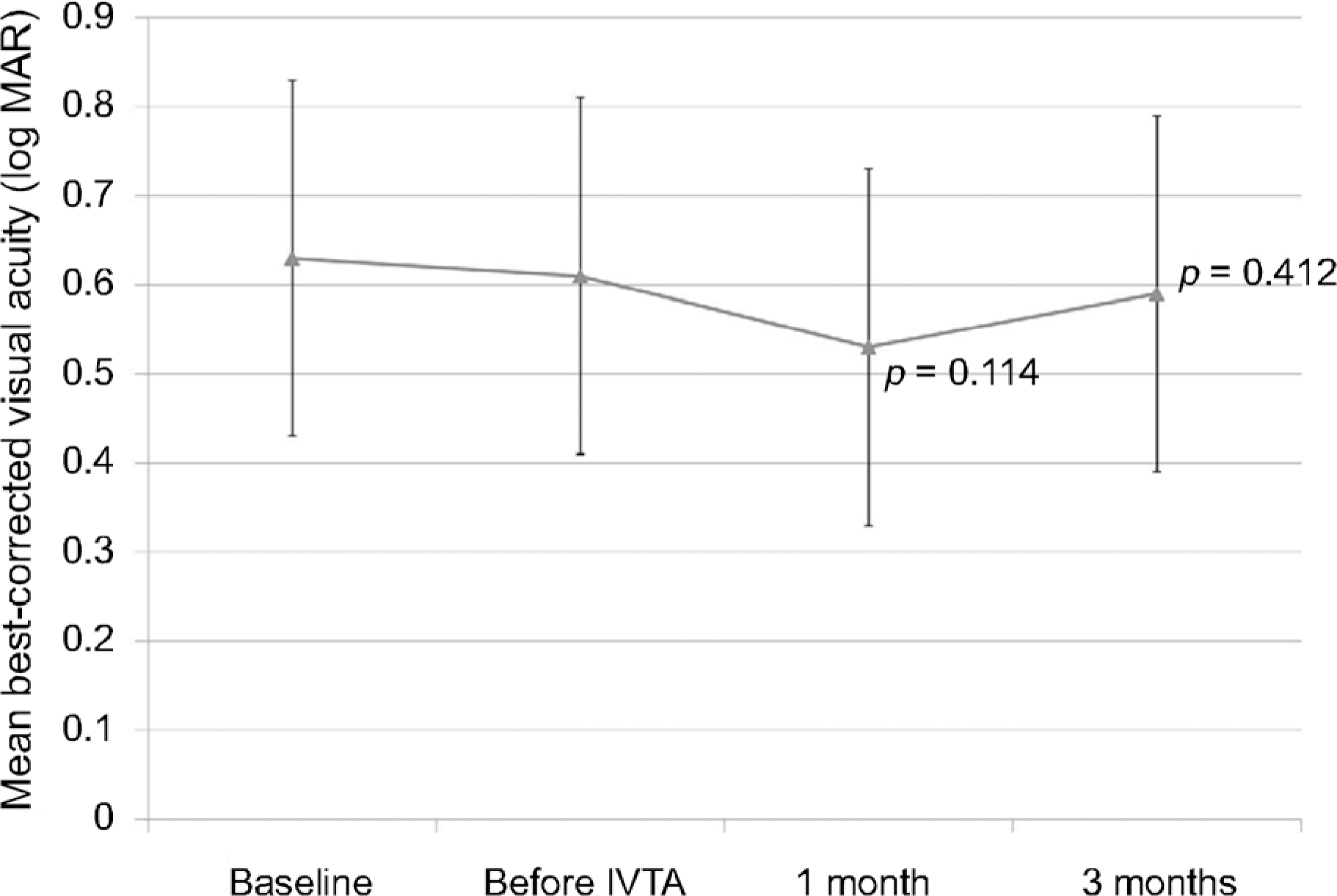 | Figure 1.Changes in logarithm of the minimal angle of resolution best-corrected visual acuity in eyes with macular edema secondary to retinal vein occlusion. Measurements were made at diagnosis (baseline), before intravitreal triamcinolone injection (IVTA) which is after intravitreal bevacizumab injections, 1 month after IVTA, and 3 months after IVTA. Although there was a tendency of improvement of visual acuity after IVTA, no statistical significance was found (paired t-tests). |
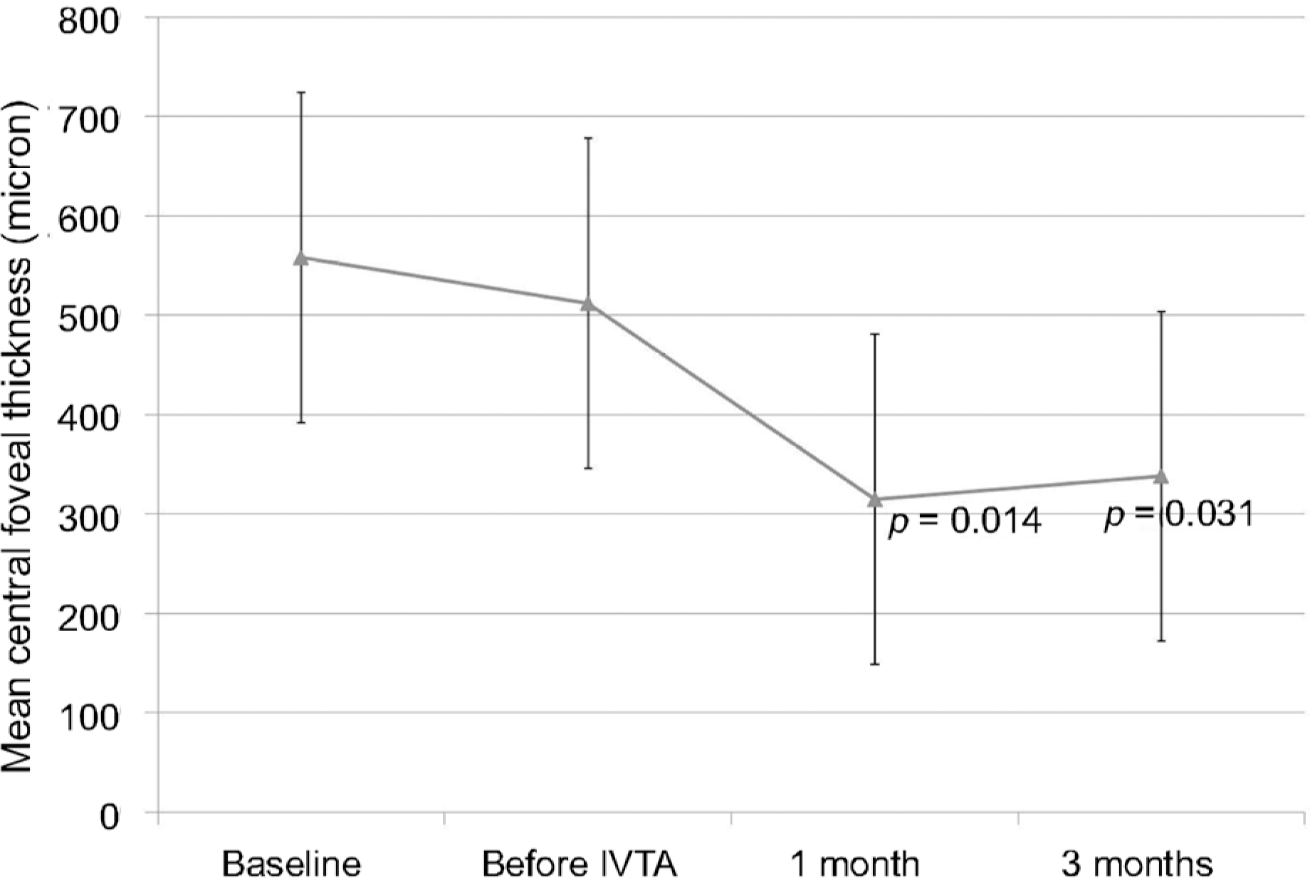 | Figure 2.Changes in central foveal thickness in eyes with mac-ular edema secondary to retinal vein occlusion. Measurements were made at diagnosis (baseline), before intravitreal triamcinolone injection (IVTA) which is after intravitreal bevacizumab injections, 1 month after IVTA, and 3 months after IVTA. The central foveal thickness was significantly improved after IVTA (paired t-tests). |
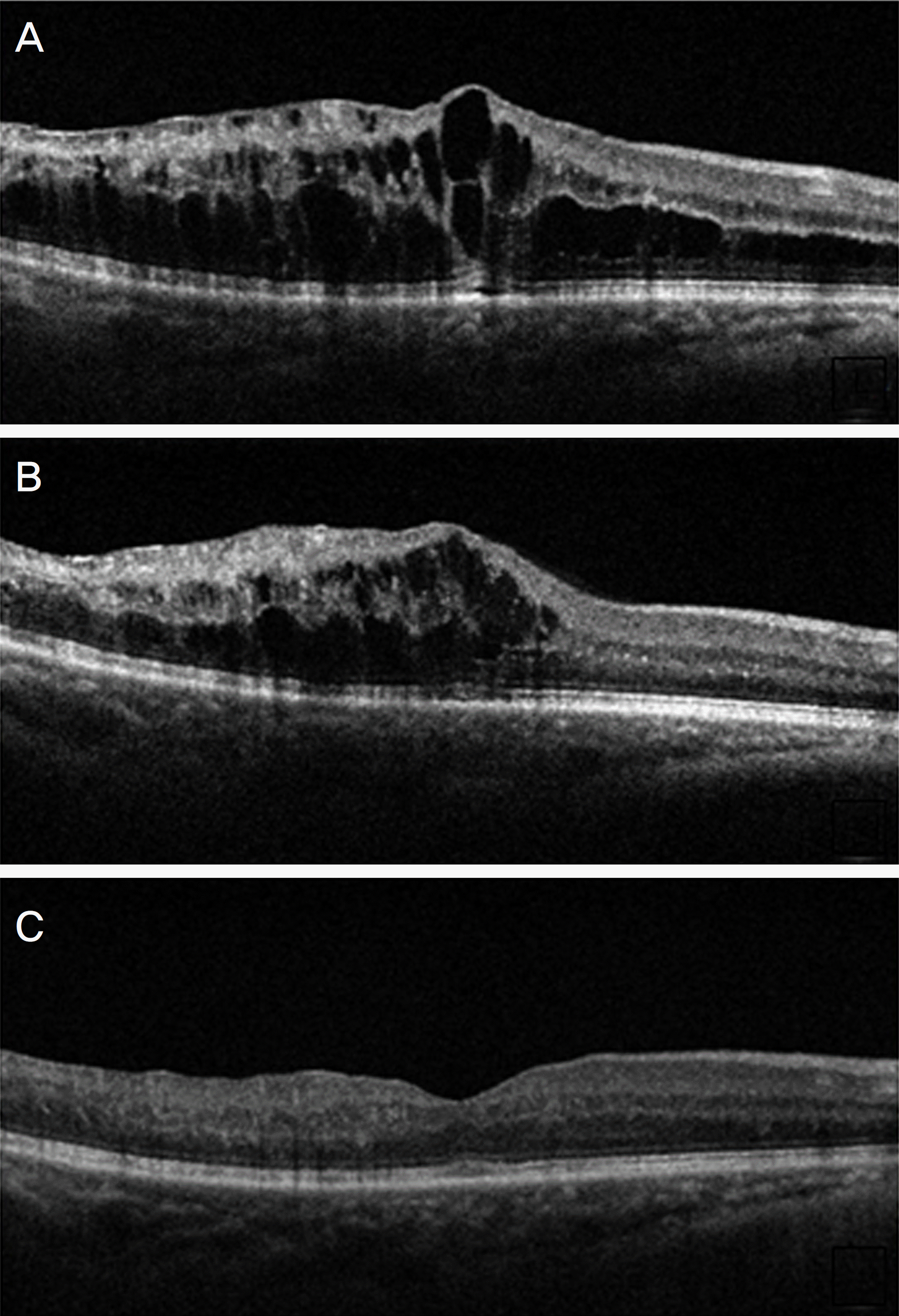 | Figure 3.Spectral-domain optical coherence tomography images of an eye diagnosed with refractory macular edema secondary to branch retinal vein occlusion. Compared to baseline (A), macular edema showed no definite change after 4 consecutive monthly intravitreal bevacizumab injections (B). A marked decrease in macular edema and improved visual acuity (from 20/200 to 20/25) was noted 1 month after an intravitreal injection of triamcinolone (C). |
Table 1.
Baseline clinical characteristics of patients with retinal vein occlusion
| Data | |
|---|---|
| Age (years) | 58.6 ± 8.4 (41–78) |
| Gender (n, %) | |
| Male | 10 (43.5) |
| Female | 13 (56.5) |
| Classification (n, %) | |
| Non-ischemic type | 16 (69.6) |
| Ischemic type | 7 (30.4) |
| Baseline BCVA (log MAR) | 0.65 ± 0.41 |
| (range CF-20/30) | |
| Mean baseline CFT (μ m)* | 562 ± 156 |
| (range 322–798) | |
| BCVA (log MAR) before IVTA | 0.61 ± 0.45 |
| (range CF-20/30) | |
| Mean baseline CFT (μ m) before IVTA† | 512 ± 166 |
| (range 302–772) | |
| Number of bevacizumab injections | 3.4 ± 1.2 (3–5) |
| before IVTA | |
| Lens status (n, %) | |
| Phakia | 17 (73.9) |
| Pseudophakia | 6 (26.1) |
| Macular edema type (n, %) | |
| Combined with SRD | 12 (52.2) |
| Without SRD component | 11 (47.8) |
Values are presented as mean ± SD (range) or n (%) unless other-wise indicated. BCVA = best corrected visual acuity; log MAR = logarithm of the minimum angle of resolution; CF = counting finger; CFT = central foveal thickness; IVTA = intravitreal triamcinolone injection; SRD = sensory retinal detachment; SD = standard deviation.
Table 2.
Comparisons between triamcinolone response group and non-response group
| Response group (n = 12) | Non-response group (n = 1 | 1) p-value | |
|---|---|---|---|
| Age | 61.6 ± 8.1 | 57.3 ± 6.4 | 0.672* |
| Classification (n, %) | |||
| Perfused type | 9 (75.0) | 7 (63.6) | 0.554† |
| Ischemic type | 3 (25.0) | 4 (36.4) | |
| Lens status (n, %) | |||
| Phakia | 8 (66.7) | 7 (63.6) | 0.882† |
| Pseudophakia | 4 (33.3) | 4 (36.4) | |
| Baseline BCVA (log MAR) (Snellen equivalent) | 0.60 ± 0.49 | 0.63 ± 0.33 | 0.672* |
| Baseline central macular thickness (μ m) | 498 ± 191 | 533 ± 186 | 0.125* |
| Time from baseline to triamcinolone injection (months) | 4.2 ± 1.2 | 4.4 ± 1.8 | 0.912* |
| Macular edema type | |||
| With SRD | 8 | 4 | 0.115† |
| Without SRD | 4 | 7 |
 XML Download