Abstract
Purpose
To investigate the clinical manifestations and characteristics of extruded conjunctival melanocytic mass. Methods: A total of 33 patients who had extruded conjunctival melanocytic mass and who underwent excisional biopsy were retrospectively reviewed.
Results
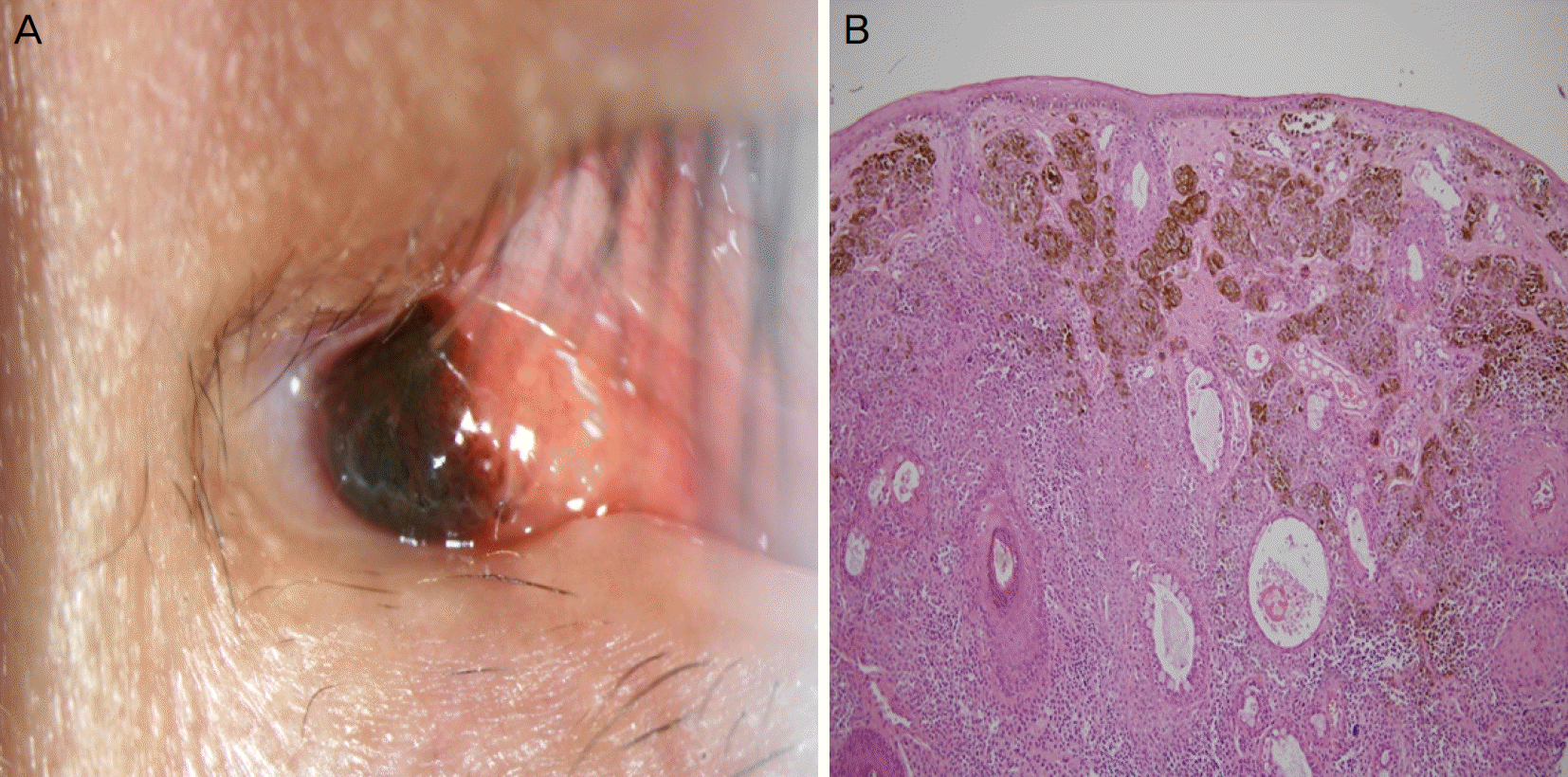
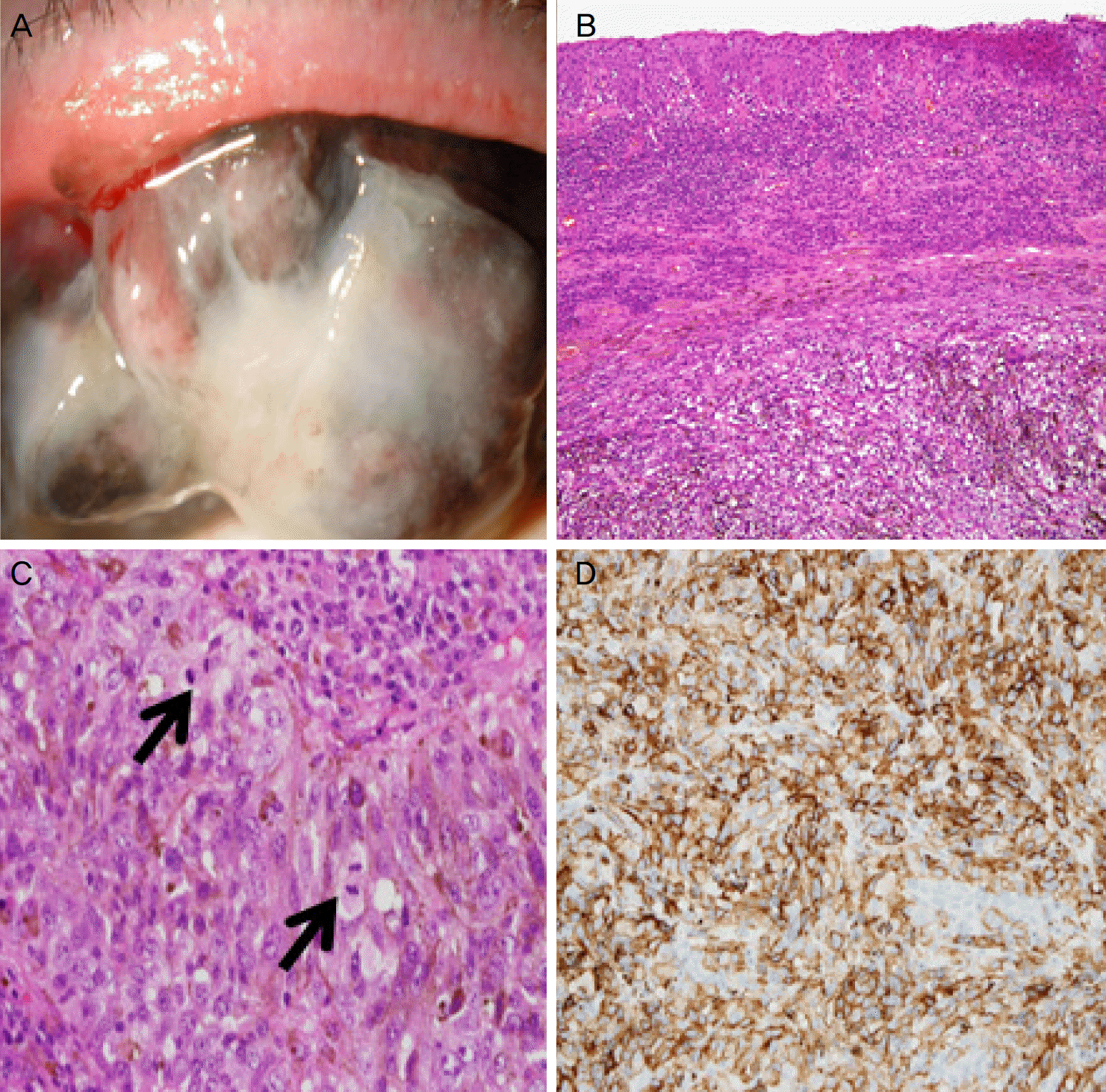
Based on the excisional biopsy results, 13 patients (40%) were diagnosed with compound nevus, nine patients (27%) with subepithelial nevus, eight patients (24%) with primary acquired melanosis without atypia, and three patients (9%) with malignant melanoma. Compound nevus was located on the temporal side of the cornea in 54% of affected cases, bulbar conjunctival in 77%, and was partially pigmented (brown) in 61%. The average size of the melanocytic mass was 24 mm when histological analysis showed melanin nevus cells in the conjunctival epithelial layer and subepithelial stromal layer. Subepithelial nevus was located on the temporal side of the cornea (56%) and in the bulbar conjunctival (78%) and had a brown color (78%). The average size of the melanocytic mass was 28 mm when histological analysis showed melanin nevus cells located only in the subepithelial stromal layer and forming nest shapes. Primary acquired melanosis without atypia was located on the temporal side of the cornea (62.5%) and bulbar conjunctival (75%) and had brown color (75%). The average size of melanin nevus cells located only in the basement membrane of the epithelial layer was 30 mm. Three of these masses were malignant melanoma, and all cases were located on the superior side of the cornea and palpebral conjunctiva. All cases were black and had an average size of 53 mm, with malignant cells observed in all layers of the conjunctiva and connective tissue.
Go to : 
References
1. Shields JA, Shields CL. Atlas of Eyelid and Conjunctival Tumors. 1st ed.Philadelphia: Lippincott Williams & Wilkins;1999. p. 199–334.
5. Grossniklaus HE, Green WR, Luckenbach M, Chan CC. Conjunctival lesions in adults: a clinical and histopathologic review. Cornea. 1987; 6:78–116.
6. Chung YS, Kim CY, Yoon YD. A case of conjunctival melanoma. J Korean Ophthalmol Soc. 2000; 41:1012–6.
7. Folberg R, McLean IW, Zimmerman LE. Conjunctival melanosis and melanoma. Ophthalmology. 1984; 91:673–8.

8. Rosenberg C, Finger PT. Cutaneous malignant melanoma abdominal to the eye, lids, and orbit. Surv Ophthalmol. 2008; 53:187–202.
9. Folberg R, Jakobiec FA, Bernardino VB, Iwamoto T. Benign conjunctival melanocytic lesions: clinicopathologic features. Ophthalmology. 1989; 96:436–61.
10. Shields CL, Fasiudden AF, Mashayekhi A, Shields JA. Conjunctival nevi: clinical features and natural course in 410 consecutive patients. Arch Ophthalmol. 2004; 122:167–75.
11. Zembowicz A, Mandal RV, Choopong P. Melanocytic lesions of the conjunctiva. Arch Pathol Lab Med. 2010; 134:1785–92.

12. Sharara NA, Alexander RA, Luthert PJ, et al. Differential immunoreactivity of melanocytic lesions of the conjunctiva. Histopathology. 2001; 39:426–31.

13. Maly A, Epstein D, Meir K, Pe'er J. Histological criteria for abdominal of atypia in melanocytic conjunctival lesions. Pathology. 2008; 40:676–81.
14. Jeoung JW, Kim TI, Lee JH, et al. Argon laser ablation of abdominal nevus. J Korean Ophthalmol Soc. 2004; 45:1989–94.
15. Park JJ, Jeong BJ, Seo HD, et al. Treatment of conjunctival nevus with argon laser. J Korean Ophthalmol Soc. 2004; 45:1995–9.
16. Sugiura M, Colby KA, Mihm MC Jr, Zembowicz A. Low-risk and high-risk histologic features in conjunctival primary acquired melanosis with atypia: clinicopathologic analysis of 29 cases. Am J Surg Pathol. 2007; 31:185–92.

17. Jakobiec FA, Folberg R, Iwamoto T. Clinicopathologic abdominal of premalignant and malignant melanocytic lesions of the conjunctiva. Ophthalmology. 1989; 96:147–66.
18. Jakobiec FA. The ultrastructure of conjunctival melanocytic tumors. Trans Am Ophthalmol Soc. 1984; 82:599–752.
19. Gloor P, Alexandrakis G. Clinical characterization of primary abdominal melanosis. Invest Ophthalmol Vis Sci. 1995; 36:1721–9.
20. Seregard S, Kock E. Conjunctival malignant melanoma in Sweden 1969–91. Acta Ophthalmol (Copenh). 1992; 70:289–96.

21. Reese AB. Tumors of the Eye. 2nd ed.New York: Harper & Row;1963. p. 214–363.
22. Moon JS, Lee JY, Chung SK, Yu YS. A conjunctival malignant melanoma involving the cornea. J Korean Ophthalmol Soc. 1998; 39:1035–9.
23. Shields JA, Shields CL, De Potter P. Surgical management of abdominal tumors: the 1994 Lynn B. McMahan Lecture. Arch Ophthalmol. 1997; 115:808–15.
24. Shields CL, Shields JA, Armstrong T. Management of conjunctival and corneal melanoma with surgical excision, amniotic membrane allograft, and topical chemotherapy. Am J Ophthalmol. 2001; 132:576–8.

25. Kim SY, Lee SB, Yang SW. A case of conjunctival malignant abdominal with extensive corneal displacement. J Korean Ophthalmol Soc. 2005; 46:1235–9.
26. Tsujii H, Mizoe J, Kamada T, et al. Clinical results of carbon ion abdominal at NIRS. J Radiat Res. 2007; 48(Suppl A):A1–13.
Go to : 
 | Figure 1.Slit-lamp photograph and Histologic feature of Compound Nevus. (A) Slit-lamp photograph shows pigmented conjunctival mass at caruncle. (B) Histologic features of the conjunctival mass (Hematoxylin and eosin stain, ×100) shows Melanin pigments (melanocyte) in the epidermis near side, cells are in the dermis as well as in the epidermis. Melanocyte formed a nest shape. Tissue shows several stromal cysts and neurotinization (cell size is getting smaller near the dermis). It is the differentiation to neural cell and differentiation means that this tissue is benign. |
 | Figure 2.Slit-lamp photograph and Histologic features of Subepithelial Nevus. (A) Slit-lamp photograph shows pigmented conjunctival mass at superior side of bulbar conjunctiva. (B) Histologic features of the conjunctival mass shows nevus cells that has melanin pigment are in the subepithelial layer but, not in the epithelium. Nevus cells formed a sheet or nest shape (Hematoxylin and eosin stain, ×400). (C) Tissue shows no atypia. Therefore this tissue is not dysplastic or malignant melanoma, but benign nevus (Hematoxylin and eosin stain, ×40). |
 | Figure 3.Slit-lamp photograph and Histologic features of primary acquired melanosis (PAM). (A) Slit-lamp photograph shows pigmented conjunctival mass at nasal bulbar conjunctiva. (B) Histologicfeatures of the conjunctival mass. Melanocyte proliferate intra-epithelial layer without nuclear atypia. Tissue show no stromal invasion and no mitosis (Hematoxylin and eosin stain, ×200). (C) HMB45 stain positive findings of conjunctival mass, ×200. |
 | Figure 4.Slit-lamp photograph and Histologic features of Malignant melanoma. (A) Slit-lamp photograph shows pigmented conjunctival mass at superior palpebral conjunctiva. (B) Histologicfeatures of the conjunctival mass. Large nuclear cells with increased cellularity. Prominent nucleoli with melanin pigments (Hematoxylin and eosin stain, ×100). (C) Histologic features of the conjunctival mass showed severe mitosis (arrows) (Hematoxylin and eosin stain, ×400). (D) HMB45 stain positive findings of conjunctival mass. |
Table 1.
Location and pigmentation of compound nevus
Table 2.
Location and pigmentation of subepithelial nevus
Table 3.
Location and pigmentation of primary acquired melanosis
Table 4.
Location and pigmentation of malignant melanoma


 PDF
PDF ePub
ePub Citation
 Print
Print


 XML Download