Abstract
Purpose
To report the good surgical results of multiple ruptured rectus muscles with avulsion of the optic nerve.
Case summary
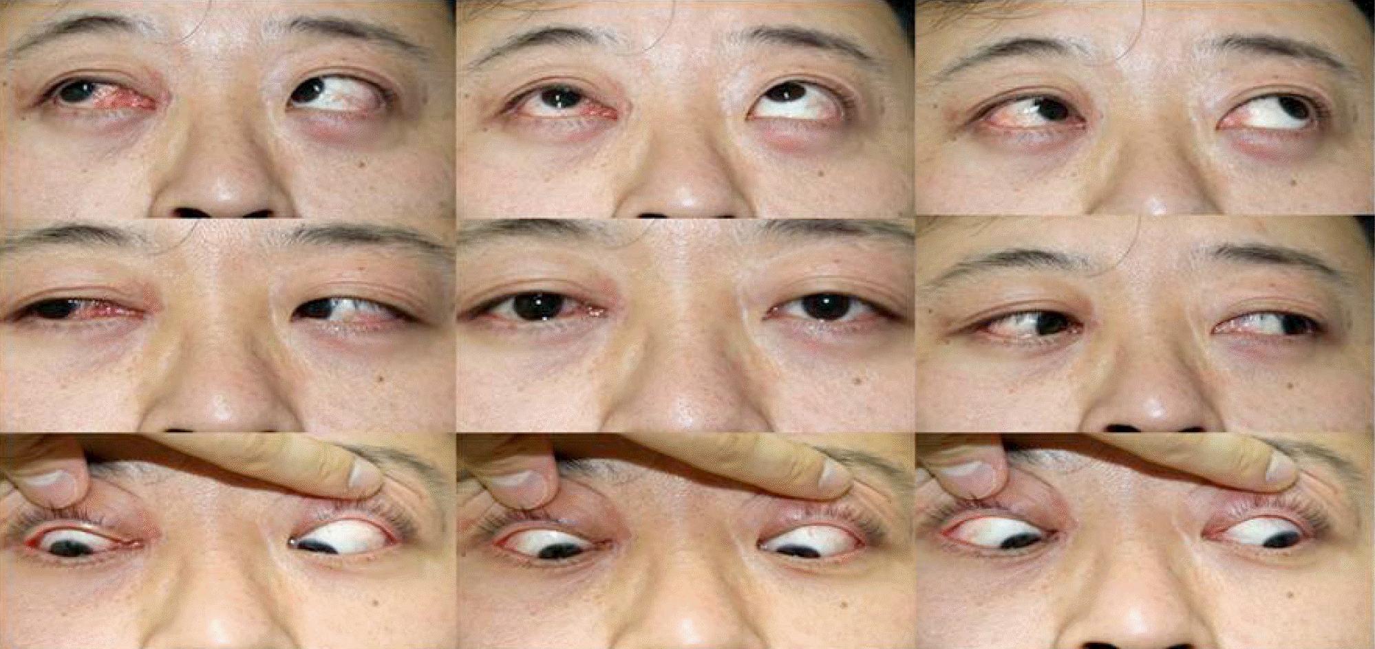
A 39-year-old male patient underwent surgical exploration after rupture of the inferior and medial rectus muscles and avulsion of the optic nerve. The disinserted muscles were attached at the primary insertion site, and a served optic nerve was not found. Six months after the injury, the patient had orthotropia in the primary position without ischemia of the anterior segment.
References
1. Richards R. Ocular motility disturbances following trauma. Adv Ophthalmic Plast Reconstr Surg. 1987; 7:133–47.
2. Paysse EA, Saunders RA, Coats DK. Surgical management of abdominal after rupture of the inferior rectus muscle. J AAPOS. 2000; 4:164–7.
3. Bloom PA, Harrad R. Medial rectus rupture; a rare condition with an unusual presentation. J R Soc Med. 1993; 86:112–3.
5. Ludwig IH, Brown MS. Flap tear of rectus muscles: an underlying cause of strabismus after orbital trauma. Ophthal Plast Reconstr Surg. 2002; 18:443–9. discussion 450.
6. Kashima T, Akiyama H, Kishi S. Longitudinal tear of the inferior rectus muscle in orbital floor fracture. Orbit. 2012; 31:171–3.

7. Fard AK, Merbs SL, Pieramici DJ. Optic nerve avulsion from a diving injury. Am J Ophthalmol. 1997; 124:562–4.

8. Foster BS, March GA, Lucarelli MJ, et al. Optic nerve avulsion. Arch Ophthalmol. 1997; 115:623–30.

9. Sanborn GE, Gonder JR, Goldberg RE, et al. Evulsion of the optic nerve: a clinicopathological study. Can J Ophthalmol. 1984; 19:10–6.
10. France TD, Simon JW. Anterior segment ischemia syndrome following muscle surgery: the AAPO&S experience. J Pediatr Ophthalmol Strabismus. 1986; 23:87–91.
11. de Smet MD, Carruthers J, Lepawsky M. Anterior segment ischemia treated with hyperbaric oxygen. Can J Ophthalmol. 1987; 22:381–3.
12. Simon JW, Price EC, Krohel GB, et al. Anterior segment ischemia following strabismus surgery. J Pediatr Ophthalmol Strabismus. 1984; 21:179–85.

13. Huerva V, Mateo AJ, Espinet R. Isolated medial rectus muscle rupture after a traffic accident. Strabismus. 2008; 16:33–7.

14. O'Toole L, Long V, Power W, O'Connor M. Traumatic rupture of the lateral rectus. Eye (Lond). 2004; 18:221–2. discussion 2.
Figure 1.
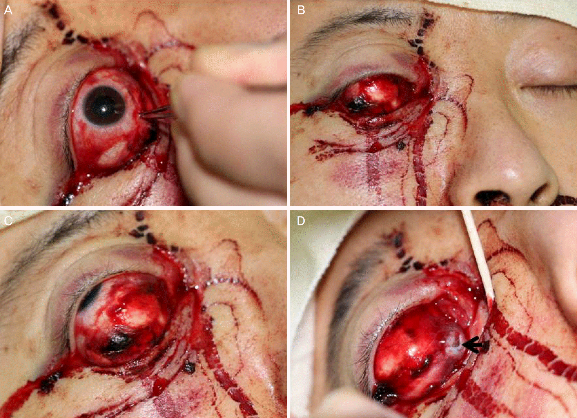
External photograph of eyeball. (A) The pupil was fixed and dilated. (B) A conjunctival laceration and the stump of ruptured inferior and medial rectus muscle was seen. (C) The right eye was fixed in extreme abduction and supraduction. There was no adduction on attempted left gaze and no infraduction on attempted down gaze. (D) The completely severed optic nerve is visible (black arrow).

Figure 2.
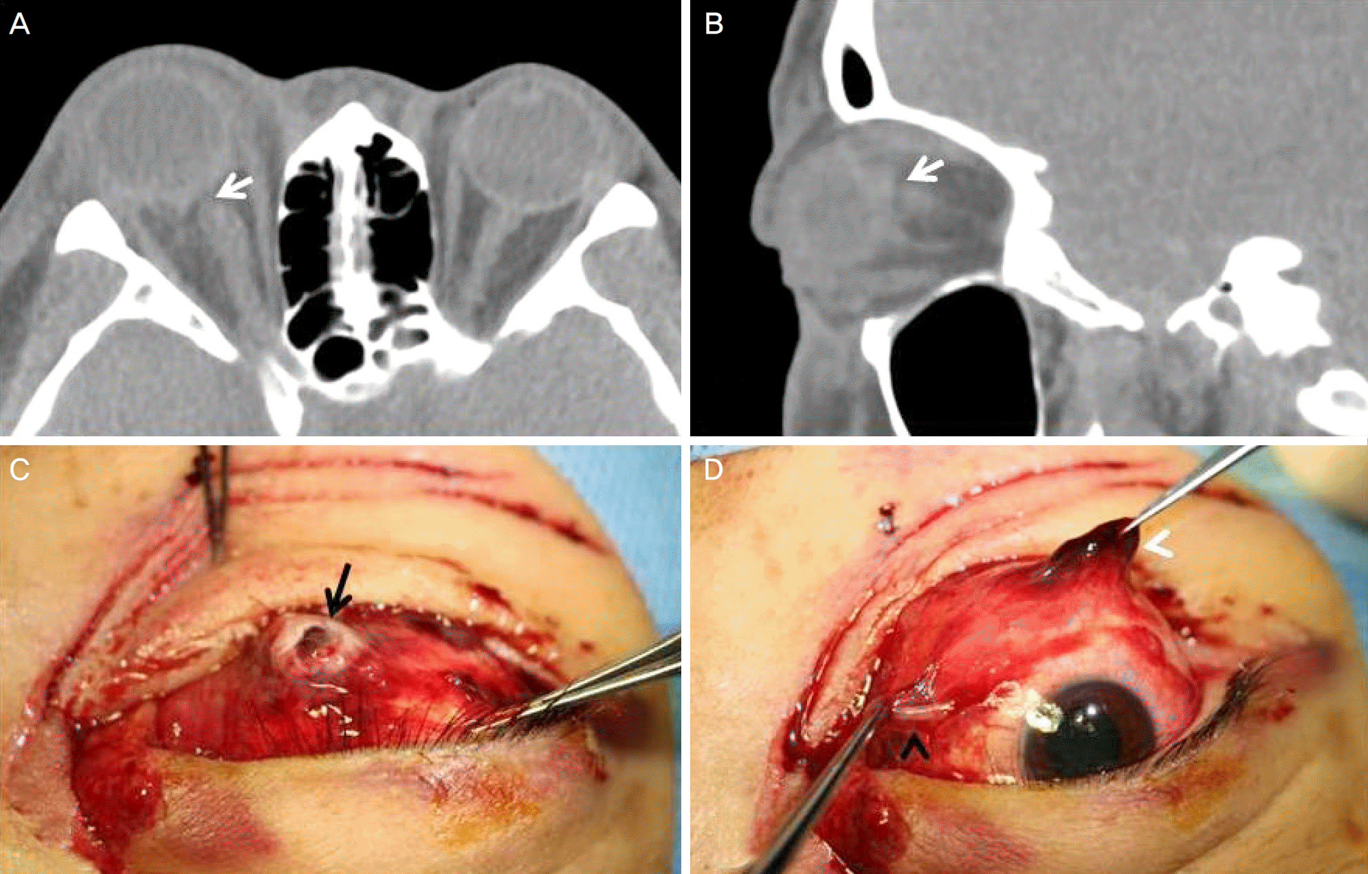
Computed tomography image and external photograph of eyeball. (A, B) The facial bone computed tomography image showed a ruptured optic nerve at the intraorbital insertion site (white arrow). (C, D) Examination under anesthesia revealed a completely severed optic nerve (black arrow), complete disinsertion of the medial (black arrowhead) and inferior rectus muscles (white arrowhead).


 PDF
PDF ePub
ePub Citation
 Print
Print


 XML Download