

 PDF
PDF ePub
ePub Citation
 Print
Print


Abstract
Purpose
To report a case of tamoxifen-induced retinopathy diagnosed using spectral domain optical coherence tomography (SD-OCT).
Case summary
A 44-year-old female presented with metamorphopsia in the left eye and binocular vision loss which started 5 months prior. She had no record of external trauma, diabetes or high blood pressure; however, she had been taking 21.9 g tamoxifen (20 mg/day) since October 2012 after a surgery of her left breast due to cancer. On the initial visit, fundus photography showed crystalline dot-like deposits in both parafoveae. Additionally, fluorescence angiography revealed a small leakage around the macular area. Optical coherence tomography (OCT) was obtained to differentiate from other diseases because fundus photography showed crystalline retinopathy. The OCT revealed a normal right eye but the left macula had a microcystic lesion. Based on the diagnosis of tamoxifen-induced retinopathy, the patient stopped taking tamoxifen. Three months after discontinuation of tamoxifen, fundus photography showed slightly decreased crystalline deposits in the parafoveal area and visual acuity of the right eye was slightly improved. However, SD-OCT showed a slightly aggravated disruption of the outer retina in both eyes.
References
1. Paganini-Hill A, Clark LJ. Eye problems in breast cancer patients treated with tamoxifen. Breast Cancer Res Treat. 2000; 60:167–72.

2. Lazzaroni F, Scorolli L, Pizzoleo CF, et al. Tamoxifen retinopathy: does it really exist? Graefes Arch Clin Exp Ophthalmol. 1998; 236:669–73.
3. Kaiser-Kupfer MI, Lippman ME. Tamoxifen retinopathy. Cancer Treat Rep. 1978; 62:315–20.
4. Nayfield SG, Gorin MB. Tamoxifen associated eye disease. A review. J Clin Oncol. 1996; 14:1018–26.
5. Hahn DK, Park YH. A case of crystalline retinopathy. J Korean Ophthalmol soc. 1995; 36:142–6.
6. Chang TS, Aylward W, Clarkson JG, Gass JD. Asymmetric can-thaxanthin retinopathy. Am J Ophthalmol. 1995; 119:801–2.
7. Bullock JD, Albert DM. Fleck retina. Appearance secondary to ox-alate crystals from methoxyflurane anesthesia. Arch Ophthalmol. 1975; 93:26–31.
8. Chang T, Gonder JR, Ventresca MR. Low-dose tamoxifen retinopathy. Can J Ophthalmol. 1992; 27:148–9.
9. Doshi RR, Fortun JA, Kim BT, et al. Pseudocystic foveal cavitation in tamoxifen retinopathy. Am J Ophthalmol. 2014; 157:1291–8.e3.
10. Jeng KW, Wheatley HM. Intravitreal triamcinolone acetonide treatment of tamoxifen maculopathy with associated cystoid abdominal edema. Retin Cases Brief Rep. 2015; 9:64–6.
11. Srikantia N, Mukesh S, Krishnaswamy M. Crystalline maculopathy: a rare complication of tamoxifen therapy. J Cancer Res Ther. 2010; 6:313–5.
12. Bourla DH, Gonzales CR, Mango CW, et al. Intravitreous vascular endothelial growth factor (VEGF) inhibitor therapy for tamoxifen induced macular edema. Semin Ophthalmol. 2007; 22:87–8.
13. Suhk HJ, Sohn JH, Yoon YH. Retinopathy and keratopathy associated with chronic tamoxifen medication. J Korean Ophthalmol soc. 2002; 43:2354–9.
14. Cho HK, Ko SM. A case of crystalline retinopathy. J Korean Ophthalmol soc. 1997; 38:1628–31.
15. Kim HD, Seo MS. Crystalline retinopathy without corneal dystrophy. J Korean Ophthalmol Soc. 2000; 41:1445–50.
Figure 1.
Fundus photography and spectral domain optical coherence tomography (SD-OCT) examination. At patient's first visit, fundus photography shows crystalline dot-like deposits in parafoveal area (A, B). SD-OCT shows normal finding in the right eye and cyst-like lesion and disruption of the outer retina in the left eye (C, D).
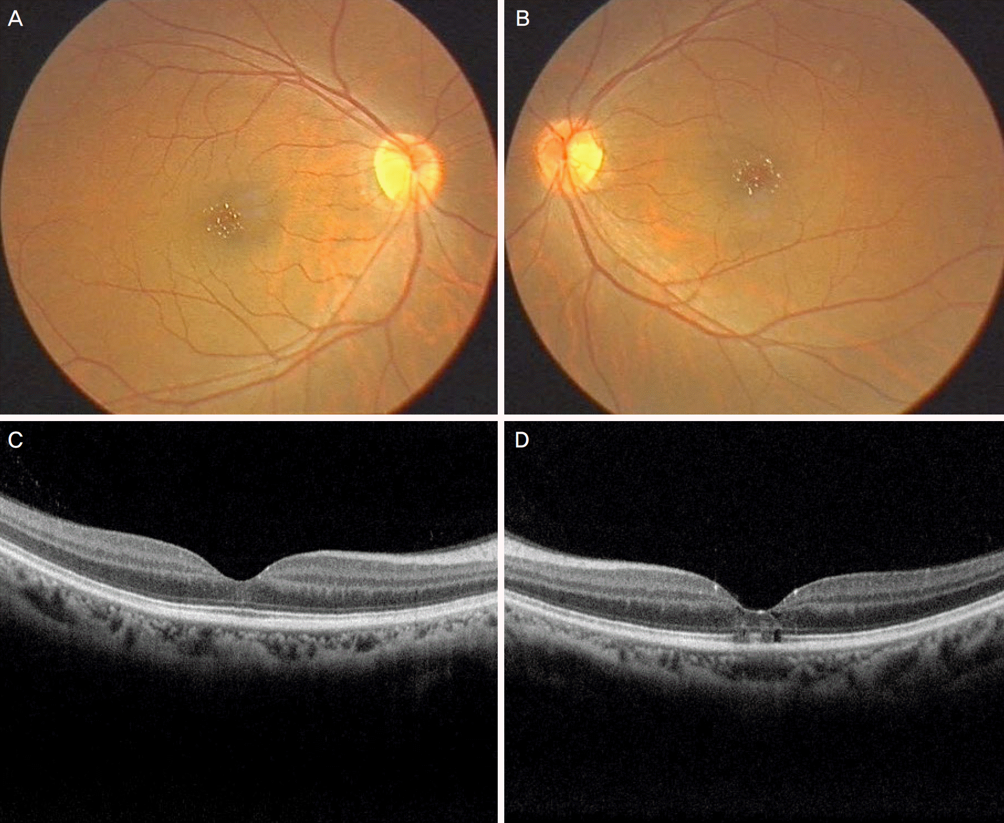
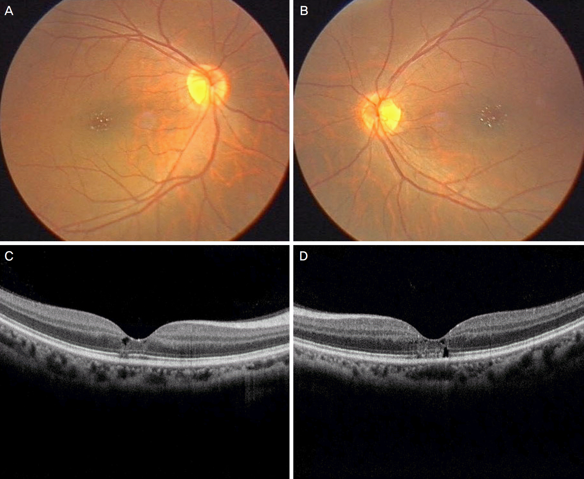
Figure 2.
Fundus photography and spectral domain optical coherence tomography (SD-OCT) examination. Three months after cessation of tamoxifen, fundus photography shows slightly decreased crystalline deposits in parafoveal area (A, B). SD-OCT shows slightly aggravated cyst-like lesion and disruption of the outer retina in both eye (C, D).
 XML Download