Abstract
Purpose
To report a case of visual deterioration and atrophied retina after pars plana vitrectomy (PPV) and silicone oil tamponade for the treatment of retinal detachment with previous encircling scleral buckling.
Case summary
A 29-year-old female visited for treatment of rhegmatogenous retinal detachment (RRD) in the right eye which was not completely resolved after encircling scleral buckling. Logarithm of minimal angle of resolution (log MAR) and best corrected visual acuity (BCVA) was 0.3. Retinal detachment from 3 to 8 O'clock without macular involvement was identified. Pars plana vitrectomy, endophotocoagulation and silicone oil tamponade were performed. During the operation, retinal dialysis and retinal break at the superonasal periphery were observed. The patient complained of central scotoma at 2 days postoperatively and hyper-reflection of the inner retina was identified on optical coherence tomography (OCT). At 2 weeks postoperatively, the OCT image revealed a thin retina and impending macular hole. After 2 months, the silicone oil was removed. Although the retina was well attached, the retina remained atrophied and the log MAR BCVA was 0.16.
References
1. Hassan TS, Sarrafizadeh R, Ruby AJ, et al. The effect of duration of macular detachment on results after the scleral buckle repair of primary, macula-off retinal detachments. Ophthalmology. 2002; 109:146–52.

2. Tani P, Robertson DM, Langworthy A. Prognosis for central vision and anatomic reattachment in rhegmatogenous retinal detachment with macula detached. Am J Ophthalmol. 1981; 92:611–20.

3. Garrity JA, Liesegang TJ. Ocular complications of atopic dermatitis. Can J Ophthalmol. 1984; 19:21–4.
4. Ha SM, Shin JP, Kim SY. Rhegmatogenous retinal detachment abdominal with atopic dermatitis. J Korean Ophthalmol Soc. 2004; 45:419–24.
5. Ingram RM. Retinal detachment associated with atopic dermatitis and cataract. Br J Ophthalmol. 1965; 49:96–7.

6. Karel I, Myska V, Kvícalov á E. Ophthalmological changes in atopic dermatitis. Acta Derm Venereol. 1965; 45:381–6.
7. Katsura H, Hida T. Atopic dermatitis. Retinal detachment abdominal with atopic dermatitis. Retina. 1984; 4:148–51.
8. Christensen UC, la Cour M. Visual loss after use of intraocular abdominal oil associated with thinning of inner retinal layers. Acta Ophthalmol. 2012; 90:733–7.
9. Caramoy A, Foerster J, Allakhiarova E, et al. Spectraldomain abdominalal coherence tomography in subjects over 60 years of age, and its implications for designing clinical trials. Br J Ophthalmol. 2012; 96:1325–30.
10. Song WK, Lee SC, Lee ES, et al. Macular thickness variations with sex, age, and axial length in healthy subjects: a spectral abdominal-optical coherence tomography study. Invest Ophthalmol Vis Sci. 2010; 51:3913–8.
11. Chen SN, Hwang JF, Chen YT. Macular thickness measurements in central retinal artery occlusion by optical coherence tomography. Retina. 2011; 31:730–7.

12. Robertson DM, McLaren JW. Photic retinopathy from the operating room microscope. Study with filters. Arch Ophthalmol. 1989; 107:373–5.
13. Budde M, Cursiefen C, Holbach LM, Naumann GO. Silicone oil– associated optic nerve degeneration. Am J Ophthalmol. 2001; 131:392–4.
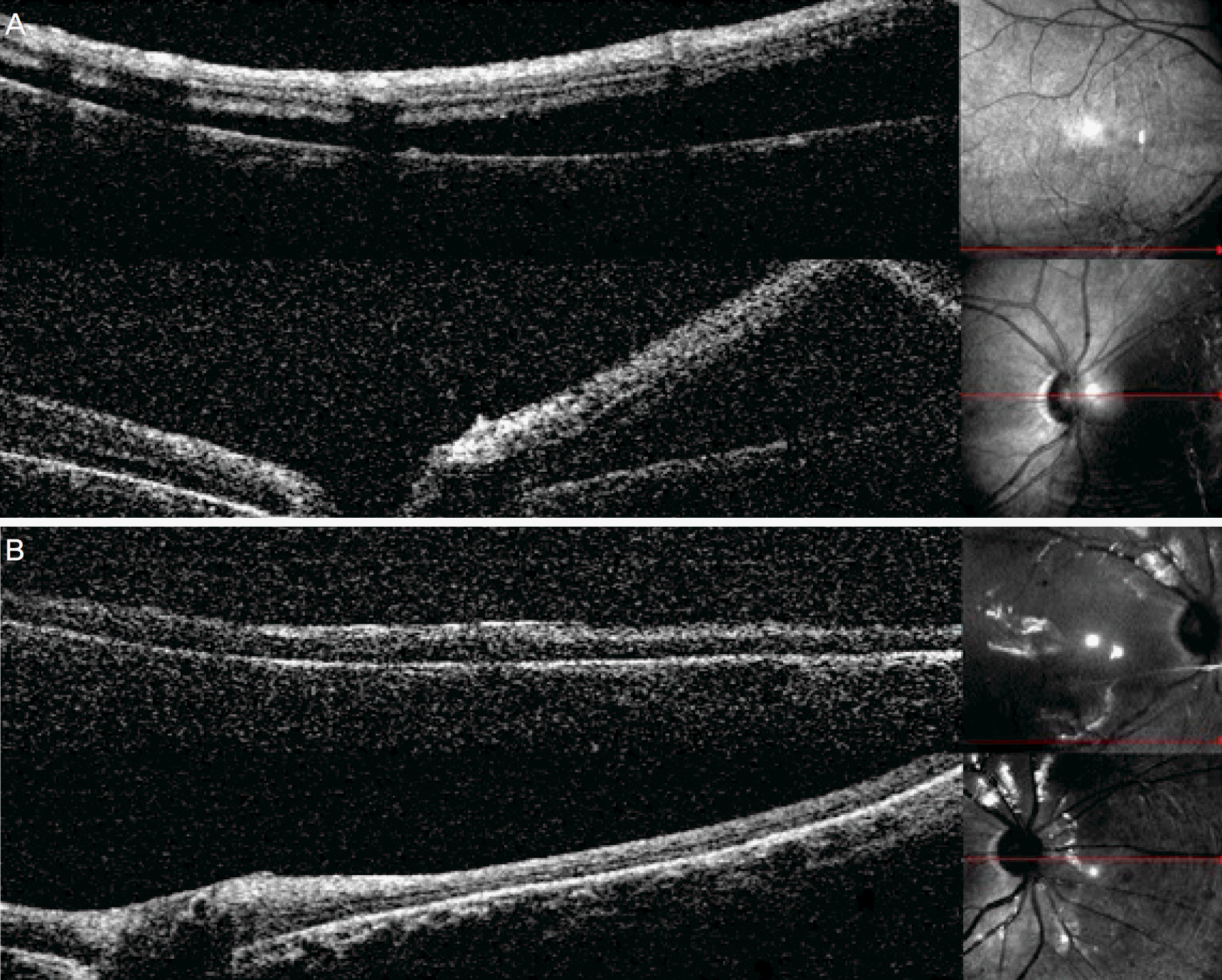
Figure 1.
Optical coherence tomography (OCT) findings of inferior and peripapillary fundus. (A) Preoperative findings show detached retina. (B) Postoperative day 2 findings reveal reattached retina.

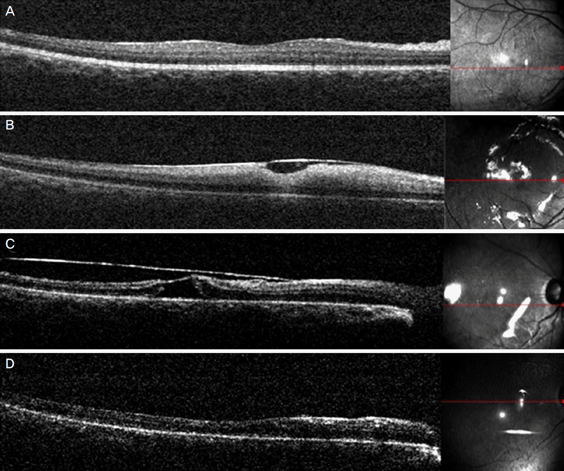
Figure 2.
Macular optical coherence tomography (OCT) findings. (A) Preoperative OCT shows normal structure of retina. (B) Hyperreflective inner retina and the silicone oil margin is identified at postoperative day 2. (C) The impending macular hole is revealed at postoperative 2 weeks. (D) Thin retina without macular hole is observed at postoperative 6 weeks.

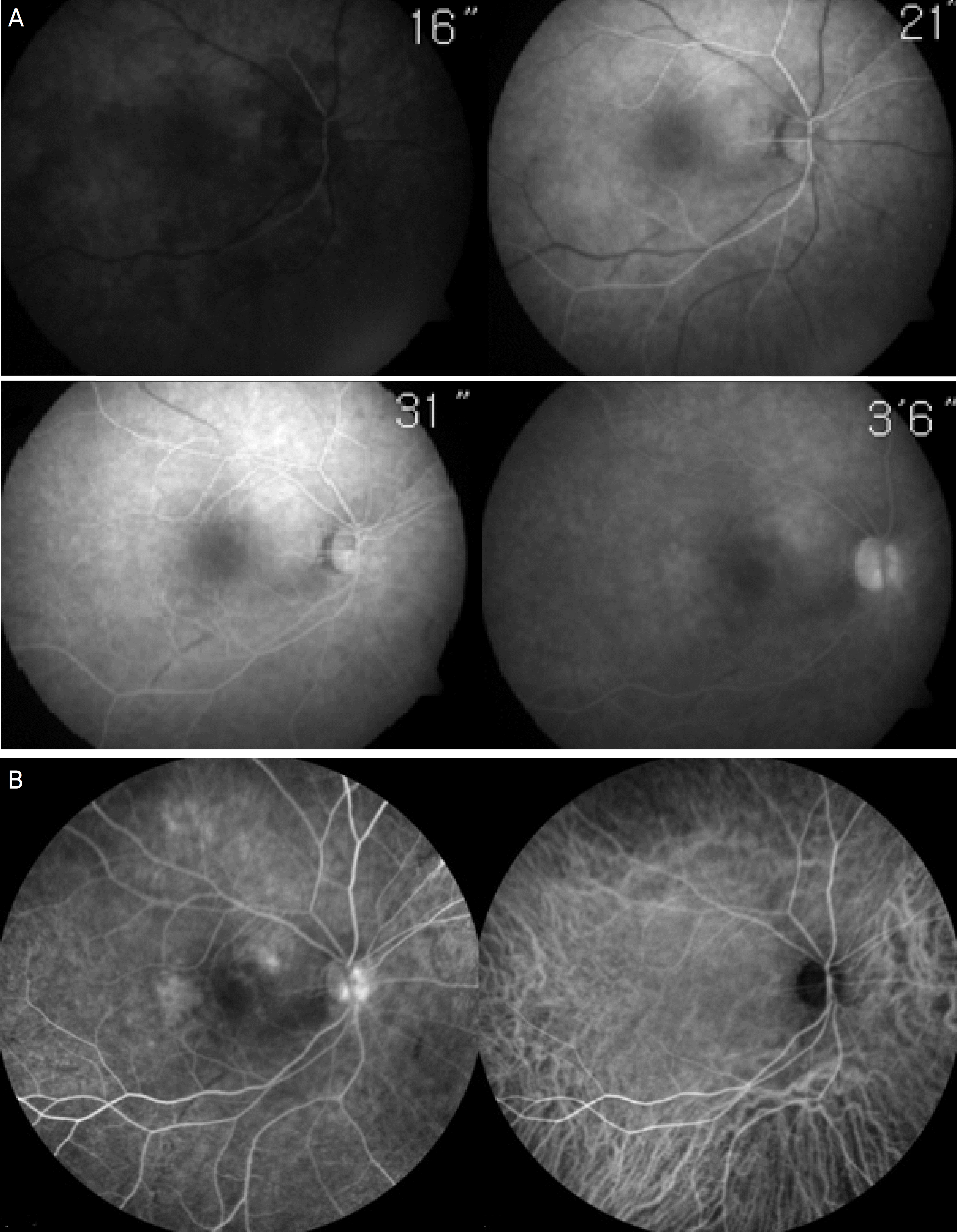
Figure 3.
Fluorescein angiography (FAG) and indocyanine green angiography (ICG) findings after removal of silicone oil. (A) FAG shows delayed arteriovenous transit time and patchy hypofluoroscein at posterior pole in choroidal phase, 2 days after removal of silicone oil. (B) Two weeks after removal of silicone oil, FAG shows slight hypofluorescein in choroidal back flush and some hyper-fluoroscein of stain but ICG findings is unremarkable.


 PDF
PDF ePub
ePub Citation
 Print
Print


 XML Download