

 PDF
PDF ePub
ePub Citation
 Print
Print


Abstract
Purpose
To report a case of endothelial keratitis occurred after reactivation of herpes simplex virus following immunosuppressant therapy for Kaposi's varicelliform eruption.
Case summary
A 23-year-old female was referred for ocular pain and blurred vision. She had atopic dermatitis and was diagnosed with Kaposi's varicelliform eruption on her face after using an immunosuppressant. Slit lamp examination revealed central corneal edema in the right eye. She was initially diagnosed with contact lens-induced keratitis. Subsequently, the contact lens was removed and topical antiviral agent used for prevention of ocular involvement. Four days after treatment, Wesseley immune ring of deep stromal haze and cells in the anterior chamber were present. She was diagnosed with endothelial keratitis caused by reactivation of herpes simplex virus after using an immunosuppressant. Topical steroid, hypertonic saline eye drops and cycloplegic eye drops were added to the treatment for the progression of endothelial keratitis. Corneal edema was decreased 2 weeks after treatment and anterior chamber cells decreased 1 month after treatment. There was no recurrence during the fol-low-up period.
Go to : 
References
1. Wheeler CE Jr, Abele DC. Eczema herpeticum, primary and recurrent. Arch Dermatol. 1966; 93:162–73.

2. Gulkilik G, Demirci G, Ozdamar AM, Muftuoglu GI. A case of herpetic keratitis after intravitreal triamcinolone injection. Cornea. 2007; 26:1000–1.
3. Wollenberg A, Zoch C, Wetzel S, et al. Predisposing factors and clinical features of eczema herpeticum: a retrospective analysis of 100 cases. J Am Acad Dermatol. 2003; 49:198–205.
4. Higgins PG, Crow KD. Recurrent Kaposi's varicelliform eruption in Darier's disease. Br J Dermatol. 1973; 88:391–4.
5. Palleschi GM, Falcos D, Giacomelli A, Caproni M. Kaposi's varicelliform eruption in pemphigus foliaceus. Int J Dermatol. 1996; 35:809–10.
6. Hayashi S, Yamada Y, Dekio S, Jidoi J. Kaposi's varicelliform eruption in a patient with mycosis fungoides. Clin Exp Dermatol. 1997; 22:41–3.
7. Brion N, Guillaume JC, Dubertret L, Touraine R. Disseminated cutaneous herpes of the adult and Sézary syndrome (author's transl). Ann Dermatol Venereol. 1981; 108:517–21.
8. Flint ID, Spencer DM, Wilkin JK. Eczema herpeticum in abdominal with familial benign chronic pemphigus. J Am Acad Dermatol. 1993; 28(2 Pt 1):257–9.
9. Nishimura M, Maekawa M, Hino Y, et al. Kaposi's varicelliform eruption. Development in a patient with a healing second-degree burn. Arch Dermatol. 1984; 120:799–800.
10. Verbov J, Munro DD, Miller A. Recurrent eczema herpeticum abdominal with ichthyosis vulgaris. Br J Dermatol. 1972; 86:638–40.
11. Higaki S, Inoue Y, Yoshida A, et al. Case of bilateral multiple herpetic epithelial keratitis manifested as dendriform epithelial edema during primary Kaposi's varicelliform eruption. Jpn J Ophthalmol. 2008; 52:127–9.
Go to :
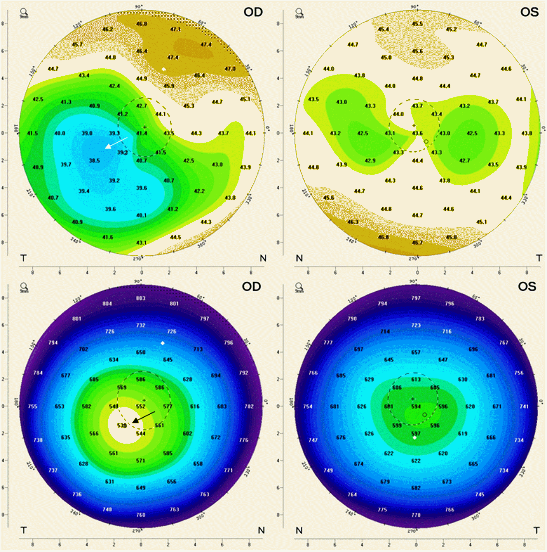 | Figure 1.Change of anerior curvature in cornea topography after keratitis. Corneal topography of right eye shows flattening of anterior cornea curvature (white arrow) and central cornea thinning (black arrow) than left eye after keratitis due to reactivation of herpes simplex virus after using an immunosuppressant in Kaposi's varicelliform eruption. OD = oculus dexter; OS = oculus sinister; N = nasal; T = temporal. |
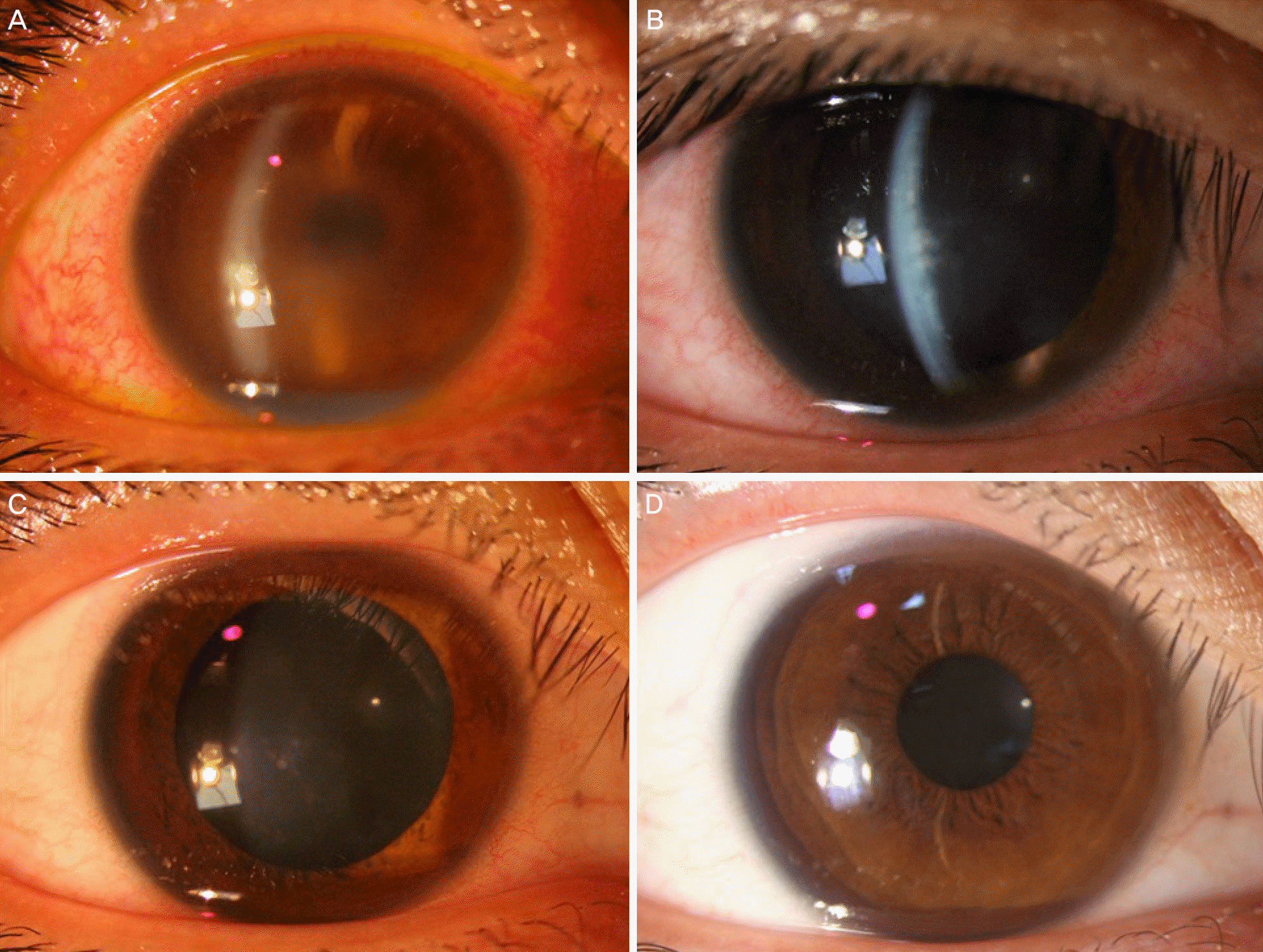 | Figure 2.Changes of the cornea after ocular involvement of Kaposi's varicelliform eruption. (A) Slit lamp examination of right eye shows Wessley immune ring of stromal haze. Anterior chamber cells are also present. (B) Decreased corneal edema in right eye is shown after instillation of 1% prednisolone and cycloplegic agent after two weeks of treatment. (C) Anterior chamber cells are not present in right eye after one month. (D) There is no recurrence during the two years of follow up period. |
 XML Download