Abstract
Purpose
Following planned posterior continuous curvilinear capsulorrhexis (PCCC) during cataract surgery in adults, we evaluated the clinical effects of visual acuity and prevention of posterior capsule opacity.
Methods
The clinical results were studied retrospectively by comparing 43 eyes of 43 patients who underwent cataract surgery with PCCC (the experimental group) and 46 eyes of 31 patients who underwent cataract surgery without PCCC (the control group). Preoperative and postoperative best corrected visual acuities (BCVAs) of patients were measured. BCVA (using log MAR) and the occurrence of posterior capsule opacity were closely monitored in both groups preoperatively, two months postoperatively, and at each group's final visit (14.6 months postoperatively for the experimental group and 15.7 months for the control group). One-piece plate intraocular lens was used in cataract surgery.
Results
Preoperative BCVA was lower in the control group but not significantly. The 2-month mean postoperative BCVA showed improvement in vision in both the control and experimental groups. In both groups, the BCVA was decreased at the final examination compared with the 2-month postoperative BCVA, and significant differences between the two groups were not observed. Under slit lamp examination, anterior hyaloid opacity was observed in 13 of 43 eyes that underwent PCCC. The decrease in BCVA in 13 eyes with anterior hyaloid opacity was significantly different (p < 0.05) compared with the 2-month post-operative BCVA.
Go to : 
References
1. Frezzotti R, Caporossi A. Pathogenesis of posterior capsular opacification. Part I. Epidemiological and clinico-statistical data. J Cataract Refract Surg. 1990; 16:347–52.
2. Ohadi C, Moreira H, McDonnell PJ. Posterior capsule opacification. Curr Opin Ophthalmol. 1991; 2:46–52.

3. Jamal SA, Solomon LD. Risk factors for posterior capsule pearling after uncomplicated extracapsular cataract extraction and pla-no-convex posterior chamber lens implantation. J Cataract Refract Surg. 1993; 19:333–8.
4. Rönbeck M, Zetterström C, Wejde G, Kugelberg M. Comparison of posterior capsule opacification development with 3 intraocular lens types: five-year prospective study. J Cataract Refract Surg. 2009; 35:1935–40.
5. Solomon KD, Legler UF, Kostick AM. Capsular opacification after cataract surgery. Curr Opin Ophthalmol. 1992; 3:46–51.

6. Hayashi K, Hayashi H, Nakao F, Hayashi F. Posterior capsule abdominal after cataract surgery in patients with diabetes mellitus. Am J Ophthalmol. 2002; 134:10–6.
7. Apple DJ, Solomon KD, Tetz MR, et al. Posterior capsule opacification. Surv Ophthalmol. 1992; 37:73–116.

8. Varga A, Sacu S, Vécsei-Marlovits PV, et al. Effect of posterior capsule opacification on macular sensitivity. J Cataract Refract Surg. 2008; 34:52–6.

9. Georgopoulos M, Menapace R, Findl O, et al. After-cataract in adults with primary posterior capsulorhexis: comparison of hydrogel and silicone intraocular lenses with round edges after 2 years. J Cataract Refract Surg. 2003; 29:955–60.
10. Ruderman JM, Mitchell PG, Kraff M. Pupillary block following Nd:YAG laser capsulotomy. Ophthalmic Surg. 1983; 14:418–9.

11. Schubert HD. A history of intraocular pressure rise with reference to the Nd:YAG laser. Surv Ophthalmol. 1985; 30:168–72.

12. Schubert HD. Vitreoretinal changes associated with rise in abdominal pressure after Nd:YAG capsulotomy. Ophthalmic Surg. 1987; 18:19–22.
13. Koch DD, Liu JF, Gill EP, Parke DW 2nd. Axial myopia increases the risk of retinal complications after neodymium-YAG laser abdominal capsulotomy. Arch Ophthalmol. 1989; 107:986–90.
14. Ober RR, Wilkinson CP, Fiore JV Jr, Maggiano JM. Rhegmatogenous retinal detachment after neodymium-YAG laser capsulotomy in phakic and pseudophakic eyes. Am J Ophthalmol. 1986; 101:81–9.

15. Georgopoulos M, Menapace R, Findl O, et al. Posterior continuous curvilinear capsulorhexis with hydrogel and silicone intraocular lens implantation: development of capsulorhexis size and capsule opacification. J Cataract Refract Surg. 2001; 27:825–32.
16. Hartmann C, Wiedemann P, Gothe K, et al. Prevention of abdominal cataract by intracapsular administration of the antimitotic daunomycin. Ophtalmologie. 1990; 4:102–6.
17. Tarsio JF, Kelleher PJ, Tarsio M, et al. Inhibition of cell abdominal on lens capsules by 4197X-ricin A immunoconjugate. J Cataract Refract Surg. 1997; 23:260–6.
18. Gimbel HV. Posterior capsule tears using phacoemulsification causes, prevention and management. Eur J Implant Refract Surg. 1990; 2:63–9.

19. Zetterström C, Kugelberg U, Oscarson C. Cataract surgery in abdominal with capsulorhexis of anterior and posterior capsules and hep-arin-surface-modified intraocular lenses. J Cataract Refract Surg. 1994; 20:599–601.
20. Galand A, van Cauwenberge F, Moosavi J. Posterior capsulorhexis in adult eyes with intact and clear capsules. J Cataract Refract Surg. 1996; 22:458–61.

21. Wormstone IM. Posterior capsule opacification: a cell biological perspective. Exp Eye Res. 2002; 74:337–47.
22. Vasavada A, Desai J. Primary posterior capsulorhexis with and without anterior vitrectomy in congenital cataracts. J Cataract Refract Surg. 1997; 23(Suppl 1):645–51.

23. Fenton S, O'Keefe M. Primary posterior capsulorhexis without abdominal vitrectomy in pediatric cataract surgery: longer-term abdominal. J Cataract Refract Surg. 1999; 25:763–7.
24. Buehl W, Findl O. Effect of intraocular lens design on posterior capsule opacification. J Cataract Refract Surg. 2008; 34:1976–85.

25. Lin CL, Wang AG, Chou JC, et al. Heparin-surface-modified abdominal lens implantation in patients with glaucoma, diabetes, or uveitis. J Cataract Refract Surg. 1994; 20:550–3.
26. Gatinel D, Lebrun T, Le Toumelin P, Chaine G. Aqueous flare abdominal by heparin-surface-modified poly(methyl methacrylate) and acrylic lenses implanted through the same-size incision in patients with diabetes. J Cataract Refract Surg. 2001; 27:855–60.
27. Larsson R, Selén G, Björdklund H, Fagerholm P. Intraocular PMMA lenses modified with surface-immobilized heparin: abdominal of biocompatibility in vitro and in vivo. Biomaterials. 1989; 10:511–6.
28. Kang S, Kim MJ, Park SH, Joo CK. Comparison of clinical results between heparin surface modified hydrophilic acrylic and abdominal acrylic intraocular lens. Eur J Ophthalmol. 2008; 18:377–83.
29. Wilhelmus KR, Emery JM. Posterior capsule opacification abdominal phacoemulsification. Ophthalmic Surg. 1980; 11:264–7.
30. Ryu CH, Kim HB, Lim SJ. Clinical result of planned posterior abdominal curvilinear capsulorrhexis in adult cataract patients: 1 year follow-up. J Korean Ophthalmol Soc. 2000; 41:2547–54.
31. Kim DE, Jo JM, Kim WS. Clinical results of primary posterior abdominal curvilinear capsulorhexis. J Korean Ophthalmol Soc. 2003; 44:2228–34.
Go to : 
 | Figure 1.Grade of lens epithelial cell (LEC) ongrowth in patients with posterior continuous curvilinear capsulorhexis. (A-E) The progress of 0–4 grade, showing aggravated LEC. |
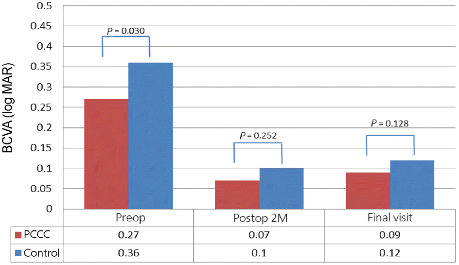 | Figure 2.Preoperative and postoperative best corrected visual acuity in patients with posterior continuous curvilinear capsulorhexis (PCCC) and control group. Results are analyzed using Mann-Whitney U-test. ‘Final visit’ means ‘6–18 months’. BCVA = best corrected visual acuity; Preop = preoperative; Postop = postoperative; M = months. |
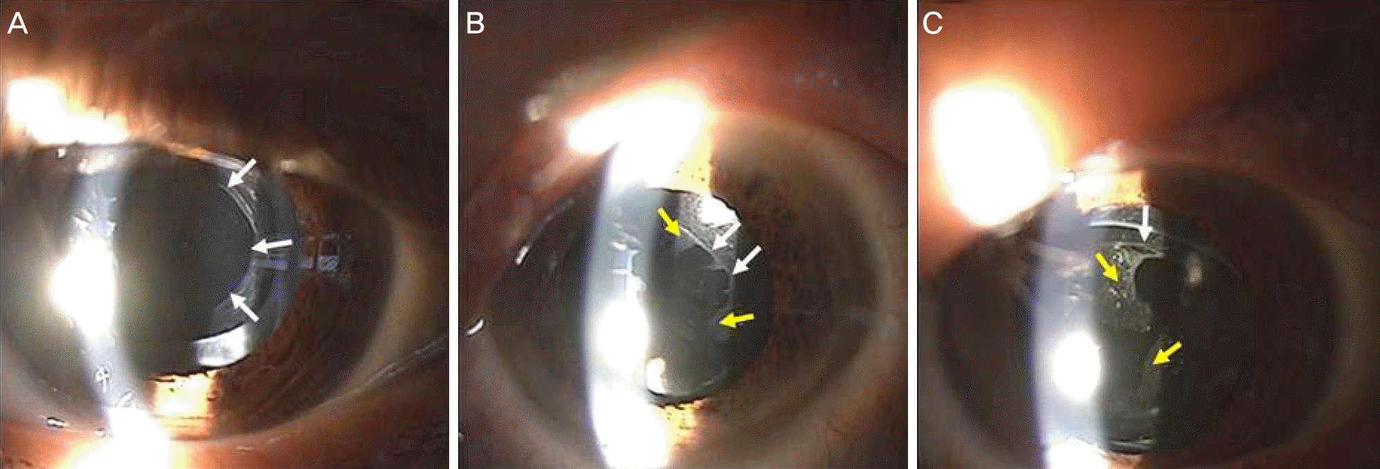 | Figure 3.Slit lamp photographs of one year postperative follow up after posterior continuous curvilinear capsulorhexis (PCCC) with each different patient. (A) Appearance of clear posterior continuous curvilinear capsulorhexis area. (B, C) lens epithelial cell (LEC) ongrowth can be seen on the PCCC. White arrows indicate PCCC line, and yellow arrows indicate LEC ongrowth. |
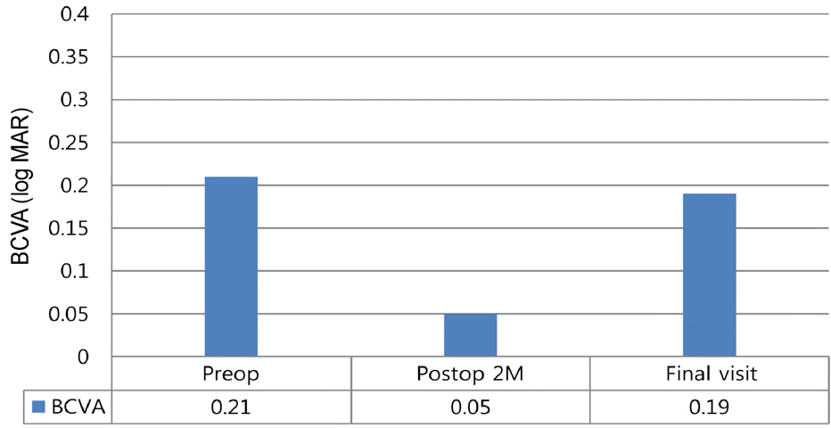 | Figure 4.Preoperative and postoperative visual acuity of 13 eyes, showing anterior hyaloid opacity and reclosure after posterior continuous curvilinear capsulorhexis. ‘Final visit’ means ‘6–18 months’. BCVA = best corrected visual acuity; Preop = preoperative; Postop = postoperative; M = months. |
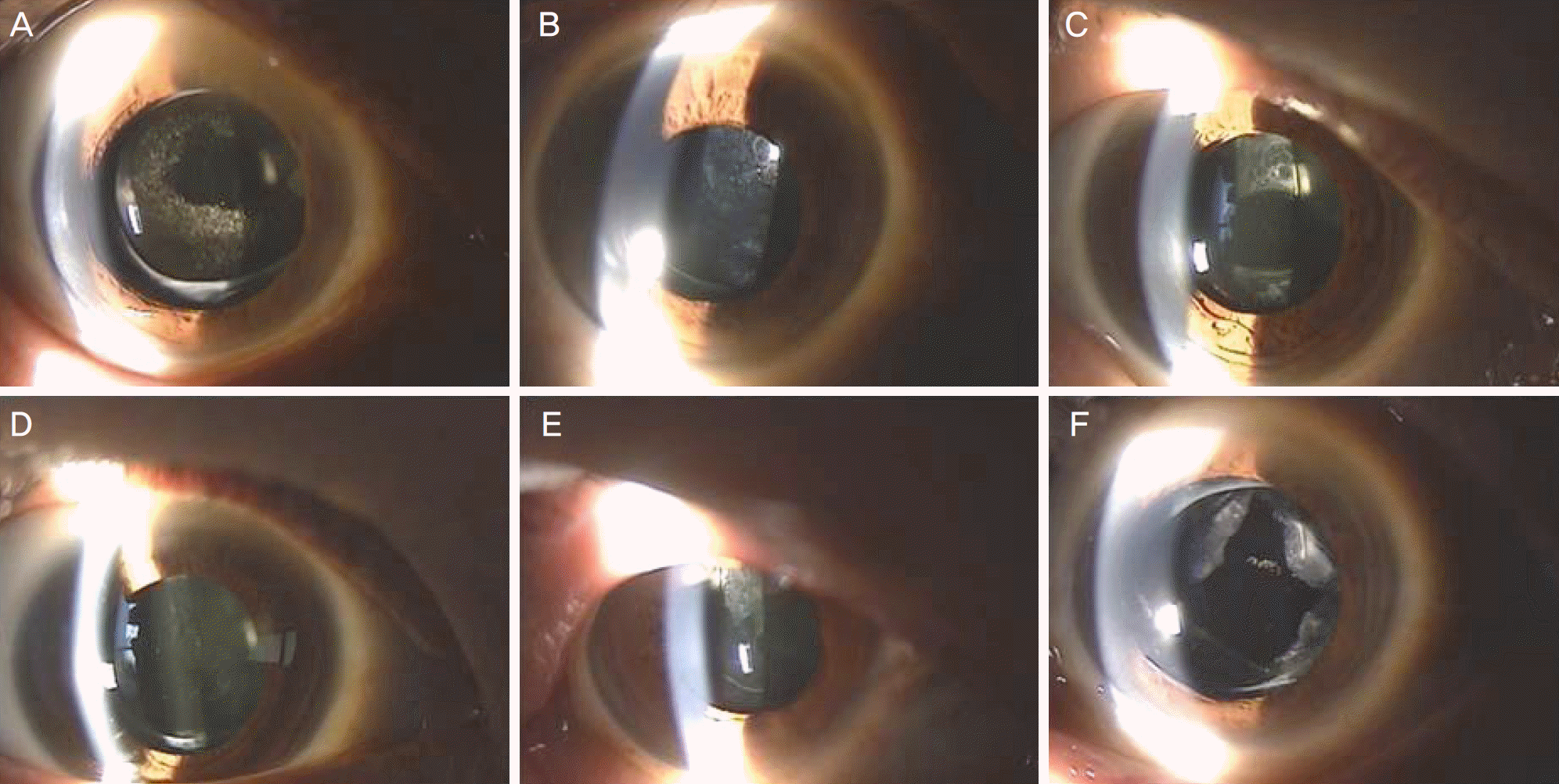 | Figure 5.Slit lamp photographs of anterior hyaloid opacity and reclosure after 12 to 18months of posterior continuous curvilinear capsulorhexis (PCCC) with each different patient. (A) 18 months after PCCC. Grade 2. (B) 12 months after PCCC. Grade 4. (C) 12 months after PCCC. Grade 2 (D) 12 months after PCCC. Grade 4 (E) 13 months after PCCC. Grade 3. (F) Appearance of after neodymium-doped yttrium aluminium garnet laser capsulotomy on total closure of PCCC. 18 months after PCCC. |
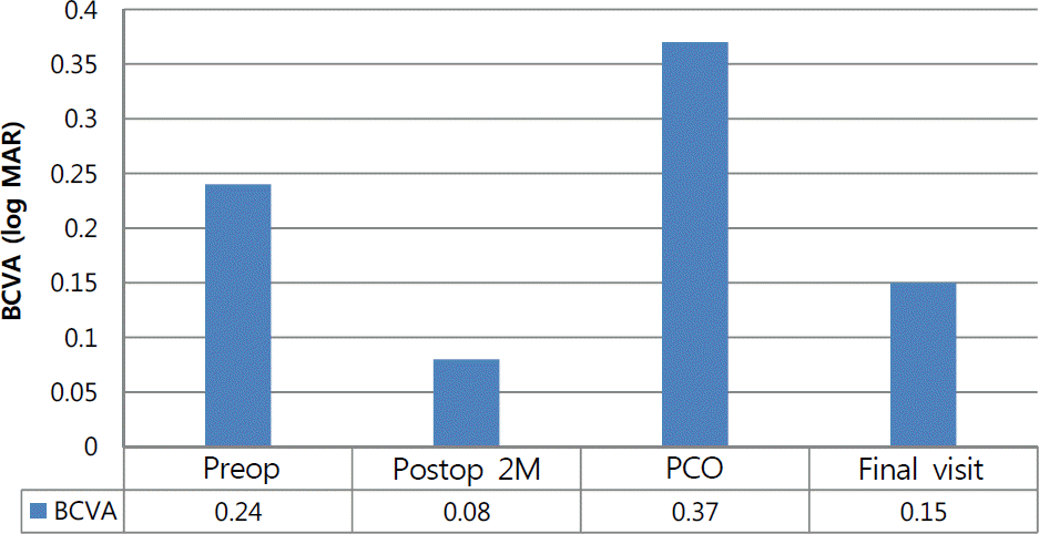 | Figure 6.Changes in visual acuity after neodymium-doped yttrium aluminium garnet laser capsulotomy on control group of eyes with posterior capsule opacity (PCO). ‘Final visit’ means ‘6–18 months’. BCVA = best corrected visual acuity; Preop = preoperative; Postop = postoperative; M = months. |
Table 1.
Study variables and spherical equivalent of PCCC and control group


 PDF
PDF ePub
ePub Citation
 Print
Print


 XML Download