

 PDF
PDF ePub
ePub Citation
 Print
Print


Abstract
Purpose:
To report a case of macular hole secondary to presumptive infectious posterior uveitis involving the fovea that sponta-neously resolved after medical treatment.
Case summary
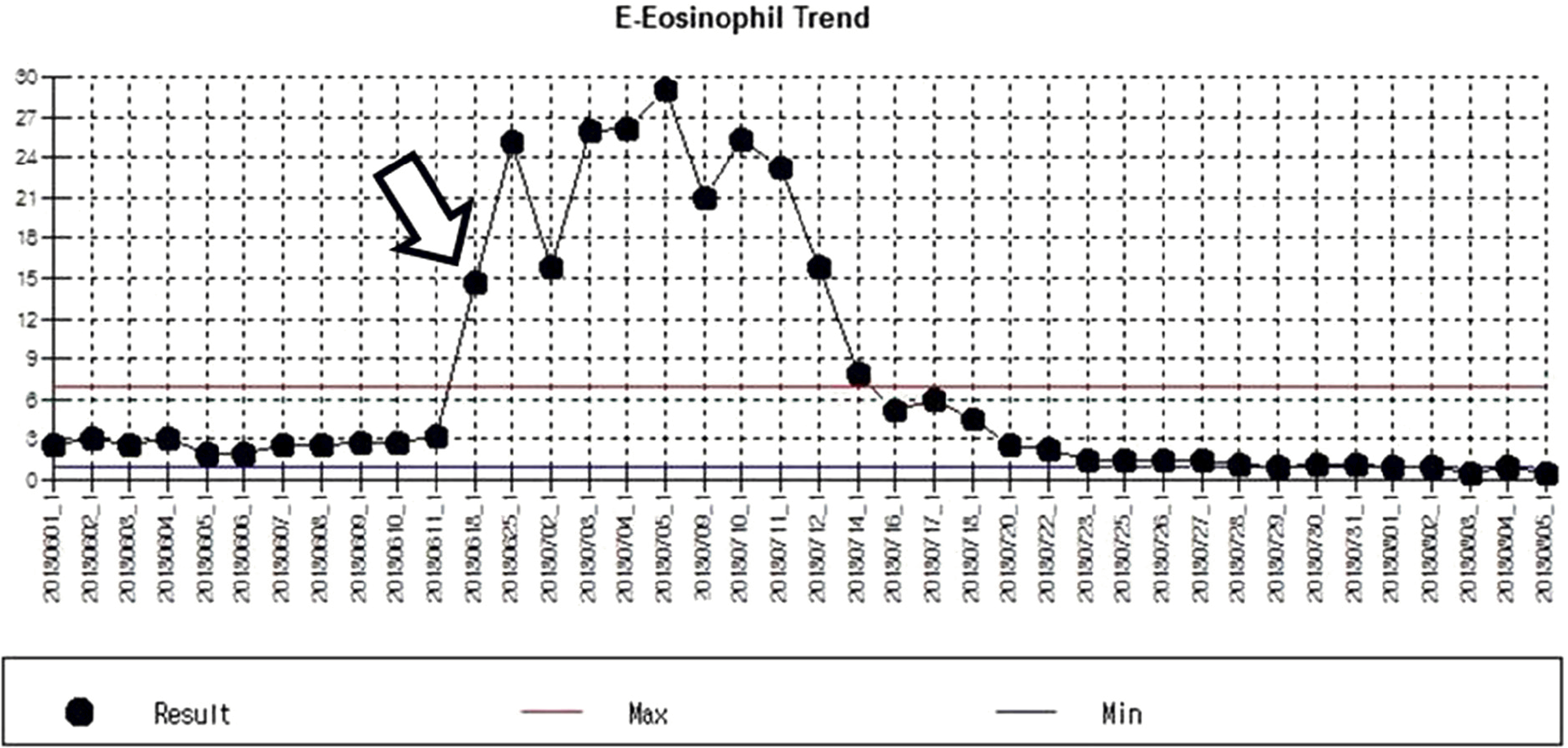
A 33-year-old male visited our clinic for decreased visual acuity in his left eye. He was treated with im-munosuppressive therapy including steroid after bone marrow transplantation. Best corrected visual acuity (BCVA) was 0.05 in the left eye. Slit lamp examination showed mild anterior vitritis, and fundus examination showed a macular hole with surrounding whitish infiltration at the fovea. Spectral domain optical coherence tomography (SD-OCT) revealed a full thickness macular hole with surrounding hyper-reflective masses with an infiltration-like appearance involving all retinal layers. Serum anti-toxocara IgG was positive (ELISA), and eosinophil count and immunoglobulin E was elevated. Under diagnosis of presumptive ocular toxocar-iasis, the patient was treated with albendazole. After medical treatment for toxocariasis, the whitish foveal infiltration became smaller and more discrete. SD-OCT revealed spontaneous closure of the macular hole, and BCVA was improved to 0.4 after a 4-month follow-up.
Go to : 
References
1. Rothova A, Suttorp-van Schulten MS. Frits Treffers W. Kijlstra A. Causes and frequency of blindness in patients with intraocular in-flammatory disease. Br J Ophthalmol. 1996; 80:332–6.

2. Bonnin N, Cornut PL, Chaise F. . Spontaneous closure of mac-ular holes secondary to posterior uveitis: case series and a literature review. J Ophthalmic Inflamm Infect. 2013; 3:34.
3. Jee D, Kim KS, Lee WK. . Clinical features of ocular toxocar-iasis in adult Korean patients. Ocul Immunol Inflamm 2015: Aug. 1–10.
4. Ahn SJ, Ryoo NK, Woo SJ. Ocular toxocariasis: clinical features, diagnosis, treatment, and prevention. Asia Pac Allergy. 2014; 4:134–41.
5. Ahn SJ, Woo SJ, Jin Y. . Clinical features and course of ocular toxocariasis in adults. PloS Negl Trop Dis. 2014; 8:e2938.
6. Smiddy WE, Flynn HW Jr. Pathogenesis of macular holes and ther-apeutic implications. Am J Ophthalmol. 2004; 137:525–37.
7. Halkiadakis I, Pantelia E, Giannakopoulos N. . Macular hole closure after peribulbar steroid injection. Am J Ophthalmol. 2003; 136:1165–7.
Go to :
 | Figure 1.Initial fundus photograph, SD-OCT image and Fluoresce in angiography of patient’s left eye. (A) Fundus photograph re-vealed a mild diffuse vitritis with overlying whitish foveal infiltration. (B) Fluorescein angiography showed centripetal impregnation of the macular lesion and disc stainning on late phase. (C-F) SD-OCT confirmed a full thickness macular hole with overlying whitish foveal infiltration. SD-OCT = spectral domain-optical coherence tomography. |
 | Figure 3.After medical etiological treatment, fundus photographs of the patient’s left eye. Fundus photograph showed gradually more discrete macular whitish infiltration and decreased in size (A, B, C). Four months after medical treatment, fundus photograph demonstrated foveal infiltration was disappeared (D). |
 | Figure 4.After medical etiological treatment, SD-OCT of the patient’s left eye. SD-OCT showed gradual spontaneous closure of the macular hole preserving the macular concave figure (A, B, C). Four months after medical treatment, SD-OCT image demonstrated that the macular hole was closed (D). SD-OCT = spectral domain-optical coherence tomography. |
 XML Download