

 PDF
PDF ePub
ePub Citation
 Print
Print


Abstract
Purpose
To evaluate the efficacy of swept source optical coherence tomography (SS-OCT) by comparing the measurement of central corneal thickness (CCT) to the measurement obtained using Orbscan II, anterior segment optical coherence tomography (AS-OCT) and ultrasound pachymetry.
Methods
One examiner measured the CCT in 65 eyes of 65 healthy subjects using Orbscan II, AS-OCT, SS-OCT and ultrasound pachymetry. The mean values and correlations were analyzed.
Results
The average CCT measurements obtained using Orbscan II, AS-OCT, SS-OCT and ultrasound pachymetry were 534.83 ± 38.46, 517.80 ± 32.48, 528.22 ± 33.71 and 528.02 ± 34.90 μ m, respectively. A significant linear correlation was observed among Orbscan II, AS-OCT, SS-OCT and ultrasound pachymetry (r > 0.894, p < 0.001). There was no significant difference between the SS-OCT and ultrasound pachymetry (p = 0.782).
Go to : 
References
1. Ou RJ, Shaw EL, Glasgow BJ. Keratectasia after laser in situ abdominal (LASIK): evaluation of the calculated residual stromal bed thickness. Am J Ophthalmol. 2002; 134:771–3.
2. Wang Z, Chen J, Yang B. Posterior corneal surface topographic changes after laser in situ keratomileusis are related to residual abdominal bed thickness. Ophthalmology. 1999; 106:406–9. discussion 409–10.
3. Grieshaber MC, Schoetzau A, Zawinka C, et al. Effect of central corneal thickness on dynamic contour tonometry and Goldmann applanation tonometry in primary open-angle glaucoma. Arch Ophthalmol. 2007; 125:740–4.

4. Jonas JB, Stroux A, Velten I, et al. Central corneal thickness corabdominal with glaucoma damage and rate of progression. Invest Ophthalmol Vis Sci. 2005; 46:1269–74.
5. Kim DH, Kim MS, Kim JH. Early corneal-thickness changes after penetrating keratoplasty. J Korean Ophthalmol Soc. 1997; 38:1355–61.
6. Solomon OD. Corneal indentation during ultrasonic pachometry. Cornea. 1999; 18:214–5.
7. Choi KS, Nam SM, Lee HK, et al. Comparison of central corneal thickness after the instillation of topical anesthetics: proparacaine versus oxybuprocaine. J Korean Ophthalmol Soc. 2005; 46:757–62.
8. Li EY, Mohamed S, Leung CK, et al. Agreement among 3 methods to measure corneal thickness: ultrasound pachymetry, Orbscan II, and Visante anterior segment optical coherence tomography. Ophthalmology. 2007; 114:1842–7.
9. Akman A, Asena L, Güngör SG. Evaluation and comparison of the new swept source OCT-based IOLMaster 700 with the IOLMaster 500. Br J Ophthalmol. 2016; 100:1201–5.
10. Baum G, Greenwood I. The application of ultrasonic locating abdominals to ophthalmology. II. Ultrasonic slit lamp in the ultrasonic visualization of soft tissues. AMA Arch Ophthalmol. 1958; 60:263–79.
11. Marsich MW, Bullimore MA. The repeatability of corneal abdominal measures. Cornea. 2000; 19:792–5.
12. Miglior S, Albe E, Guareschi M, et al. Intraobserver and interobserver reproducibility in the evaluation of ultrasonic pachymetry measurements of central corneal thickness. Br J Ophthalmol. 2004; 88:174–7.
13. Ling T, Ho A, Holden BA. Method of evaluating ultrasonic pachometers. Am J Optom Physiol Opt. 1986; 63:462–6.
14. Copt RP, Thomas R, Mermoud A. Corneal thickness in ocular abdominal, primary open-angle glaucoma, and normal tension glaucoma. Arch Ophthalmol. 1999; 117:14–6.
15. Al-Farhan HM, Al-Otaibi WM. Comparison of central corneal thickness measurements using ultrasound pachymetry, ultrasound biomicroscopy, and the Artemis-2 VHF scanner in normal eyes. Clin Ophthalmol. 2012; 6:1037–43.
16. Khaja WA, Grover S, Kelmenson AT, et al. Comparison of central corneal thickness: ultrasound pachymetry versus abdominal optical coherence tomography, specular microscopy, and Orbscan. Clin Ophthalmol. 2015; 9:1065–70.
17. Jonuscheit S, Doughty MJ. Regional repeatability measures of corneal thickness: Orbscan II and ultrasound. Optom Vis Sci. 2007; 84:52–8.
18. Choi SH, Kim JH, Han NS, et al. Comparison of corneal thickness measurements with optical low coherence reflectometry, orbscan system and ultrasound pachymeter. J Korean Ophthalmol Soc. 2006; 47:19–24.
19. Thomas J, Wang J, Rollins AM, Sturm J. Comparison of corneal thickness measured with optical coherence tomography, abdominal pachymetry, and a scanning slit method. J Refract Surg. 2006; 22:671–8.
20. Bao F, Wang Q, Cheng S, et al. Comparison and evaluation of abdominal corneal thickness using 2 new noncontact specular microscopes and conventional pachymetry devices. Cornea. 2014; 33:576–81.
21. Nam SM, Im CY, Lee HK, et al. Accuracy of RTVue optical abdominal tomography, Pentacam, and ultrasonic pachymetry for the measurement of central corneal thickness. Ophthalmology. 2010; 117:2096–103.
22. Uçakhan OO, Ozkan M, Kanpolat A. Corneal thickness abdominals in normal and keratoconic eyes: Pentacam comprehensive eye scanner versus noncontact specular microscopy and ultrasound pachymetry. J Cataract Refract Surg. 2006; 32:970–7.
23. Christensen A, Nárvaez J, Zimmerman G. Comparison of central corneal thickness measurements by ultrasound pachymetry, konan noncontact optical pachymetry, and orbscan pachymetry. Cornea. 2008; 27:862–5.
24. Kunert KS, Peter M, Blum M, et al. Repeatability and agreement in optical biometry of a new swept-source optical coherence abdominal-based biometer versus partial coherence interferometry and optical low-coherence reflectometry. J Cataract Refract Surg. 2016; 42:76–83.
25. Yaylali V, Kaufman SC, Thompson HW. Corneal thickness abdominals with the Orbscan Topography System and ultrasonic pachymetry. J Cataract Refract Surg. 1997; 23:1345–50.
26. Kang PS, Yang YS, Kim JD. Comparison of corneal thickness measurements with the orbscan and ultrasonic pachymetry. J Korean Ophthalmol Soc. 2000; 41:1697–703.
27. Liu Z, Huang AJ, Pflugfelder SC. Evaluation of corneal thickness and topography in normal eyes using the Orbscan corneal topography system. Br J Ophthalmol. 1999; 83:774–8.
28. Bechmann M, Thiel MJ, Neubauer AS, et al. Central corneal abdominal measurement with a retinal optical coherence tomography device versus standard ultrasonic pachymetry. Cornea. 2001; 20:50–4.
29. Wong AC, Wong CC, Yuen NS, Hui SP. Correlational study of abdominal corneal thickness measurements on Hong Kong Chinese using optical coherence tomography, Orbscan and ultrasound pachymetry. Eye (Lond). 2002; 16:715–21.
30. Shim HS, Choi CY, Lee HG, et al. Utility of the anterior segment optical coherence tomography for measurements of central corneal thickness. J Korean Ophthalmol Soc. 2007; 48:1643–8.
31. Zhao PS, Wong TY, Wong WL, et al. Comparison of central abdominal thickness measurements by visante anterior segment abdominal coherence tomography with ultrasound pachymetry. Am J Ophthalmol. 2007; 143:1047–9.
32. Fukuda R, Usui T, Miyai T, et al. Corneal thickness and volume measurements by swept source anterior segment optical coherence tomography in normal subjects. Curr Eye Res. 2013; 38:531–6.
33. Prakash G, Agarwal A, Jacob S, et al. Comparison of four-ier-domain and time-domain optical coherence tomography for assessment of corneal thickness and intersession repeatability. Am J Ophthalmol. 2009; 148:282–90.e2.
34. Unterhuber A, Povazay B, Hermann B, et al. In vivo retinal optical coherence tomography at 1040 nm-enhanced penetration into the choroid. Opt Express. 2005; 13:3252–8.
Go to :
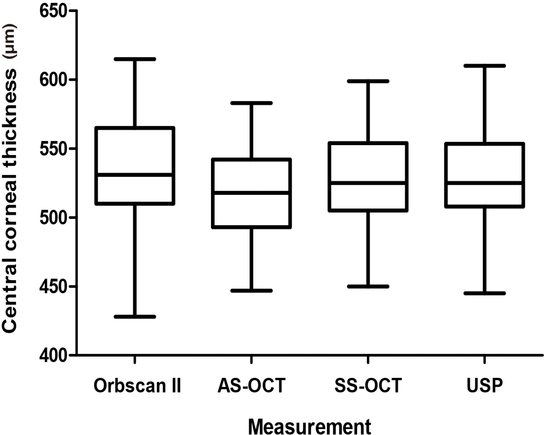 | Figure 1.Box plots showing mean central corneal thickness measured by Orbscan II, anterior segment optical coherence tomography (AS-OCT), swept source optical coherence tomography (SS-OCT), and ultrasound pachymetry (USP). The horizontal lines within the box represent medians, the ends of the boxes represent the first and third quartiles, and the whiskers represent the smallest and largest non-outlier values. |
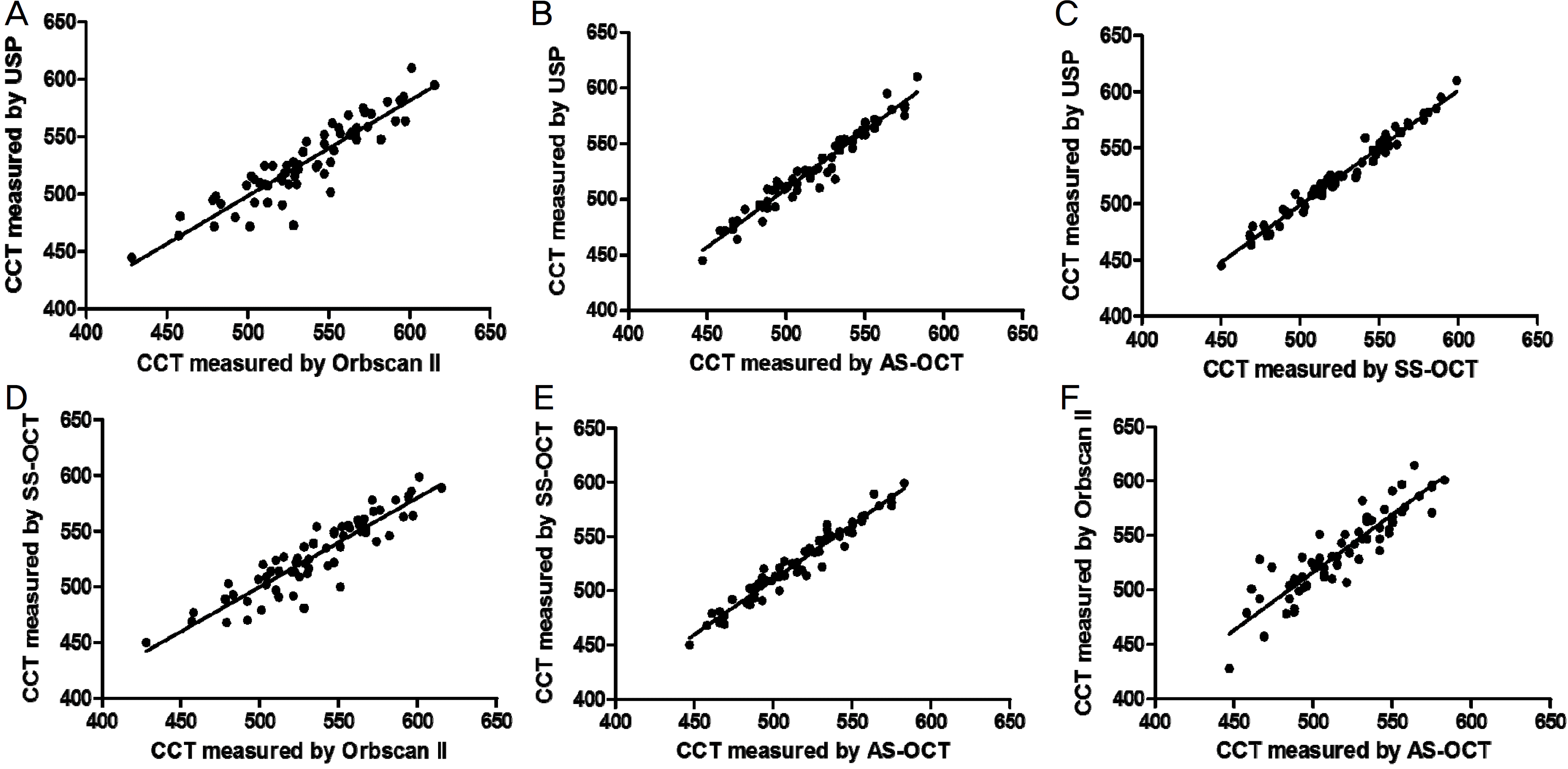 | Figure 2.The correlation plot of the central corneal thickness (CCT) measured by ultrasound pachymetry (USP), Orbscan II, anterior segment optical coherence tomography (AS-OCT), and swept source optical coherence tomography (SS-OCT). (A) Correlation between USP and Orbscan II (r2 = 0.834, p < 0.001), The best-fit line (y = 0.83× + 83.28) is designated by solid line. (B) Correlation between USP and AS-OCT (r2 = 0.947, p < 0.001), The best-fit line (y = 1.05× −13.48) is designated by solid line.(C) Correlation between USP and SS-OCT (r2 = 0.973, p < 0.001), The best-fit line (y = 1.02× – 11.42) is designated by solid line.(D) Correlation between SS-OCT and Orbscan II (r2 = 0.830, p < 0.001), The best-fit line (y = 0.80× + 99.58) is designated by solid line. (E) Correlation between SS-OCT and AS-OCT (r2 = 0.948, p < 0.001). The best-fit line (y = 1.01× + 4.87) is designated by solid line. (F) Correlation between Orbscan II and AS-OCT (r2 = 0.899, p < 0.001). The best-fit line (y = 1.06× – 14.66) is designated by solid line. |
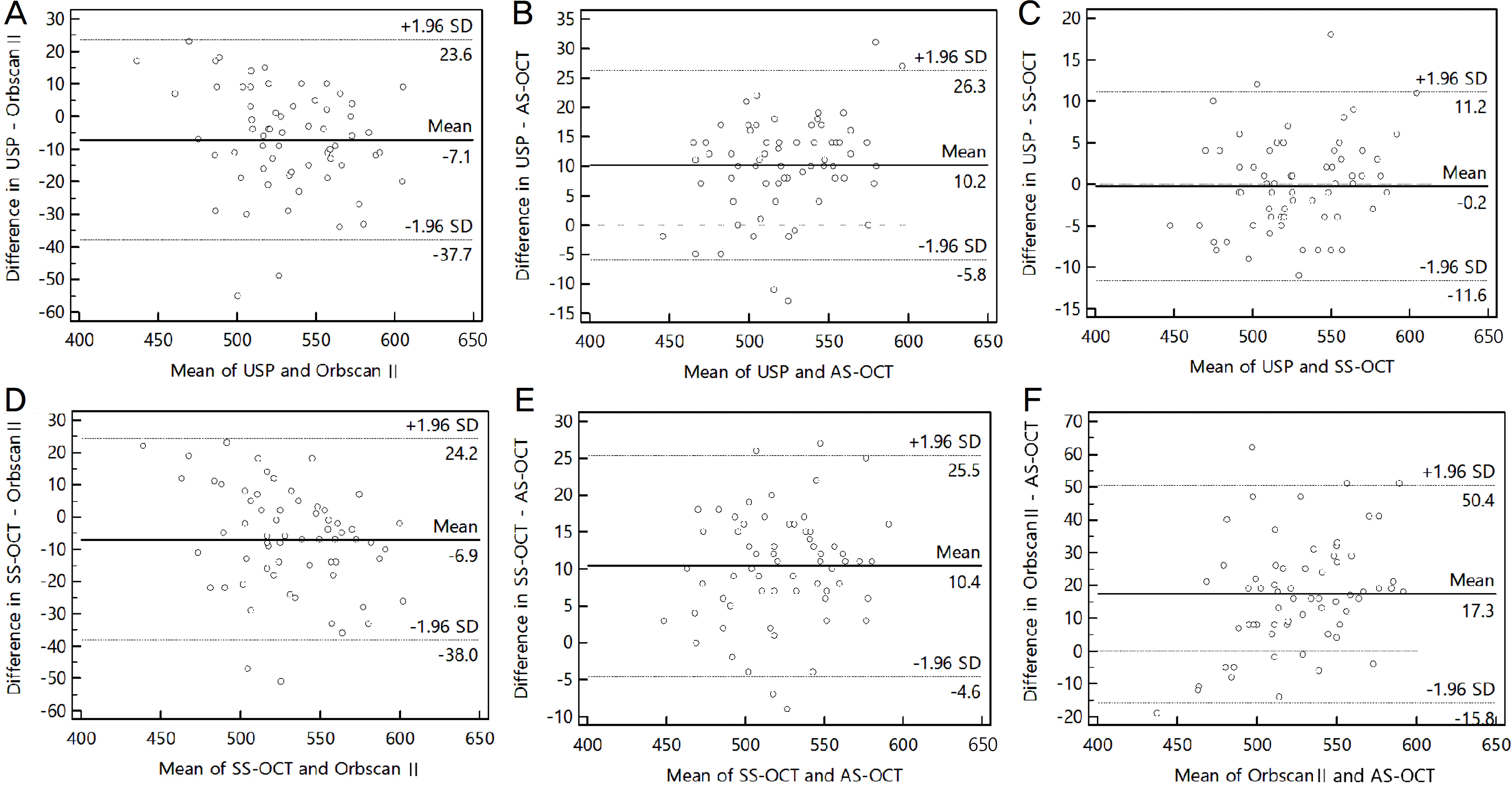 | Figure 3.Bland-Altman plots of the difference in central corneal thickness (CCT, μ m). Bland-Altman plots comparing measurement of CCT (A) between ultrasound pachymetry (USP) and Orbscan II, (B) between USP and anterior segment optical coherence tomography (AS-OCT), (C) between USP and swept source optical coherence tomography (SS-OCT), (D) between SS-OCT and Orbscan II, (E) between SS-OCT and AS-OCT, (F) between Orbscan II and AS-OCT. The middle solid line represents the mean difference in CCT values and the upper and lower dashed lines represent the 95% limits of agreement. SD = standard deviation. |
Table 1.
Pairwise comparison of central corneal thickness measurements
| Comparison | Mean difference ± SD | p-value* |
|---|---|---|
| Ultrasound pachymetry and Orbscan II | 6.82 ± 15.92 | 0.001 |
| Ultrasound pachymetry and AS-OCT | 10.22 ± 8.18 | <0.001 |
| Ultrasound pachymetry and SS-OCT | 0.20 ± 5.80 | 0.782 |
| SS-OCT and Orbscan II | 6.62 ± 16.26 | 0.002 |
| SS-OCT and AS-OCT | 10.42 ± 7.68 | <0.001 |
| Orbscan II and AS-OCT | 17.03 ± 17.34 | <0.001 |
 XML Download