Abstract
Purpose
To evaluate the efficacy of strip meniscometry test for dye eye syndrome (DES) by measuring the correlation between strip meniscometry and conventional test measurements.
Methods
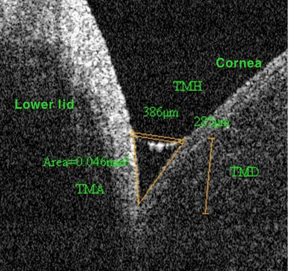
All subjects were examined using the Schirmer test, tear breakup time (TBUT) and strip meniscometry using SMTube (Echo Electricity Co., Ltd., Fukushima, Japan). Tear meniscus height (TMH), tear meniscus depth (TMD) and tear meniscus area (TMA) were measured using Fourier-domain optical coherence tomography. The DES group (n = 46 eyes) was compared with the normal group (n = 30 eyes) and correlation was assessed using Spearman's correlation coefficient.
Results
Strip meniscometry measurement was significantly correlated with Schirmer score (r = 0.6080, p < 0.01), TBUT (r = 0.5980, p < 0.01), TMH (r = 0.6210, p < 0.01), TMD (r = 0.6080, p < 0.01) and TMA (r = 0.6370, p < 0.01). Strip meniscometry was significantly lower in the DES group (4.58 ± 1.94 mm) than the normal group (7.07 ± 2.61 mm, p < 0.05).
Conclusions
Strip meniscometry was significantly correlated with other conventional test measurements for dry eye syndrome. Strip meniscometry is less time consuming and a less invasive method than the Schirmer test. Strip meniscometry could be an efficient tool to evaluate patients with dry eye syndrome in a clinical setting.
Go to : 
References
1. The definition and classification of dry eye disease: report of the Definition and Classification Subcommittee of the International Dry Eye WorkShop (2007). Ocul Surf. 2007; 5:75–92.
2. Kim WJ, Kim HS, Kim MS. Current trends in the recognition and treatment of dry eye: a survey of ophthalmologists. J Korean Ophthalmol Soc. 2007; 48:1614–22.

3. Nichols KK, Nichols JJ, Zadnik K. Frequency of dry eye abdominal test procedures used in various modes of ophthalmic practice. Cornea. 2000; 19:477–82.
4. Nichols KK, Nichols JJ, Mitchell GL. The lack of association abdominal signs and symptoms in patients with dry eye disease. Cornea. 2004; 23:762–70.
5. Nichols KK, Mitchell GL, Zadnik K. The repeatability of clinical measurements of dry eye. Cornea. 2004; 23:272–85.

6. Lamberts DW, Foster CS, Perry HD. Schirmer test after topical anesthesia and the tear meniscus height in normal eyes. Arch Ophthalmol. 1979; 97:1082–5.

7. Holly FJ. Physical chemistry of the normal and disordered tear film. Trans Ophthalmol Soc U K. 1985; 104(Pt 4):374–80.
8. Ibrahim OM, Dogru M, Takano Y, et al. Application of visante abdominalal coherence tomography tear meniscus height measurement in the diagnosis of dry eye disease. Ophthalmology. 2010; 117:1923–9.
9. Dogru M, Ishida K, Matsumoto Y, et al. Strip meniscometry: a new and simple method of tear meniscus evaluation. Invest Ophthalmol Vis Sci. 2006; 47:1895–901.

10. Methodologies to diagnose and monitor dry eye disease: report of the Diagnostic Methodology Subcommittee of the International Dry Eye WorkShop (2007). Ocul Surf. 2007; 5:108–52.
11. Sahai A, Malik P. Dry eye: prevalence and attributable risk factors in a hospital-based population. Indian J Ophthalmol. 2005; 53:87–91.

12. Calonge M, Diebold Y, Sáez V, et al. Impression cytology of the abdominal surface: a review. Exp Eye Res. 2004; 78:457–72.
14. Feldman F, Wood MM. Evaluation of the Schirmer tear test. Can J Ophthalmol. 1979; 14:257–9.
15. Wright JC, Meger GE. A review of the Schirmer test for tear production. Arch Ophthalmol. 1962; 67:564–5.

16. Pflugfelder SC, Tseng SC, Sanabria O, et al. Evaluation of subjective assessments and objective diagnostic tests for diagnosing tear-film disorders known to cause ocular irritation. Cornea. 1998; 17:38–56.

17. Kojima T, Ishida R, Dogru M, et al. A new noninvasive tear stabdominal analysis system for the assessment of dry eyes. Invest Ophthalmol Vis Sci. 2004; 45:1369–74.
18. Ibrahim OM, Dogru M, Ward SK, et al. The efficacy, sensitivity, and specificity of strip meniscometry in conjunction with tear abdominal tests in the assessment of tear meniscus. Invest Ophthalmol Vis Sci. 2011; 52:2194–8.
Go to : 
 | Figure 1.Optical coherence tomography image of the lower tear meniscus showing the tear meniscus height, depth and area. Measurements of tear meniscus height (TMH), tear meniscus depth (TMD), tear meniscus area (TMA) were performed using RTVue software. |
 | Figure 2.The image of Strip meniscometry tube® (SMTube; Echo Electricity Co., Ltd., Fukushima, Japan). The strip ni-trocellulose membranes have a pore size of 8 μ m. Natural blue dye 1 is printed at the tip of the strip. A millimeter and a numeric scale of up to 35 mm is printed on the both sides. |
Table 1.
Test measurements for dry eye diagnosis
Table 2.
Comparison of Group A and Group B
| Parameter | Group A*(n = 46 eyes) | Group B†(n = 30 eyes) |
|---|---|---|
| Schirmer test (mm) | 17.37 ± 10.44 | 27.73 ± 8.19 |
| Strip meniscometry (mm) | 4.58 ± 1.94 | 7.07 ± 2.61 |
| TBUT (sec) | 5.59 ± 2.17 | 11.90 ± 1.86 |
| Tear meniscus height (μ m) | 230.0 ± 98.54 | 302.9 ± 96.30 |
| Tear meniscus depth (μ m) | 177.2 ± 66.32 | 225.8 ± 74.06 |
| Tear meniscus area (mm2) | 0.029 ± 0.025 | 0.050 ± 0.029 |
Table 3.
Correlation of strip meniscometry test with tear function parameters
| Parameter | Correlation coefficient (r) | p-value* |
|---|---|---|
| Schirmer test | 0.6080 | p < 0.01 |
| Tear breakup time | 0.5980 | p < 0.01 |
| Tear meniscus height | 0.6210 | p < 0.01 |
| Tear meniscus depth | 0.6080 | p < 0.01 |
| Tear meniscus area | 0.6370 | p < 0.01 |


 PDF
PDF ePub
ePub Citation
 Print
Print


 XML Download