

 PDF
PDF ePub
ePub Citation
 Print
Print


Abstract
Purpose
Although a number of clinical parameters are well known to affect dry eye (DE) disease, it is unknown which factor mostly affects the discomfort of DE. Blepharitis is recognized as one of the leading causes of evaporative-type DE disease, but there have been no large-scale study to investigate the effect of blepharitis on DE symptoms. The purpose of this study was to evaluate the factors influencing subjective ocular discomfort in DE patients with blepharitis and to determine which parameter is most highly related to severity of blepharitis.
Methods
This investigation was a cross-sectional, clinical study. The test population consisted of DE patients suffering from moderate blepharitis. Seventy-three subjects aged 22 to 81 years (mean age 56.36) were enrolled, 49 of whom completed the investigation on a total of 49 eyes. A detailed assessment was conducted, including history taking, visual analog scale (VAS) pain scoring, ocular surface disease index (OSDI) questionnaire, blepharitis severity grading (score 0–4), conjunctival, corneal fluorescein staining (score 0–4), and tear break up time (TBUT) assessment.
Results
The results revealed significant correlations between subjective symptoms and blepharitis severity. Significant increases in overall VAS score, OSDI score (p = 0.031, p = 0.006) were recorded in DE patients with severe blepharitis. Conjunctival erosion was significantly related to VAS score (p = 0.016). Other parameters were not significantly related with VAS and OSDI scores. Additionally, conjunctival erosion was related with blepharitis severity (p < 0.0001), and corneal erosion was not correlated with blepharitis severity. TBUT also did not show any statistical correlation with blepharitis.
Conclusions
Our results showed that blepharitis severity is the main factor influencing subjective pain and discomfort in DE patients, although blepharitis severity was not related with the known clinical parameters of DE such as corneal erosion and TBUT. This study indicates that targeting treatment for blepharitis can significantly improve quality of life for patients suffering from DE disease.
Go to : 
References
1. Uchino M, Schaumberg DA, Dogru M, et al. Prevalence of dry eye disease among Japanese visual display terminal users. Ophthalmology. 2008; 115:1982–8.

2. Guo B, Lu P, Chen X, et al. Prevalence of dry eye disease in Mongolians at high altitude in China: the Henan eye study. Ophthalmic Epidemiol. 2010; 17:234–41.
3. Han SB, Hyon JY, Woo SJ, et al. Prevalence of dry eye disease in an elderly Korean population. Arch Ophthalmol. 2011; 129:633–8.
4. Yamada M, Mizuno Y, Shigeyasu C. Impact of dry eye on work productivity. Clinicoecon Outcomes Res. 2012; 4:307–12.
5. Cuevas M, González-García MJ, Castellanos E, et al. Correlations among symptoms, signs, and clinical tests in evaporative-type dry eye disease caused by Meibomian gland dysfunction (MGD). Curr Eye Res. 2012; 37:855–63.
6. The definition and classification of dry eye disease: report of the Definition and Classification Subcommittee of the International Dry Eye WorkShop (2007). Ocul Surf. 2007; 5:75–92.
7. Mathers WD. Ocular evaporation in meibomian gland dysfunction and dry eye. Ophthalmology. 1993; 100:347–51.
8. McDonald JE. Surface phenomena of the tear film. Am J Ophthalmol. 1969; 67:56–64.
9. Schein OD, Tielsch JM, Munõz B, et al. Relation between signs and symptoms of dry eye in the elderly. A population-based perspective. Ophthalmology. 1997; 104:1395–401.
10. Hay EM, Thomas E, Pal B, et al. Weak association between subjective symptoms or and objective testing for dry eyes and dry mouth: results from a population based study. Ann Rheum Dis. 1998; 57:20–4.
11. Nichols KK, Nichols JJ, Mitchell GL. The lack of association abdominal signs and symptoms in patients with dry eye disease. Cornea. 2004; 23:762–70.
12. Bron AJ. The Doyne Lecture. Reflections on the tears. Eye (Lond). 1997; 11(Pt 5):583–602.
13. Bron AJ, Benjamin L, Snibson GR. Meibomian gland disease. Classification and grading of lid changes. Eye (Lond). 1991; 5(Pt 4):395–411.
14. Andrews JS. The Meibomian secretion. Int Ophthalmol Clin. 1973; 13:23–8.
15. McCann LC, Tomlinson A, Pearce EI, Diaper C. Tear and abdominal gland function in blepharitis and normals. Eye Contact Lens. 2009; 35:203–8.
16. Kaido M, Kawashima M, Ishida R, Tsubota K. Relationship of abdominal pain sensitivity with dry eye symptoms in dry eye with short tear break-up time. Invest Ophthalmol Vis Sci. 2016; 57:914–9.
17. Guillon M, Maissa C, Wong S. Symptomatic relief associated with eyelid hygiene in anterior blepharitis and MGD. Eye Contact Lens. 2012; 38:306–12.
18. Hykin PG, Bron AJ. Age-related morphological changes in lid margin and meibomian gland anatomy. Cornea. 1992; 11:334–42.
19. Blazer D, Burchett B, Service C, George LK. The association of age and depression among the elderly: an epidemiologic exploration. J Gerontol. 1991; 46:M210–5.
20. Yezierski RP. The effects of age on pain sensitivity: preclinical studies. Pain Med. 2012; 13(Suppl 2):S27–36.
21. Chia EM, Mitchell P, Rochtchina E, et al. Prevalence and abdominals of dry eye syndrome in an older population: the Blue Mountains Eye Study. Clin Experiment Ophthalmol. 2003; 31:229–32.
22. Tamer C, Oksuz H, Sogut S. Androgen status of the non-autoimmune dry eye subtypes. Ophthalmic Res. 2006; 38:280–6.
Go to :
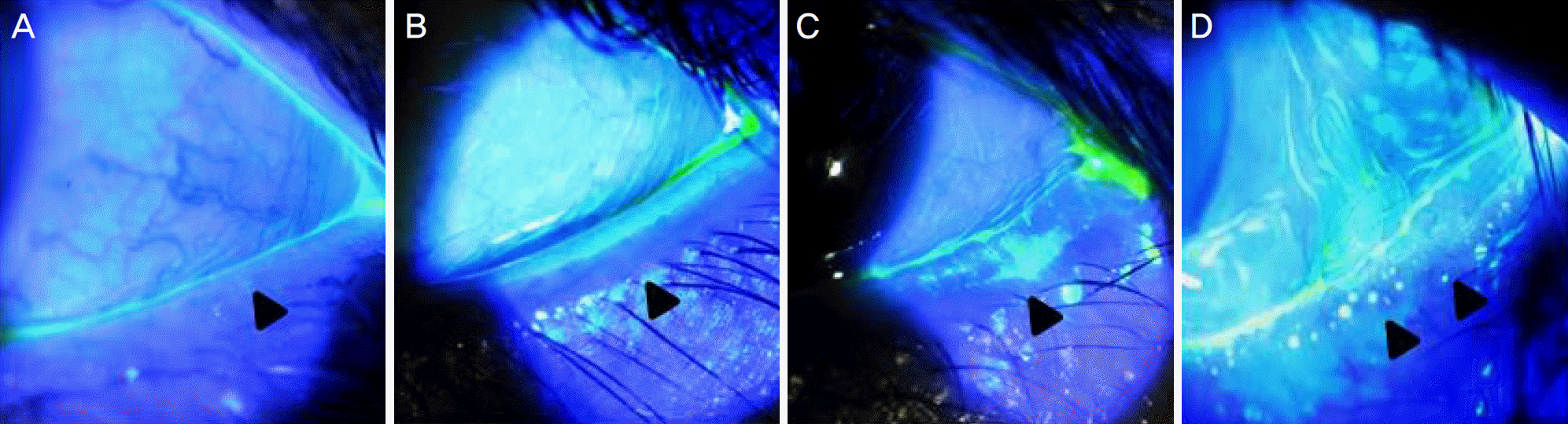 | Figure 1.Blepharitis– Obstructive meibomian gland dysfunction Grading. This is slit lamp photographs of clinical scale for blepharitis grading (especially temporal side). (A) Grade 1 blepharitis: 0–20% few capped meibomian glands. (B) Grade 2 blepahritis: 20–30% several blocked glands, and the secretions appeared thick. (C) Grade 3 blepharitis: approximately half the glands were blocked or stenosed. (D) Grade 4 blepharitis. more than half the glands were blocked/stenosed in combination with viscous secretions, tears. Black arrowheads mean the meibomian gland orifice. |
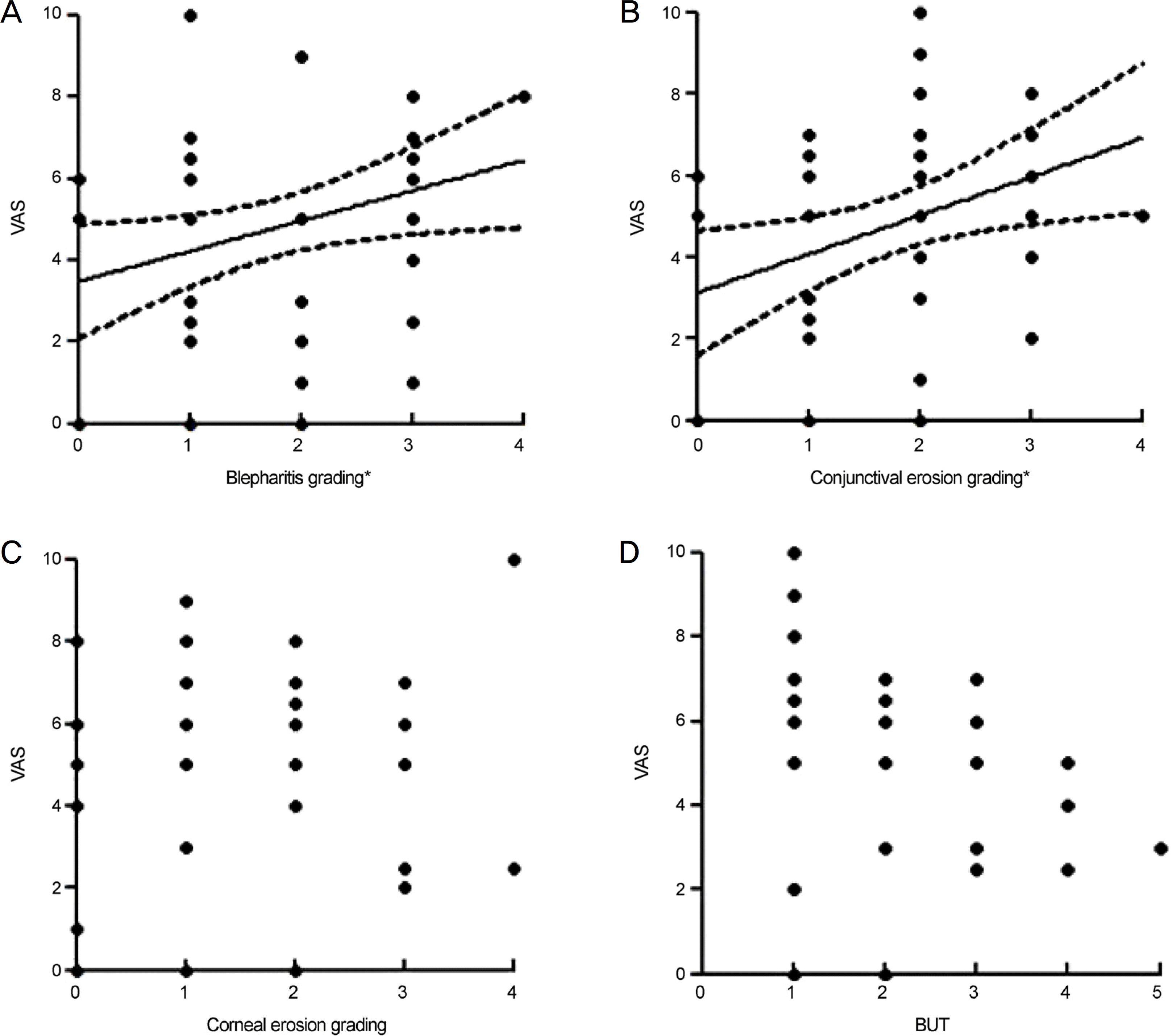 | Figure 2.Linear regression analysis of visual analogue scale (VAS) pain scoring and dry eye related ocular surface parameters. (A) Blepharitis grading was correlated with VAS score (r = 0.309, p = 0.031). (B) Conjunctival erosion grading was also related to VAS score (r = 0.342, p = 0.016). (C) Corneal erosion grading was not significantly related to VAS score (p = 0.357). (D) Fluorescein tear breakup time (BUT) showed no statistical correlation with VAS score (p = 0.520). * p-value < 0.05. |
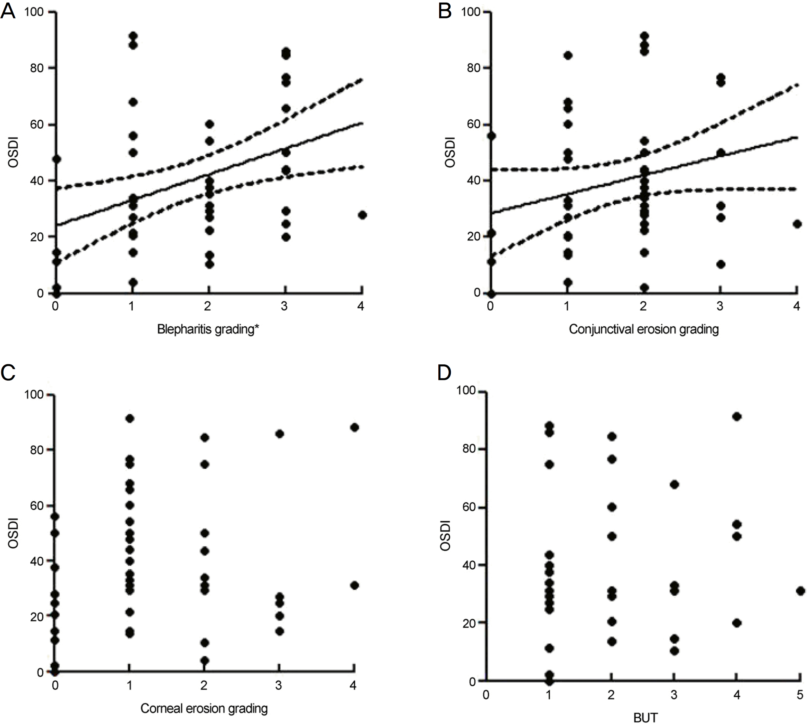 | Figure 3.Linear regression analysis of ocular surface disease index questionnaire score (OSDI) and dry eye related ocular surface parameters and other parameters. (A) Blepharitis grading was correlated with OSDI score (r = 0.389, p = 0.006). (B) Conjunctival erosion grading was not related to OSDI score (p = 0.085). (C) Corneal erosion grading was not significantly related to OSDI score (p = 0.276). (D) Fluorescein tear breakup time (BUT) showed no statistical correlation with OSDI score (p = 0.735). * p-value < 0.05. |
Table 1.
Demographic data and clinical results
| Blepharitis Grading 0–2 | Blepharitis Grading 3–4 | p-value | |
|---|---|---|---|
| Eyes (N) | 33 | 16 | |
| Male/Female | 5/28 (33) | 5/11 (16) | <0.0001† |
| Age (years) | 52.15 ± 1.76 | 60.88 ± 1.52 | 0.0026† |
| VAS score (0–10) | 4.30 ± 0.45 | 5.87 ± 0.53 | 0.0418* |
| OSDI (0–100) | 33.54 ± 3.91 | 50.31 ± 6.39 | 0.0233* |
| Conjunctival erosion (0–4) | 1.55 ± 0.15 | 2.25 ± 0.21 | 0.0103* |
| Corneal erosion (0–4) | 1.38 ± 0.20 | 1.43 ± 0.26 | 0.8533 |
| FTBUT (sec) | 2.13 ± 0.24 | 1.90 ± 0.38 | 0.6197 |
Table 2.
Pearson's correlation anlaysis of VAS, OSDI and dry eye related ocular surface parameters
| VAS | p-value | OSDI | p-value | |
|---|---|---|---|---|
| Blepharitis | 0.309 | 0.031* | 0.389 | 0.006† |
| Conjunctival erosion | 0.342 | 0.016* | 0.249 | 0.085 |
| Corneal erosion | 0.136 | 0.357 | 0.161 | 0.276 |
| FTBUT | –0.114 | 0.520 | 0.060 | 0.735 |
Table 3.
Pearson's correlation anlaysis among dry eye related ocular surface parameters
| Blepharitis | p-value | Conjunctival erosion | p-value | Corneal erosion | p-value | FTBUT | p-value | |
|---|---|---|---|---|---|---|---|---|
| Blepharitis | 0.486 | 0.000* | 0.036 | 0.809 | 0.095 | 0.593 | ||
| Corneal erosion | 0.036 | 0.809 | 0.239 | 0.101 | –0.155 | 0.381 | ||
| Conjunctival erosion | 0.486 | 0.000* | 0.239 | 0.101 | –0.078 | 0.661 | ||
| FTBUT | 0.095 | 0.593 | –0.078 | 0.661 | –0.155 | 0.381 |
 XML Download