

 PDF
PDF ePub
ePub Citation
 Print
Print


Abstract
Purpose
To report a case of acute angle closure after cataract surgery using an accommodative intraocular lens (IOL), WIOL-CF®(GELMED, Praha, Czech).
Case summary
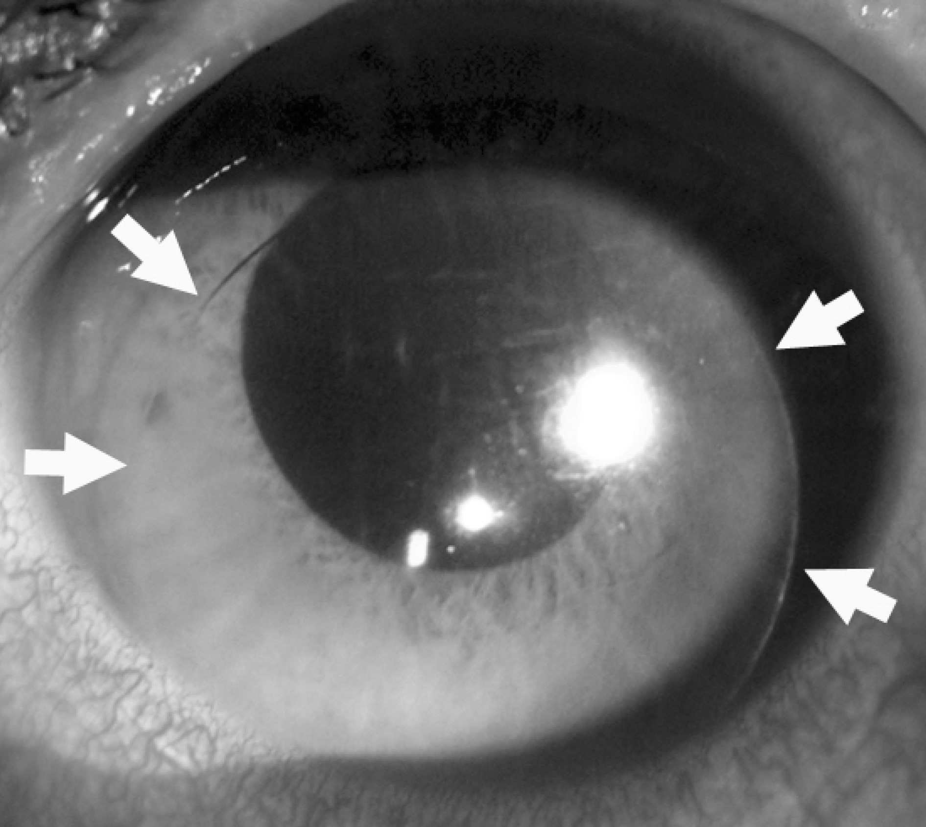
A 46-year-old male patient underwent phacoemulsification and implantation of WIOL-CF® into the capsular bag. Seven months after the surgery, a sudden increase in intraocular pressure (IOP) associated with angle closure was observed. Ultrabiomicroscopy revealed a dislocated WIOL-CF® that was pushing the peripheral iris anteriorly. Despite the use of IOP-lowering medication and peripheral laser iridotomy, IOP was not controlled. After the use of cycloplegics, the angle was widened and IOP decreased; however, after nine days, the WIOL-CF® was completely dislocated into the anterior chamber and so was removed.
Go to : 
References
1. Studeny P, Krizova D, Urminsky J. Clinical experience with the WIOL-CF accommodative bioanalogic intraocular lens: Czech national observational registry. Eur J Ophthalmol. 2016; 26:230–5.

2. Pallikaris IG, Portaliou DM, Kymionis GD, et al. Outcomes after accommodative bioanalogic intraocular lens implantation. J Refract Surg. 2014; 30:402–6.
3. Lee HS, Park SH, Kim MS. Clinical results and some problems of multifocal apodized diffractive intraocular lens implantation. J Korean Ophthalmol Soc. 2008; 49:1235–41.
4. Cheon MH, Lee JE, Kim JH, et al. One-year outcome of monocular implant of aspheric multifocal IOL. J Korean Ophthalmol Soc. 2010; 51:822–28.
5. Kang KT, Kim YC. Dislocation of polyfocal full-optics abdominal intraocular lens after neodymium-doped yttrium aluminum garnet capsulotomy in vitrectomized eye. Indian J Ophthalmol. 2013; 61:678–80.
Go to :
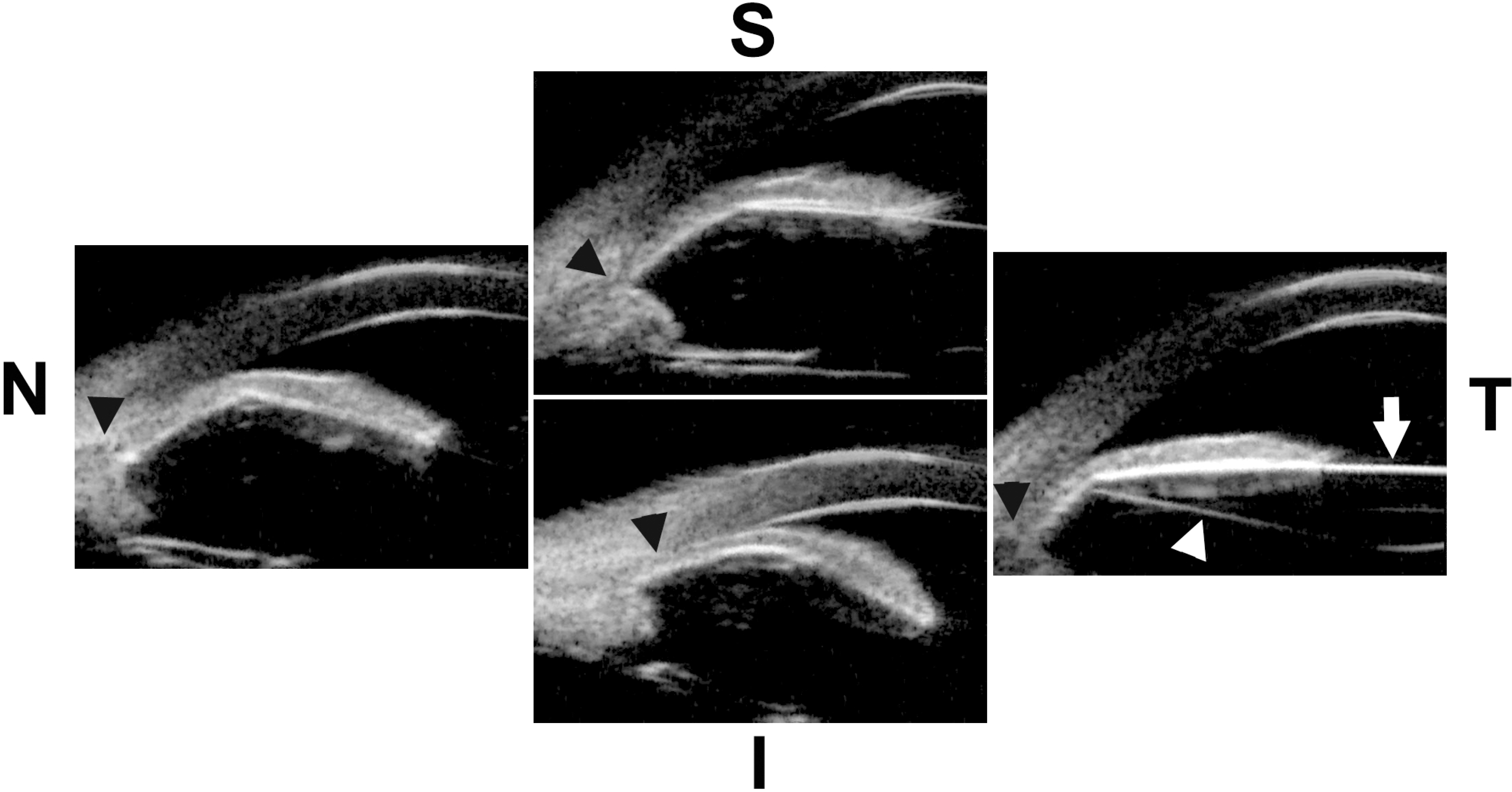 | Figure 2.Ultrasound biomicroscopic findings of superior (S), nasal (N), inferior (I), and temporal (T) angle at the time of acute angle closure. White arrow indicates anterior surface of intraocular lens (IOL), white arrowhead indicates posterior surface of IOL, and black arrowheads indicate anterior angle. IOL was dislocated from capsular bag and dislocated IOL was pushing peripheral iris anteriorly. Anterior angles were closed in all quadrants due to anterior displacement of IOL. |
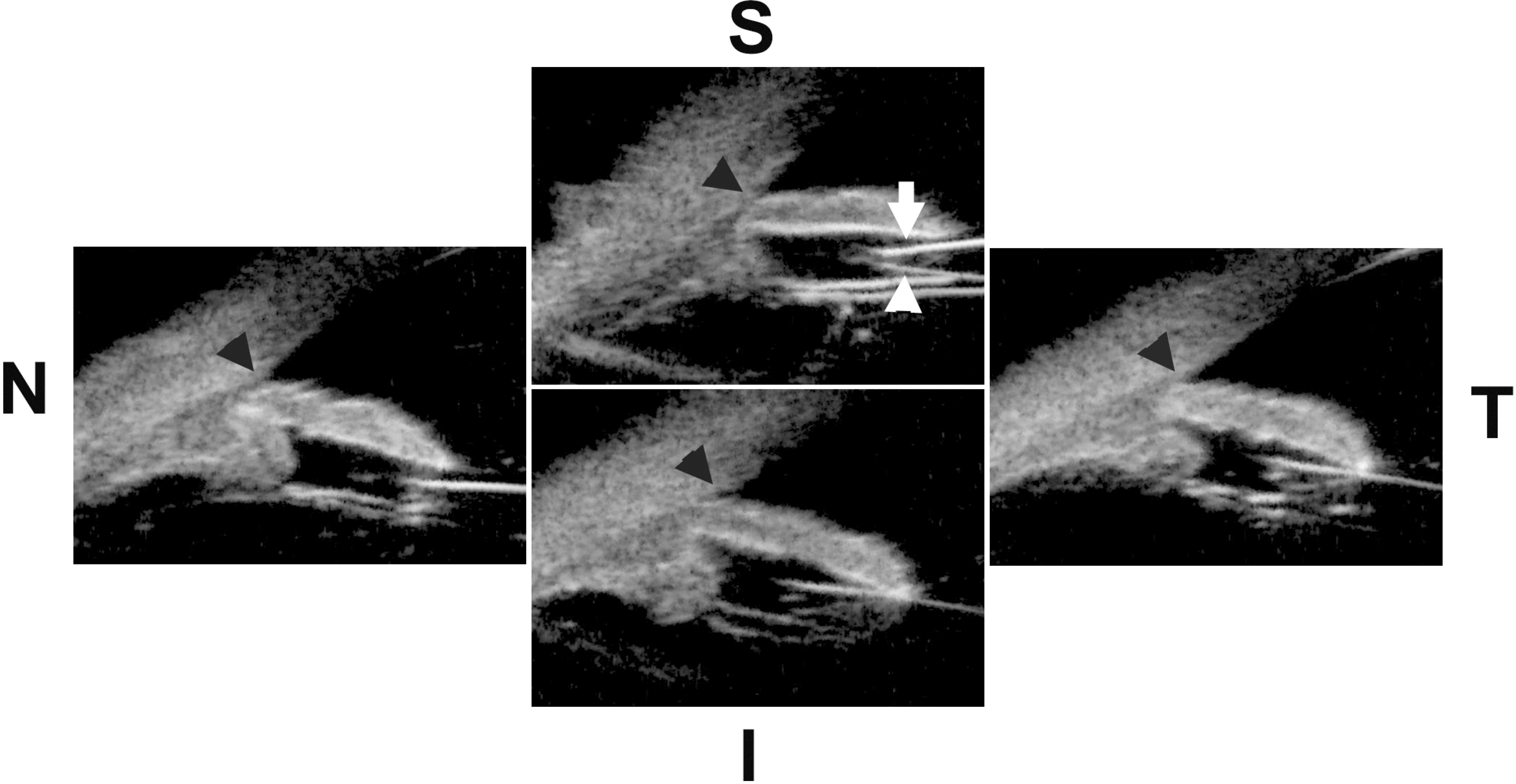 | Figure 3.Ultrasound biomicroscopic findings of superior (S), nasal (N), inferior (I), and temporal (T) angle after the instillation of cycloplegics. White arrow indicates anterior surface of intraocular lens (IOL), white arrowhead indicates posterior surface of IOL, and black arrowheads indicate anterior angle. IOL moved posteriorly and anterior angle was widened compared to the previous state. |
 XML Download