

 PDF
PDF ePub
ePub Citation
 Print
Print


Abstract
Purpose
We report a case of amaurosis fugax associated with ipsilateral internal carotid artery agenesis.
Case summary
A 50-year-old woman presented with amaurosis fugax in her left eye; the frequency of episodes of the condition had recently increased to once a month. She had a history of hypertension and dyslipidemia, and was under medical therapy. The visual acuity of both eyes was 20/20. Slit-lamp examination was normal except for pseudophakia. Ophthalmoscopy revealed a myopic tigroid fundus and a myopic tilted disc. No abnormalities were evident in fluorescein fundus angiography. Brain computed tomography showed that the left bony carotid canal was absent, and magnetic resonance angiography showed that the left internal carotid artery was also absent. She was diagnosed with left internal carotid artery agenesis. Other neurological and hematological parameters were within normal ranges. The amaurosis fugax spontaneously disappeared and has not recurred over the past 12 months. Our case, although rare, suggests that amaurosis fugax may be associated with internal carotid artery agenesis.
Go to : 
References
1. Current management of amaurosis fugax. The Amaurosis Fugax Study Group. Stroke. 1990; 21:201–8.
2. Burde RM. Amaurosis fugax. An overview. J Clin Neuroophthalmol. 1989; 9:185–9.
3. Hayreh SS, Zimmerman MB. Amaurosis fugax in ocular vascular occlusive disorders: prevalence and pathogeneses. Retina. 2014; 34:115–22.
4. Kim NR, Chin HS. Progression of impending central retinal vein occlusion to the ischemic variant following intravitreal bevacizumab. Korean J Ophthalmol. 2010; 24:179–81.

5. Lee DH, Lee SJ, Yoon IN. Clinical progress in impending central retinal vein occlusion. Korean J Ophthalmol. 2010; 24:83–8.
6. Shaw HE Jr, Osher RH, Smith JL. Amaurosis fugax associated with SC hemoglobinopathy and lupus erythematosus. Am J Ophthalmol. 1979; 87:281–5.
7. Heckmann JG, Gaul C, Neundörfer B, et al. Vasospastic amaurosis fugax. J Neurol Neurosurg Psychiatry. 2003; 74:149.
8. Jehn A, Frank Dettwiler B, Fleischhauer J, et al. Exercise-induced vasospastic amaurosis fugax. Arch Ophthalmol. 2002; 120:220–2.
9. Jo YJ, Yun YJ, Kwag JY, Kim JY. Valsalva maneuver-induced amaurosis fugax. J Korean Ophthalmol Soc. 2010; 51:779–83.
10. Afifi AK, Godersky JC, Menezes A, et al. Cerebral hemiatrophy, hypoplasia of internal carotid artery, and intracranial aneurysm. A rare association occurring in an infant. Arch Neurol. 1987; 44:232–5.
11. Graham CB 3rd, Wippold FJ 2nd, Capps GW. Magnetic resonance imaging in internal carotid artery agenesis with computed abdominal and angiographic correlation-case reports. Angiology. 1999; 50:847–54.
12. Cali RL, Berg R, Rama K. Bilateral internal carotid artery agenesis: a case study and review of the literature. Surgery. 1993; 113:227–33.
13. Mellado JM, Merino X, Ramos A, et al. Agenesis of the internal carotid artery with a trans-sellar anastomosis: CT and MRI abdominal in late-onset congenital hypopituitarism. Neuroradiology. 2001; 43:237–41.
14. Czarnecki EJ, Silbergleit R, Mehta BA, Sanders WP. Absence of the supraclinoid internal carotid artery in association with abdominal aneurysms. Neuroradiology. 1998; 40:11–4.
15. Jung BO, Lee JI, Choi JH, et al. Congenital hypoplasia of the abdominal internal carotid artery: a case report. J Korean Neurosurg Soc. 2002; 31:282–4.
16. Ito S, Miyazaki H, Iino N, et al. Unilateral agenesis and hypoplasia of the internal carotid artery: a report of three cases. Neuroradiology. 2005; 47:311–5.
17. Na SK, Park TH, Ahn JY, Kim MK. A case of unilateral agenesis of internal carotid artery with throbbing headache. J Neurocrit Care. 2008; 1:174–6.
18. Lasjaunias P, Berenstein A, Brugge KT. Clinical vascular anatomy and variations. 2nd ed.Berlin: Springer;2001. p. 414–24.
19. Naeini RM, De J, Satow T, Benndorf G. Unilateral agenesis of abdominal carotid artery with ophthalmic artery arising from posterior communicating artery. AJR Am J Roentgenol. 2005; 184:571–3.
20. Savastano S, Feltrin GP, Chiesura-Corona M, Miotta D. Cerebral ischemia due to congenital malformations of brachiocephalic ar-teries-case reports. Angiology. 1992; 43:76–83.
21. Sliwka U, Schmidt P, Reul J, Noth J. Agenesis of the internal carotid artery: color Doppler, CT, and MR angiography findings. J Clin Ultrasound. 1988; 26:213–6.
22. Halstuk KS, Littooy FN, Baker WH. Absent common carotid abdominal associated with amaurosis fugax: a case report. Surgery. 1985; 97:502–6.
Go to :
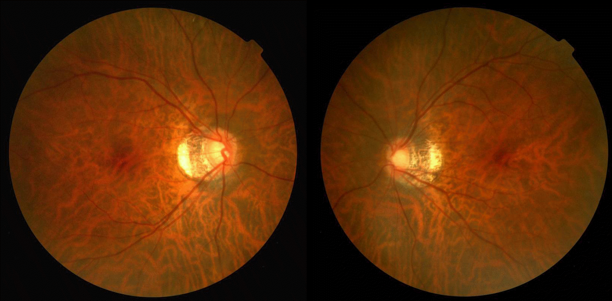 | Figure 1.Fundus photographs of the both eyes. Both eyes show myopic tigroid fundus and myopic tilted disc with peripapillary atrophy. |
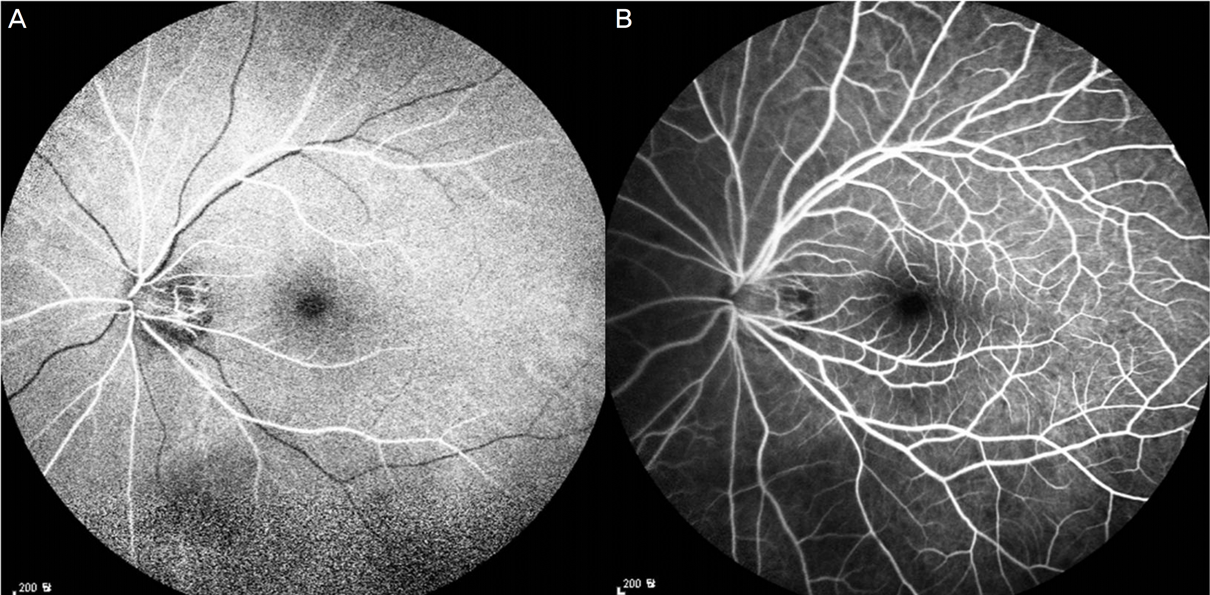 | Figure 2.Fluorescein fundus angiography of the left eye. (A) Retinal artery filling was started at 10 seconds after injection of the sodium fluorescein dye into the antecubital vein. (B) Retinal vein filling was completed at 18 seconds after injection. |
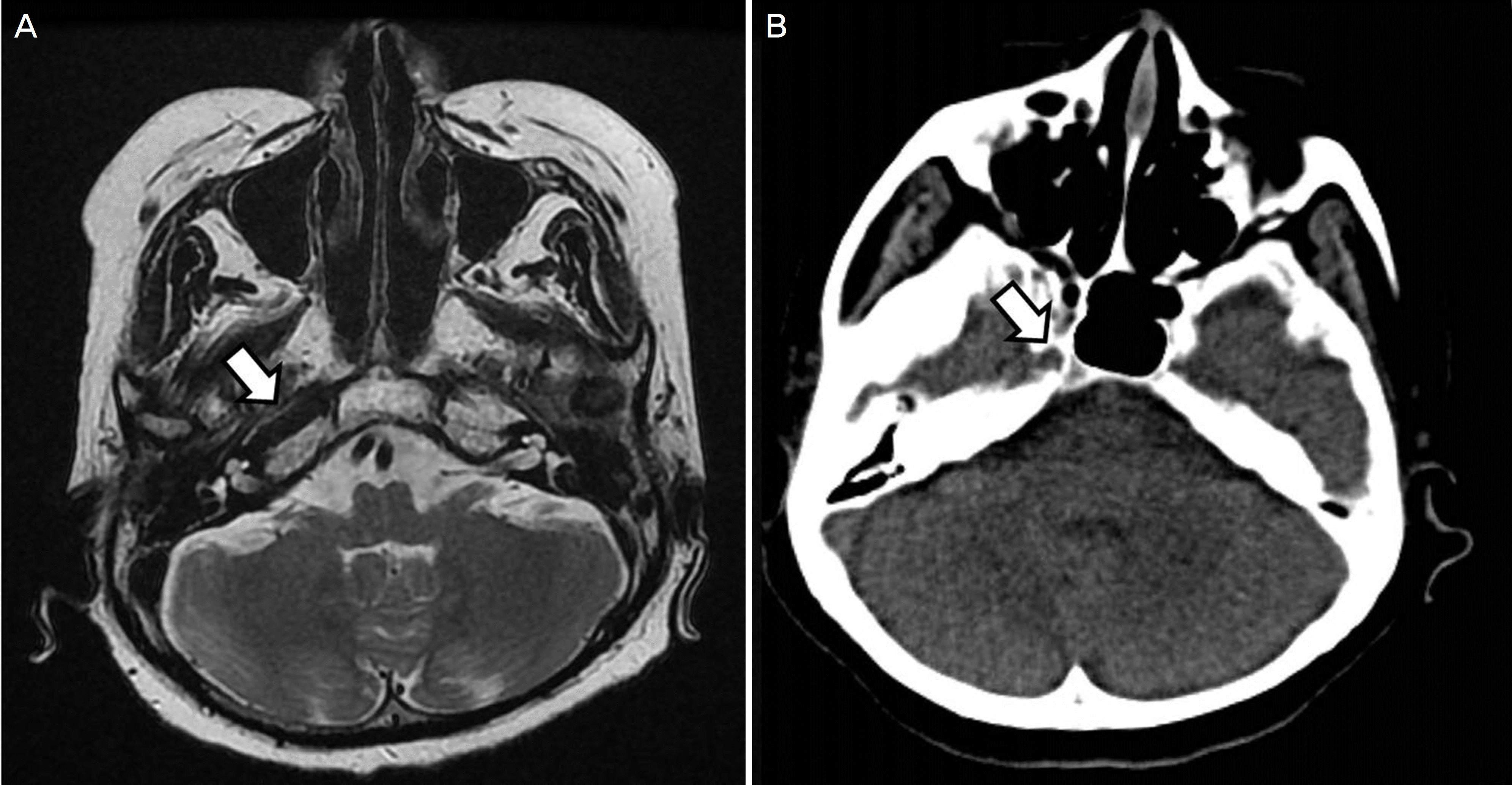 | Figure 3.Magnetic resonance imaging (MRI) and computed tomography (CT). (A) MRI and (B) CT of the skull base showing a carotid canal on the right (arrows), but absent on the left. |
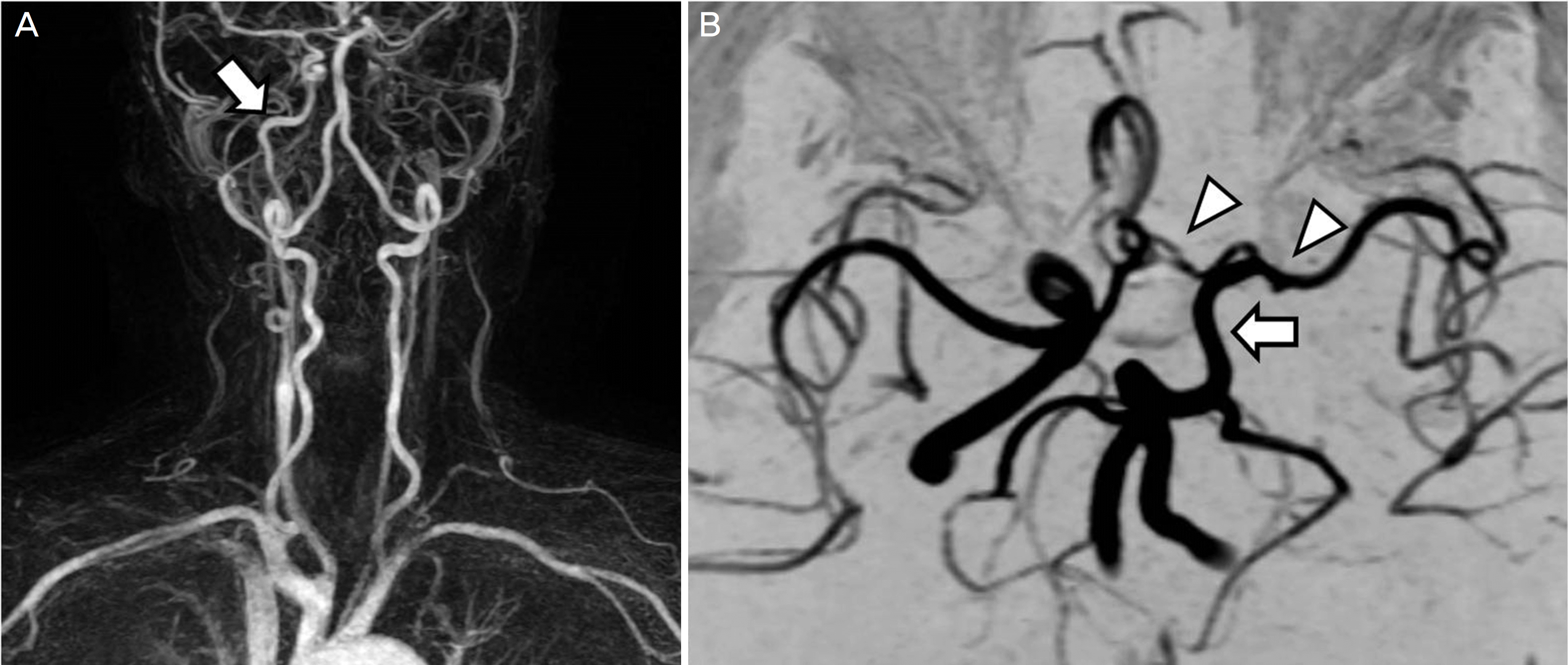 | Figure 4.Magnetic resonance angiography (MRA). (A) MRA reveals the right internal carotid artery (arrow) and the absence of the left internal carotid artery. (B) At the circle of Willis, the left middle cerebral artery and anterior cerebral artery (arrowheads) are filled via posterior communicating artery (arrow). |
 XML Download