

 PDF
PDF ePub
ePub Citation
 Print
Print


Abstract
Purpose
To report a case of eyeball displacement into the ethmoid sinus followed by early surgical intervention and good visual recovery.
Case summary
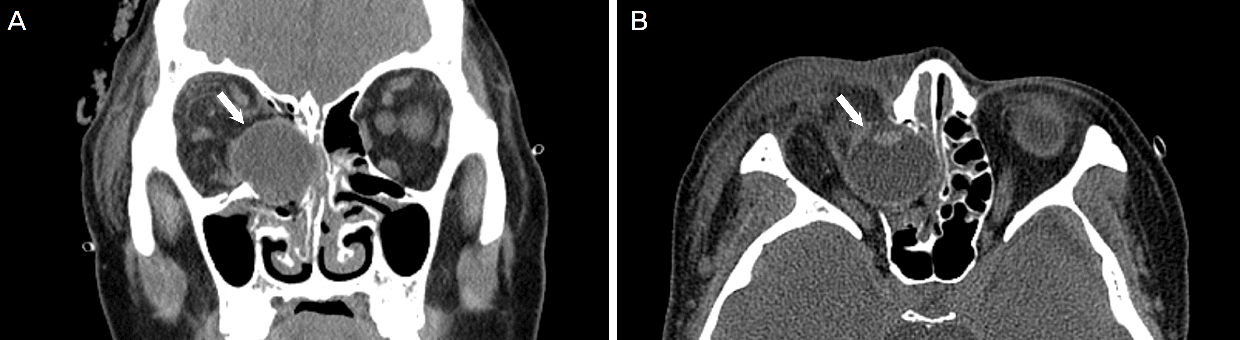
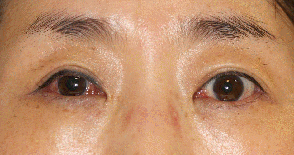
A 46-year-old female visited our hospital after she injured the right side of her face. Her visual acuity could not be measured and computed tomography revealed displacement of the right eyeball into the ethmoid sinus, as well as right medial orbital wall fracture and rupture of the right medial rectus muscle. She underwent surgical reduction of the herniated eyeball and surgical correction of the medial orbital wall fracture within 20 hours after the accident. Eighteen months after the surgery, visual acuity of the right eye improved from light perception to 20/28, and her color vision and visual field of the right eye improved to normal range.
Conclusions
Displacement of the eyeball in the orbital wall fracture is very rare, and eyeball displacement into the ethmoid sinus is even rarer. We achieved good visual outcome through early surgical intervention. The early anatomical reduction of the displacement and wall fracture may promote improved final visual outcome in other similar cases.
Go to : 
References
1. Pelton RW, Rainey AM, Lee AG. Traumatic subluxation of the globe into the maxillary sinus. AJNR Am J Neuroradiol. 1998; 19:1450–1.
2. Haggerty CJ, Roman P. Repositioning of a traumatically displaced globe with maxillary antrostomy: review of the literature and abdominal recommendations. J Oral Maxillofac Surg. 2013; 71:1915–22.
3. Okabe H, Kimura K, Sonoda S, Sakamoto T. Displacement of globe into ethmoid sinus by orbital medial wall fracture with good recovery of vision. Jpn J Ophthalmol. 2005; 49:426–8.

4. Abrishami M, Aletaha M, Bagheri A, et al. Traumatic subluxation of the globe into the maxillary sinus. Ophthal Plast Reconstr Surg. 2007; 23:156–8.
5. Kreiner B, Amer R, Sharfi G, et al. Traumatic displacement of the globe into the paranasal sinuses: case report and guidelines for treatment. J Oral Maxillofac Surg. 2008; 66:826–30.
6. Risco JM, Stratas BA, Knott RH. Prolapse of the globe into the ethmoid sinus. Am J Ophthalmol. 1984; 97:659–60.
7. Tranfa F, Di Matteo G, Di Salle F, et al. Traumatic displacement of the globe into the ethmoid sinus. Am J Ophthalmol. 2000; 130:253–4.
Go to :
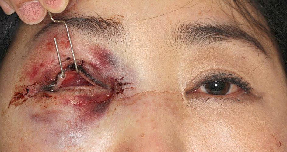 | Figure 1.Eyelid photo of the patient. Eyeball could not be found in the palpebral fissure and only conjunctiva was found. |
 XML Download