Abstract
Purpose
To evaluate clinical outcomes after combined descemet-stripping endothelial keratoplasty (DSEK) and intraocular lens (IOL) exchange in a Korean population.
Methods
The medical records of 15 patients (15 eyes) with pseudophakic bullous keratopathy who underwent combined DSEK and IOL exchange from January 2011 to January 2015 and who were followed up for more than 12 months were reviewed retrospectively.
Results
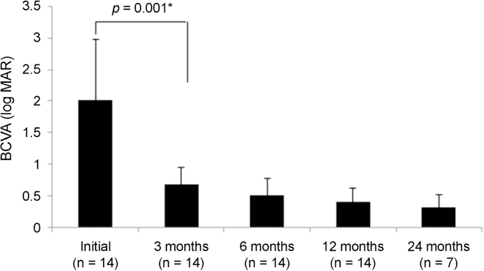
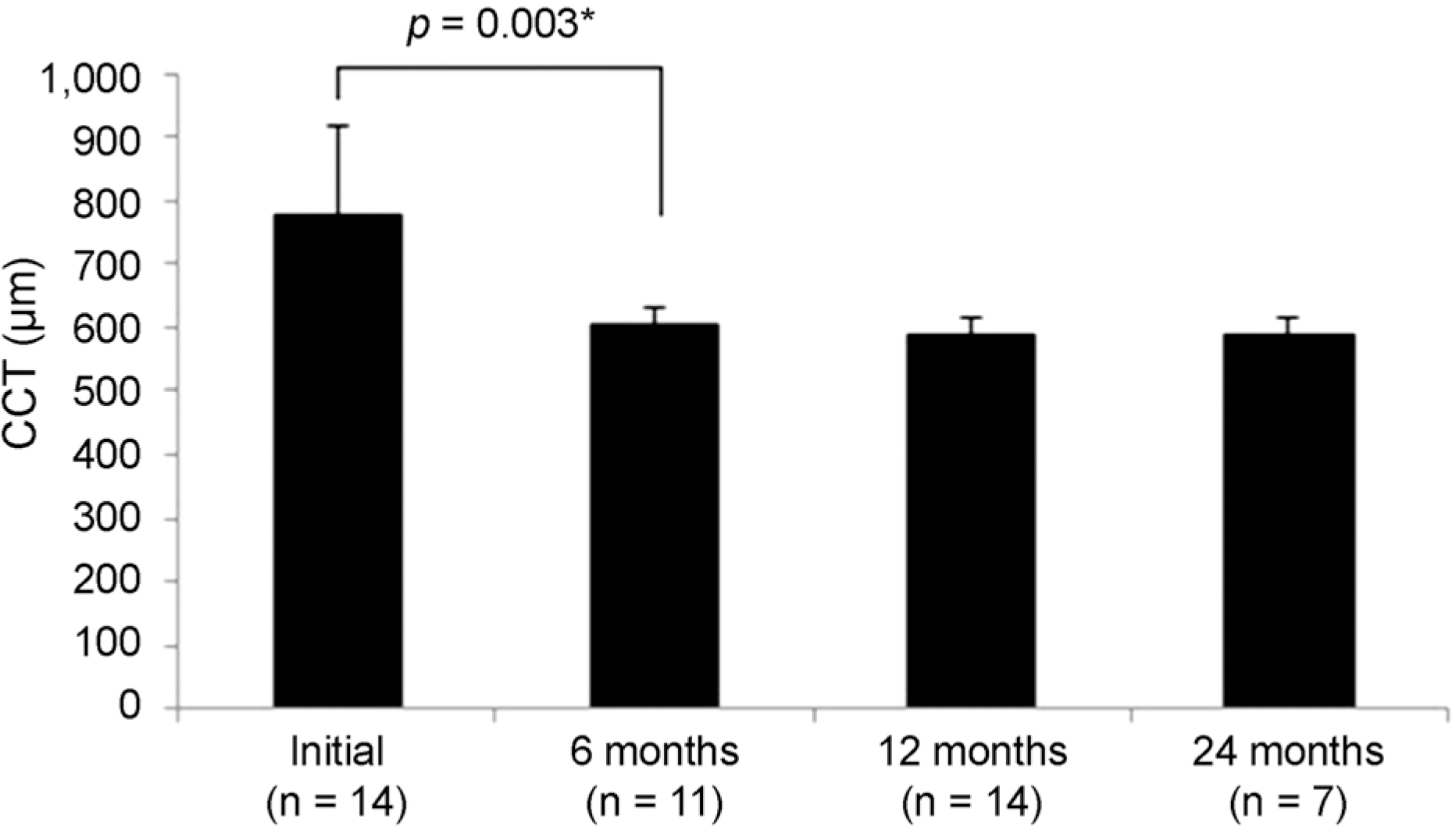
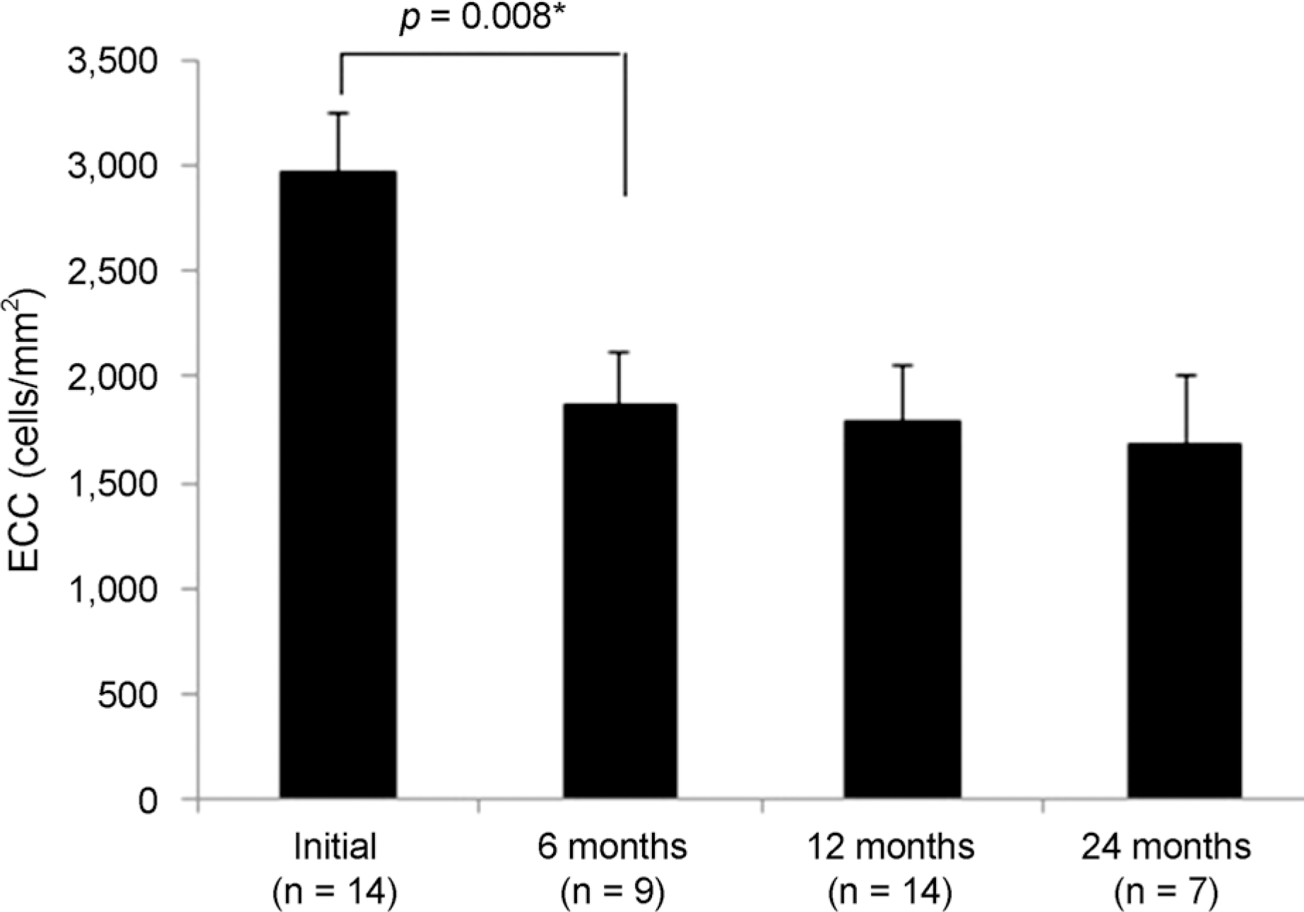
In 14 eyes with successful results after surgery, the best corrective visual acuity (BCVA) was significantly improved from 2.01 ± 0.96 (log MAR, mean) to 0.68 ± 0.26 at 3 months (p = 0.001) except for one eye that received reoperation on the endothelial disc detachment. The BCVA at postoperative 6 and 12 months gradually increased (0.51 ± 0.26 and 0.40 ± 0.22 log MAR, mean). Central corneal thickness was significantly improved from 777 ± 139 μ m to 605 ± 28 μ m at 6 months (p = 0.003) and was maintained at 12 months. The mean endothelial cell count was 2,973 ± 281/mm2 in the donor lenticules and 1,790 ± 265/mm2 at 12 months. Endothelial cell loss was 40%. The target refraction was −0.81 ± 0.16 D and the 12 months postoperative spherical equivalent was −0.28 ± 0.36 D. Complications included intraocular pressure elevation in one eye and pupillary capture in one eye.
Go to : 
References
1. Gorovoy MS, Price FW. New technique transforms corneal transplantation. Cataract Refract Surg Today. 2005; 11:55–8.
2. Koenig SB, Covert DJ, Dupps WJ Jr, Meisler DM. Visual acuity, refractive error, and endothelial cell density six months after Descemet stripping and automated endothelial keratoplasty (DSAEK). Cornea. 2007; 26:670–4.

3. Terry MA. Endothelial keratoplasty: history, current state, and future directions. Cornea. 2006; 25:873–8.
4. Shimazaki J, Amano S, Uno T, et al. National survey on bullous keratopathy in Japan. Cornea. 2007; 26:274–8.

5. Gonçalves ED, Campos M, Paris F, et al. Bullous keratopathy: etio-pathogenesis and treatment. Arq Bras Oftalmol. 2008; 71(6 Suppl):61–4.
6. Balázs E, Balázs K, Módis L Jr, Berta A. Penetrating keratoplasty for pseudophakic bullous keratopathy. Acta Chir Hung. 1997; 36:11–3.
7. Barkana Y, Segal O, Krakovski D, et al. Prediction of visual abdominal after penetrating keratoplasty for pseudophakic corneal edema. Ophthalmology. 2003; 110:286–90.
8. Wylegala E, Tarnawska D. Management of pseudophakic bullous keratopathy by combined Descemet-stripping endothelial abdominal and intraocular lens exchange. J Cataract Refract Surg. 2008; 34:1708–14.
9. Chan CC, Crandall AS, Ahmed II. Ab externo scleral suture loop fixation for posterior chamber intraocular lens decentration: abdominal results. J Cataract Refract Surg. 2006; 32:121–8.
10. Djalilian AR, Anderson SO, Fang-Yen M, et al. abdominal results of transsclerally sutured posterior chamber lenses in penetrating keratoplasty. Cornea. 1998; 17:359–64.
11. Chen ES, Shamie N, Terry MA. Descemet-stripping endothelial keratoplasty: improvement in vision following replacement of a healthy endothelial graft. J Cataract Refract Surg. 2006; 34:1044–6.

12. Price MO, Price FW Jr. Descemet's stripping with endothelial keratoplasty: comparative outcomes with microkeratome-dissected and manually dissected donor tissue. Ophthalmology. 2006; 113:1936–42.
13. Covert DJ, Koenig SB. New triple procedure: Descemet's stripping and automated endothelial keratoplasty combined with abdominal and intraocular lens implantation. Ophthalmology. 2007; 114:1272–7.
14. Holz HA, Meyer JJ, Espandar L, et al. Corneal profile analysis abdominal Descemet stripping endothelial keratoplasty and its relationship to postoperative hyperopic shift. J Cataract Refract Surg. 2008; 34:211–4.
15. Suto C, Hori S, Fukuyama E, Akura J. Adjusting intraocular lens power for sulcus fixation. J Cataract Refract Surg. 2003; 29:1913–7.

16. Allan BD, Terry MA, Price FW Jr, et al. Corneal transplant abdominal rate and severity after endothelial keratoplasty. Cornea. 2007; 26:1039–42.
18. Lee K, Hwang KY, Kim MS. Influence of endothelial cell loss abdominal preservation on graft survival in imported donor cornea. J Korean Ophthalmol Soc. 2013; 54:862–8.
19. Ide T, Yoo SH, Goldman JM, et al. Descemet-stripping automated endothelial keratoplasty: effect of inserting forceps on DSAEK abdominal tissue viability by using an in vitro delivery model and vital dye assay. Cornea. 2007; 26:1079–81.
Go to : 
 | Figure 1.Changes in best corrected visual acuity (BCVA) after combined descemet-stripping endothelial keratoplasty and intraocular lens exchange. The BCVA improved at post-operative 3 months. * Wilcoxon signed rank test. |
 | Figure 2.Changes in central corneal thickness (CCT) after combined descemet-stripping endothelial keratoplasty and intraocular lens exchange. * Wilcoxon signed rank test. |
 | Figure 3.Changes in endothelial cell count (ECC) after combined descemet-stripping endothelial keratoplasty and intraocular lens exchange. * Wilcoxon signed rank test. |
Table 1.
Characteristics of the patient who underwent combined descemet-stripping endothelial keratoplasty and intraocular lens (IOL) exchange
HTN = hypertension; IOP = intraocular pressure; ACL = anterior chamber lens; Prev. = previous; RRD = rhegmatogenous retinal detachment; Vit. eye = vitrectomized eye; DM = diabetes melitus; ERM = epiretinal membrane; CME = cystoid macular edema; CSC = chronic central serous chorioretinopathy; PKP = penetrating keratoplasty.
Table 2.
Pre- and postoperative best corrected visual acuity (BCVA, log MAR), keratometry and refractive error (D) at 12 months after operation
| Case | Preop BCVA |
Postop BCVA |
Preop Mean K (D) | Postop Mean K (D) | Target power (D) |
Refractive error (D) |
S/E (D) | |||
|---|---|---|---|---|---|---|---|---|---|---|
| 3 months | 6 months | 12 months | Spherical | Cylinder | ||||||
| 1 | 1.7 | 0.7 | 0.4 | 0.3 | 44.5 | 44.5 | −0.79 | +0.25 | −1.0 | −0.25 |
| 2 | 0.7 | 0.2 | 0.2 | 0.1 | 42.625 | 42.75 | −0.85 | +0.5 | −1.25 | −0.125 |
| 3 | 1.7 | 1.0 | 1.0 | 0.7 | 42.375 | 42.5 | −0.94 | +0.5 | −1.5 | −0.25 |
| 4 | 0.7 | 0.3 | 0.2 | 0.1 | 43.5 | 43.5 | −0.87 | +0.5 | −1.75 | −0.375 |
| 5 | 0.7 | 0.4 | 0.3 | 0.3 | 42.625 | 42.5 | −0.82 | −0.5 | −0.5 | −0.75 |
| 6 | 1.3 | 1.0 | 1.0 | 0.7 | 42.625 | 42.75 | −1.22 | 0 | −1.0 | −0.5 |
| 7 | 3.0 | 1.0 | 0.7 | 0.7 | 43.25 | 43.25 | −0.7 | +0.5 | −2.0 | −0.5 |
| 8 | 3.0 | 1.0 | 0.7 | 0.7 | 43 | 43.25 | −0.95 | 0 | −1.0 | −0.5 |
| 9 | 3 | 0.7 | 0.5 | 0.5 | 44.5 | 44.5 | −0.58 | +0.5 | −1.5 | −0.25 |
| 10 | 3 | 0.7 | 0.5 | 0.4 | 45.875 | 46 | −0.63 | +0.75 | −1.25 | 0.125 |
| 11 | 3 | 0.7 | 0.5 | 0.3 | 44.875 | 45 | −0.81 | +1.0 | −1.0 | 0.5 |
| 12 | 1.7 | 0.7 | 0.5 | 0.4 | 43.25 | 43.5 | −0.71 | +0.25 | −2.0 | −0.75 |
| 13 | 1.7 | 0.5 | 0.3 | 0.2 | 48.375 | 48 | −0.64 | +0.5 | −0.5 | 0.25 |
| 14 | 3 | 0.7 | 0.4 | 0.3 | 45.75 | 46 | −0.82 | 0 | −1.0 | −0.5 |
| Mean | 2.01 ± 0.96 | 0.68 ± 0.26 | 0.51 ± 0.26 | 0.40 ± 0.22 | 44.08 ± 1.70 | 44.21 ± 1.82 | −0.81 ± 0.16 | 0.33 ± 0.37 | −1.23 ± 0.47 | −0.28 ± 0.36 |
| p-value | – | 0.001* | 0.001* | 0.001* | – | 0.143 | – | – | – | 0.001* |
Table 3.
Pre- and postoperative central corneal thickness (CCT), donor and postoperative endothelial cell count
| Case | Preop CCT (μ m) |
Postop CCT (μ m) |
Donor ECC |
Postop ECC |
||
|---|---|---|---|---|---|---|
| 6 months | 12 months | 6 months | 12 months | |||
| 1 | 678 | N/D | 587 | 2,631 | N/D | 1,686 |
| 2 | 654 | N/D | 585 | 3,150 | 2,405 | 2,324 |
| 3 | 724 | N/D | 607 | 3,297 | N/D | 1,443 |
| 4 | 705 | 582 | 594 | 2,278 | 1,542 | 1,499 |
| 5 | 1,184 | 542 | 521 | 3,003 | N/D | 2,141 |
| 6 | 799 | 621 | 615 | 3,040 | 1,675 | 1,536 |
| 7 | 654 | 633 | 608 | 3,055 | 1,854 | 1,774 |
| 8 | 752 | 624 | 595 | 3,352 | 1,799 | 1,693 |
| 9 | 907 | 589 | 578 | 2,950 | N/D | 2,119 |
| 10 | 842 | 612 | 588 | 3,254 | 2,004 | 1,992 |
| 11 | 707 | 593 | 598 | 2,842 | 1,671 | 1,585 |
| 12 | 684 | 632 | 604 | 2,975 | 1,974 | 1,832 |
| 13 | 798 | 634 | 623 | 2,777 | N/D | 1,624 |
| 14 | 785 | 594 | 575 | 3,024 | 1,876 | 1,813 |
| Mean | 777 ± 139 | 605 ± 28 | 591 ± 24 | 2,973 ± 281 | 1,866 ± 251 | 1,790 ± 265 |
| p-value | – | 0.003* | 0.001* | – | 0.008* | 0.001* |


 PDF
PDF ePub
ePub Citation
 Print
Print


 XML Download