

 PDF
PDF ePub
ePub Citation
 Print
Print


Abstract
Purpose
To evaluate the repeatability of non-invasive tear film break-up time and identify its relationships with dry eye parameters.
Methods
A total of 100 participants (50 with dry eye, and 50 in the control group) were enrolled prospectively. Noninvasive keratograph first (NIKf-BUT) and average (NIKav-BUT) break-up times were evaluated 2 times using Keratograph 4 (Oculus, Wetzler, Germany), and then tear film break-up time with fluorescein (FBUT) was measured. The correlation analyses were performed between non-invasive parameters (NIKf-BUT and NIKav-BUT) and FBUT. Intraobserver agreements of NIKf-BUT and NIKav-BUT were assessed using intraclass correlation coefficients (ICC). The receiver operating characteristic (ROC) curve technique was used to evaluate the non-invasive method in the diagnosis of dry eye.
Results
The correlation analyses revealed positive correlation between NIKav-BUT and FBUT in both groups (dry eye; r = 0.66, p < 0.001 and control group; r = 0.77, p < 0.001). The ICCs of NIKf-BUT and NIKav-BUT were 0.72 and 0.94 in the dry eye, respectively, and 0.70 and 0.91 in the control group. NIKav-BUT was not different from FBUT in either group. The areas under the ROC curves of NIKf-BUT and NIKav-BUT were 0.917 and 0.980, respectively.
Go to : 
References
1. The definition and classification of dry eye disease: report of the Definition and Classification Subcommittee of the International Dry Eye WorkShop (2007). Ocul Surf. 2007; 5:75–92.
2. Hyon JY, Kim HM, Lee D, et al. Korean guidelines for the abdominal and management of dry eye: development and validation of clinical efficacy. Korean J Ophthalmol. 2014; 28:197–206.
3. Norn MS. Desiccation of the precorneal film. I. Corneal wet-ting-time. Acta Ophthalmol (Copenh). 1969; 47:865–80.
4. Mengher LS, Bron AJ, Tonge SR, Gilbert DJ. Effect of fluorescein instillation on the precorneal tear film stability. Curr Eye Res. 1985; 4:9–12.

5. Patel S, Murray D, McKenzie A, et al. Effects of fluorescein on tear breakup time and on tear thinning time. Am J Optom Physiol Opt. 1985; 62:188–90.
6. Mengher LS, Bron AJ, Tonge SR, Gilbert DJ. A non-invasive instrument for clinical assessment of the precorneal tear film stability. Curr Eye Res. 1985; 4:1–7.
7. Cox SM, Nichols KK, Nichols JJ. Agreement between automated and traditional measures of tear film breakup. Optom Vis Sci. 2015; 92:e257–63.
8. Hong J, Sun X, Wei A, et al. Assessment of tear film stability in dry eye with a newly developed keratograph. Cornea. 2013; 32:716–21.
9. Jiang Y, Ye H, Xu J, Lu Y. Noninvasive Keratograph assessment of tear film break-up time and location in patients with age-related cataracts and dry eye syndrome. J Int Med Res. 2014; 42:494–502.
10. Schiffman RM, Christianson MD, Jacobsen G, et al. Reliability and validity of the Ocular Surface Disease Index. Arch Ophthalmol. 2000; 118:615–21.
11. Korb DR. Survey of preferred tests for diagnosis of the tear film and dry eye. Cornea. 2000; 19:483–6.
12. Nichols KK, Mitchell GL, Zadnik K. The repeatability of clinical measurements of dry eye. Cornea. 2004; 23:272–85.
13. Lee JH, Kee CW. The significance of tear film break-up time in the diagnosis of dry eye syndrome. Korean J Ophthalmol. 1988; 2:69–71.
14. Goto E, Tseng SC. Differentiation of lipid tear deficiency dry eye by kinetic analysis of tear interference images. Arch Ophthalmol. 2003; 121:173–80.
15. Guillon JP. Use of the Tearscope Plus and attachments in the abdominal examination of the marginal dry eye contact lens patient. Adv Exp Med Biol. 1998; 438:859–67.
16. Best N, Drury L, Wolffsohn JS. Clinical evaluation of the Oculus Keratograph. Cont Lens Anterior Eye. 2012; 35:171–4.
17. Cho P, Douthwaite W. The relation between invasive and non-invasive tear break-up time. Optom Vis Sci. 1995; 72:17–22.
Go to :
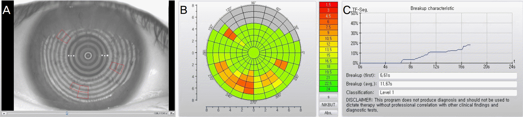 | Figure 1.Representative image of non-invasive keratograph tear film break-up time (NIK-BUT) using Oculus Keratograph 4 (Oculus, Wetzler, Germany). (A) A real time image recorded the entire course of break up process. Placido rings were reflected from surface of cornea and their distortions were recorded as the red-framed rectangular break-up units. (B) The final report was summarized as tear film break-up colour-code map. (C) Noninvasive keratograph first break-up time (NIKf-BUT), non-invasive keratograph average break-up time (NIKav-BUT) and break-up progress of break-up units were provided. |
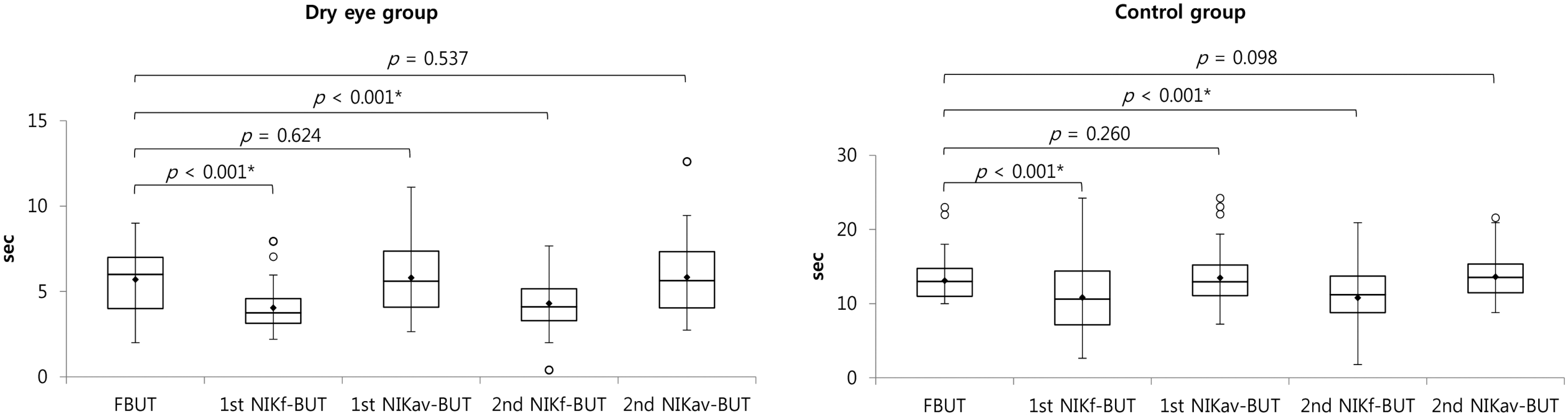 | Figure 2.Distribution of non-invasive keratograph tear film break-up time (NIK-BUT) and tear film break-up time with fluorecein (FBUT) using box and whisker plot in the dry eye and control groups. The boxes include 50% of the measured values between 1st and 3rd quartiles and the median (horizontal line). The upper and lower fences indicate 1.5 times the interquartile range (IQR) from 3rd and 1st quartiles. The outliers which are more than 1.5 IQR from the box are shown as circles. The mean of each parameter are shown as diamond. Comparison between FBUT and NIK-BUTs was performed by paired sample t-test. NIKf-BUT = non-invasive keratograph first break-up time; NIKav-BUT = non-invasive keratograph average break-up time.* p-value < 0.05 by paired sample t-test. |
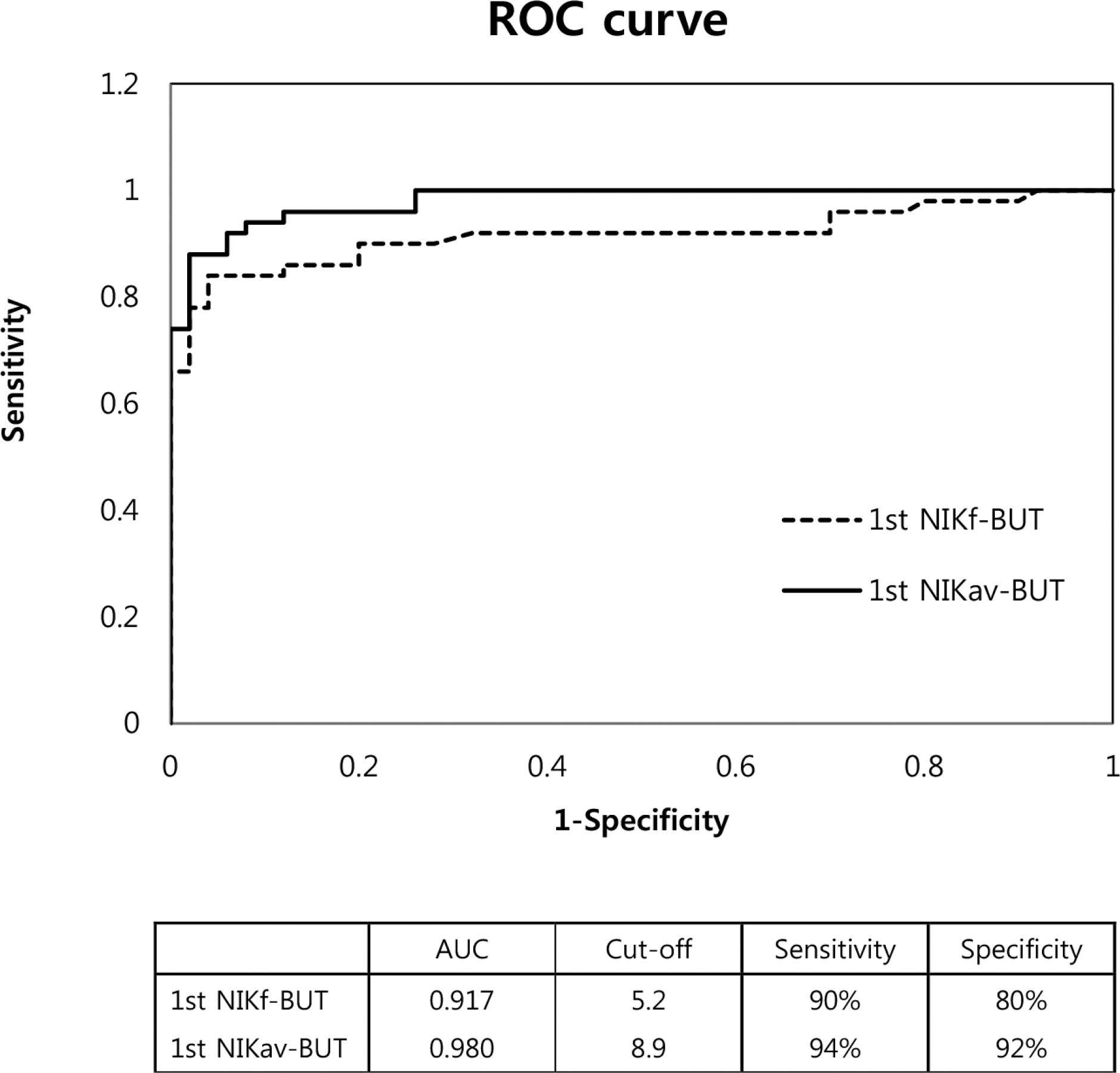 | Figure 3.Receiver operating characteristic curve (ROC) of non-invasive keratograph tear film break-up time (NIK-BUT). The area under the curve (AUC) is 0.917 in 1st non-invasive keratograph first break-up time (NIKf-BUT) and 0.980 in 1st non-invasive keratograph average break-up time (NIKav-BUT). The cutoff value derived from ROC curve was provided. The difference of AUC between NIKf-BUT and NIKav-BUT was 0.063 and was significant (p = 0.032). |
Table 1.
Demographics and ocular parameters of the study subjects
| Dry eye group | Control group | p-value | |
|---|---|---|---|
| Eyes (n) | 50 | 50 | |
| Age (years) | 54.6 ± 10.9 | 52.8 ± 11.8 | 0.347* |
| Gender (male:female) | 18:32 | 23:27 | 0.309† |
| FBUT (seconds) | 5.7 ± 1.7 | 13.1 ± 2.9 | <0.001* |
| 1st NIKf-BUT (sec) | 4.1 ± 1.3 | 10.8 ± 4.9 | <0.001* |
| 1st NIKav-BUT (sec) | 5.8 ± 2.0 | 13.5 ± 3.7 | <0.001* |
| 2nd NIKf-BUT (sec) | 4.3 ± 1.5 | 10.8 ± 4.2 | <0.001* |
| 2nd NIKav-BUT (sec) | 5.8 ± 2.1 | 13.6 ± 2.9 | <0.001* |
| Schirmer test (mm/5 min) | 7.0 ± 3.6 | 13.2 ± 5.6 | <0.001* |
| OSDI score | 40.1 ± 14.2 | 25.1 ± 13.3 | <0.001* |
Values are presented as mean ± SD unless otherwise indicated. For the NIKf-BUT, the intraclass correlation coefficient between first and second examination was 0.717 in the dry eye and 0.700 in the control group. For the NIKav-BUT, the intraclass correlation coefficient between first and second examination was 0.943 in the dry eye and 0.912 in the control group.
Table 2.
Pearson's correlation coefficients for all reference methods
| FBUT | 1st NIKf | 1st NIKav | 2nd NIKf | 2nd NIKav | Schirmer | OSDI | |
|---|---|---|---|---|---|---|---|
| FBUT | 1 | 0.440† | 0.660† | 0.592† | 0.689† | 0.289* | −0.046 |
| 1st NIKf | 0.612† | 1 | 0.687† | 0.567† | 0.601† | 0.111 | −0.118 |
| 1st NIKav | 0.768† | 0.832† | 1 | 0.638† | 0.893† | 0.228 | 0.003 |
| 2nd NIKf | 0.503† | 0.545† | 0.558† | 1 | 0.685† | 0.374† | −0.052 |
| 2nd NIKav | 0.724† | 0.654† | 0.860† | 0.730† | 1 | 0.206 | 0.063 |
| Schirmer | 0.377† | 0.230 | 0.311* | 0.247 | 0.337* | 1 | −0.112 |
| OSDI | −0.162 | −0.309* | −0.265 | −0.270 | −0.233 | −0.045 | 1 |
Values in light gray background area represent correlation in the normal control group, and the other values represent correlation in the dry eye group.
 XML Download