

 PDF
PDF ePub
ePub Citation
 Print
Print


Abstract
Methods
A retrospective study of 11 eyes of 11 patients with a diagnosis of ONSM between 2002 and 2015 at Severance Hospital.
Results
The mean age at symptom onset was 47.6 years. Ten females and 1 male participated in the study and all tumors were unilateral. Patients typically presented with visual loss and proptosis. Three patients complained of limited extraocular movements and seven patients exhibited visual field defects. Three patients who had a greater growth rate with intracranial involvement and two patients who had decreased vision received treatments. Five patients maintained good vision and visual field during the follow-up period. However, one patient who underwent surgical treatment presented significant visual loss and deterioration of visual field defect. One out of two patients who received three-dimensional conformal radiotherapy (3D-CRT) experienced improvement in visual field, and the other showed no change in visual field defect but remained stable with decreased tumor size. One out of two patients who underwent gamma-knife surgery showed aggravated visual field defect and the other presented with visual loss.
Conclusions
ONSM is typically a slow-growing tumor. Deterioration of visual loss and visual field defect can occur after treatment of ONSM. Therefore, management should be considered carefully and should be limited to cases in which progression of the disease is advanced or tumor growth is fast. 3D-CRT can be considered in patients in need of treatment.
Go to : 
References
1. Dutton JJ. Optic nerve sheath meningiomas. Surv Ophthalmol. 1992; 37:167–83.

2. Wright JE, Call NB, Liaricos S. Primary optic nerve meningioma. Br J Ophthalmol. 1980; 64:553–8.
3. Harold Lee HB, Garrity JA, Cameron JD, et al. Primary optic nerve sheath meningioma in children. Surv Ophthalmol. 2008; 53:543–58.
4. Bosch MM, Wichmann WW, Boltshauser E, Landau K. Optic nerve sheath meningiomas in patients with neurofibromatosis type 2. Arch Ophthalmol. 2006; 124:379–85.
5. Castel A, Boschi A, Renard L, De Potter P. Optic nerve sheath meningiomas: clinical features, functional prognosis and controversial treatment. Bull Soc Belge Ophtalmol. 2000; 275:73–8.
6. O'Donnell B. Surgical approaches to the orbit. Orbit. 2007; 26:81–2.
7. Turbin RE, Thompson CR, Kennerdell JS, et al. A long-term visual outcome comparison in patients with optic nerve sheath meningioma managed with observation, surgery, radiotherapy, or surgery and radiotherapy. Ophthalmology. 2002; 109:890–9. discussion 899–900.
8. Turbin RE, Pokorny K. Diagnosis and treatment of orbital optic nerve sheath meningioma. Cancer Control. 2004; 11:334–41.
9. Berman D, Miller NR. New concepts in the management of optic nerve sheath meningiomas. Ann Acad Med Singapore. 2006; 35:168–74.
10. Saeed P, Rootman J, Nugent RA, et al. Optic nerve sheath meningiomas. Ophthalmology. 2003; 110:2019–30.
11. Eddleman CS, Liu JK. Optic nerve sheath meningioma: current abdominal and treatment. Neurosurg Focus. 2007; 23:E4.
12. Liu JK, Forman S, Moorthy CR, Benzil DL. Update on treatment modalities for optic nerve sheath meningiomas. Neurosurg Focus. 2003; 14:e7.
13. Miller NR. New concepts in the diagnosis and management of abdominal nerve sheath meningioma. J Neuroophthalmol. 2006; 26:200–8.
14. Leksell L. Stereotactic Radiosurgery. J Neurol Neurosurg Psychiatry. 1983; 46:797–803.
15. Siddiqui JD, Loeffler JS, Murphy MA. Radiation optic neuropathy after proton beam therapy for optic nerve sheath meningioma. J Neuroophthalmol. 2013; 33:165–8.
16. Moyer PD, Golnik KC, Breneman J. Treatment of optic nerve sheath meningioma with three-dimensional conformal radiation. Am J Ophthalmol. 2000; 129:694–6.
17. Lee AG, Woo SY, Miller NR, et al. Improvement in visual function in an eye with a presumed optic nerve sheath meningioma after treatment with three-dimensional conformal radiation therapy. J Neuroophthalmol. 1996; 16:247–51.
18. Vagefi MR, Larson DA, Horton JC. Optic nerve sheath meningioma: visual improvement during radiation treatment. Am J Ophthalmol. 2006; 142:343–4.
19. Lee EK, Kim SJ, Paek SH, et al. Clinical features and management outcome of optic nerve sheath meningioma in Korea. J Korean Ophthalmol Soc. 2011; 52:74–85.
Go to :
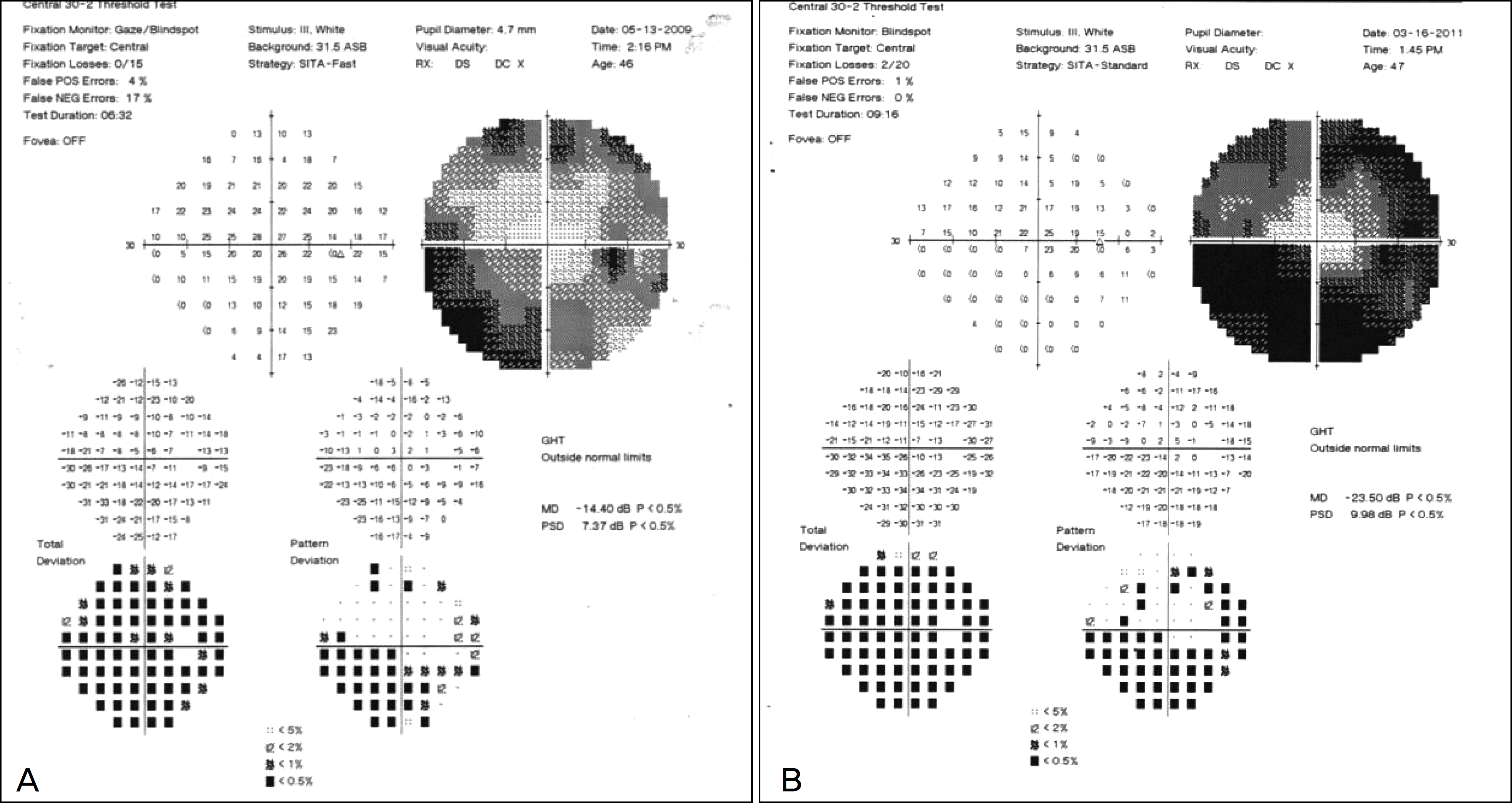 | Figure 1.Case 1.(A) This 45-year-old woman presented with decreased vision of the right eye. Her visual field examination shows inferior scotoma. (B) Her visual field showed increased visual field defect after 22 months. |
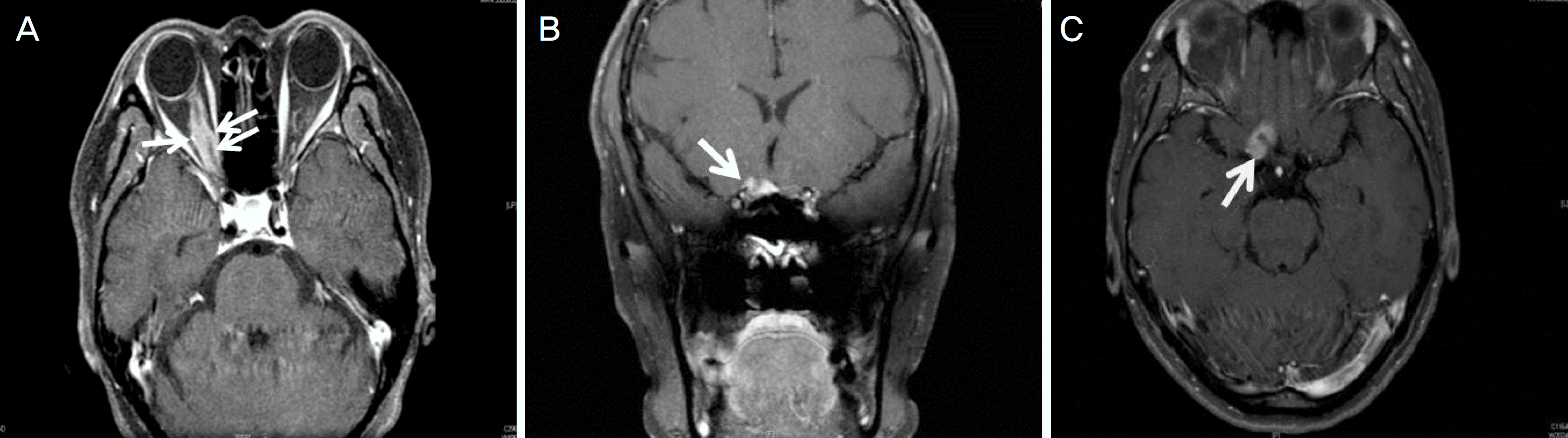 | Figure 2.Case 1. (A) Her coronal view of ganolinium-enhanced T1 magnetic resonance sequences show tubular-apical expansion optic nerve sheath meningioma (arrows) at first visit. (B, C) After 22 months, her tumor showed intracranial involvement (arrows). |
 | Figure 3.Case 5. (A) This 40-year-old woman presented with decreased vision of the left eye. Her visual field examination shows inferior scotoma at left eye. (B) Her coronal view of ganolinium-enhanced T1 magnetic resonance sequences show tubular-form meningioma (arrow). The length of tumor was 25.12 mm in coronal view. (C) After Gamma-Knife surgery, her tumor sized decreased. |
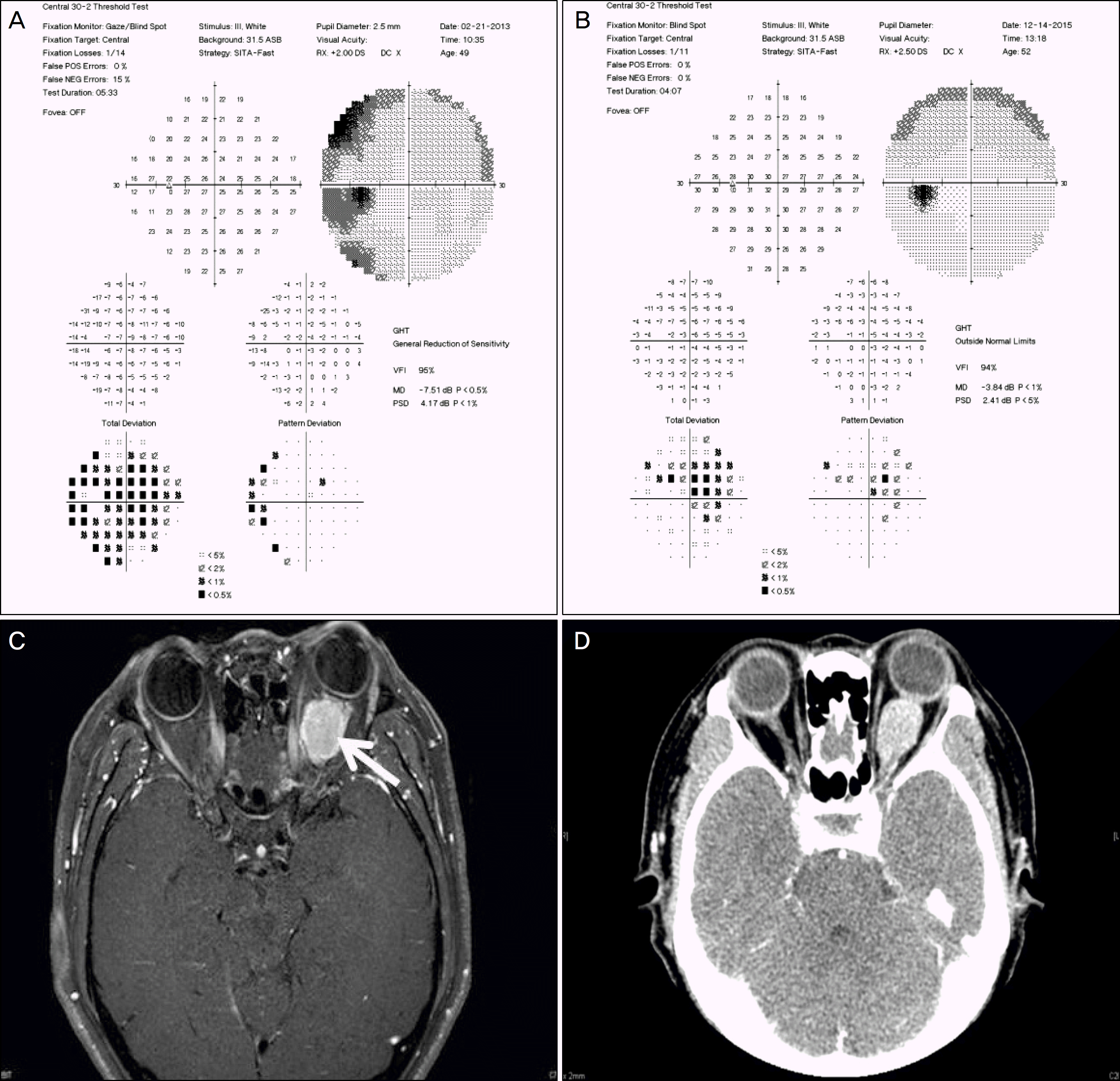 | Figure 4.Case 4. (A) This 49-year-old woman presented with decreased vision and exophthalmos of her left eye. Her initial visual field examination showed temporal visual field defect of the left eye. (B) After three-dimensional conformal radiotherapy, her visual field remained unchanged. (C) Her gadolinium-enhanced T1 magnetic resonance sequences shows globular form optic nerve sheath meningioma, tumor size was 22.96 × 16.72 mm (arrow). (D) After three-dimensional conformal radiotherapy, tumor size slightly decreased (22.22 × 15.47 mm) on her orbital computed tomography using contrast and her exophthalmos decreased. |
 | Figure 5.Case 9. (A) This 40-year-old woman presented with decreased vision of her left eye. Initial best corrected visual acuity was 0.7 and visual field examination showed no significant scotoma on her left eye. (B) She was diagnosed with optic nerve sheath meningioma of the left eye. The tumor size was 12.96 × 8.67 mm. (C) Her visual field examination was normal and unchanged. |
Table 1.
Demographics of patients
Table 2.
Treatment results
 XML Download