Abstract
Case summary
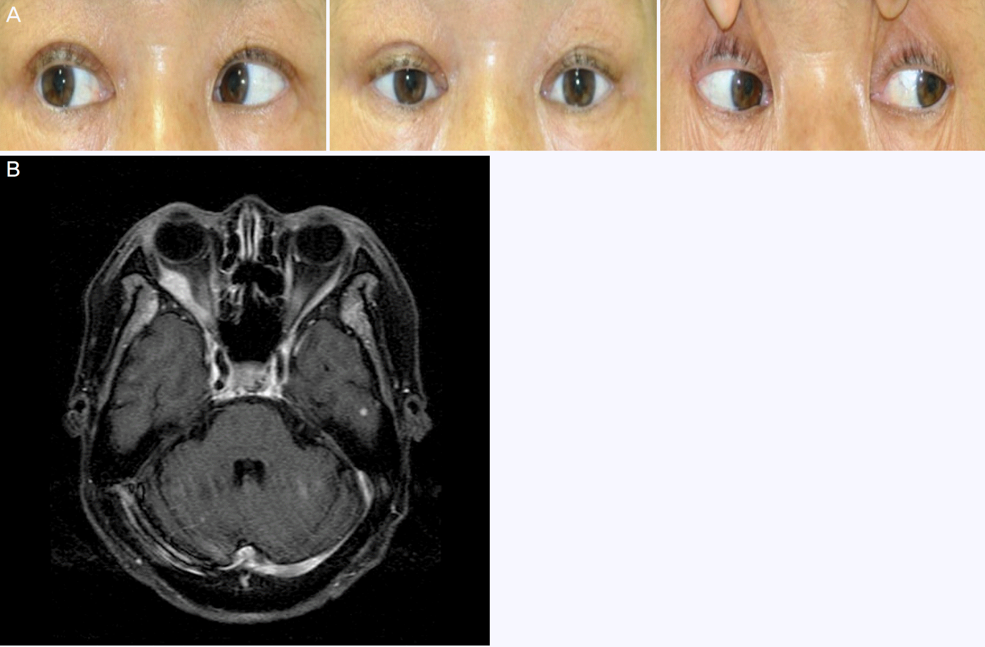
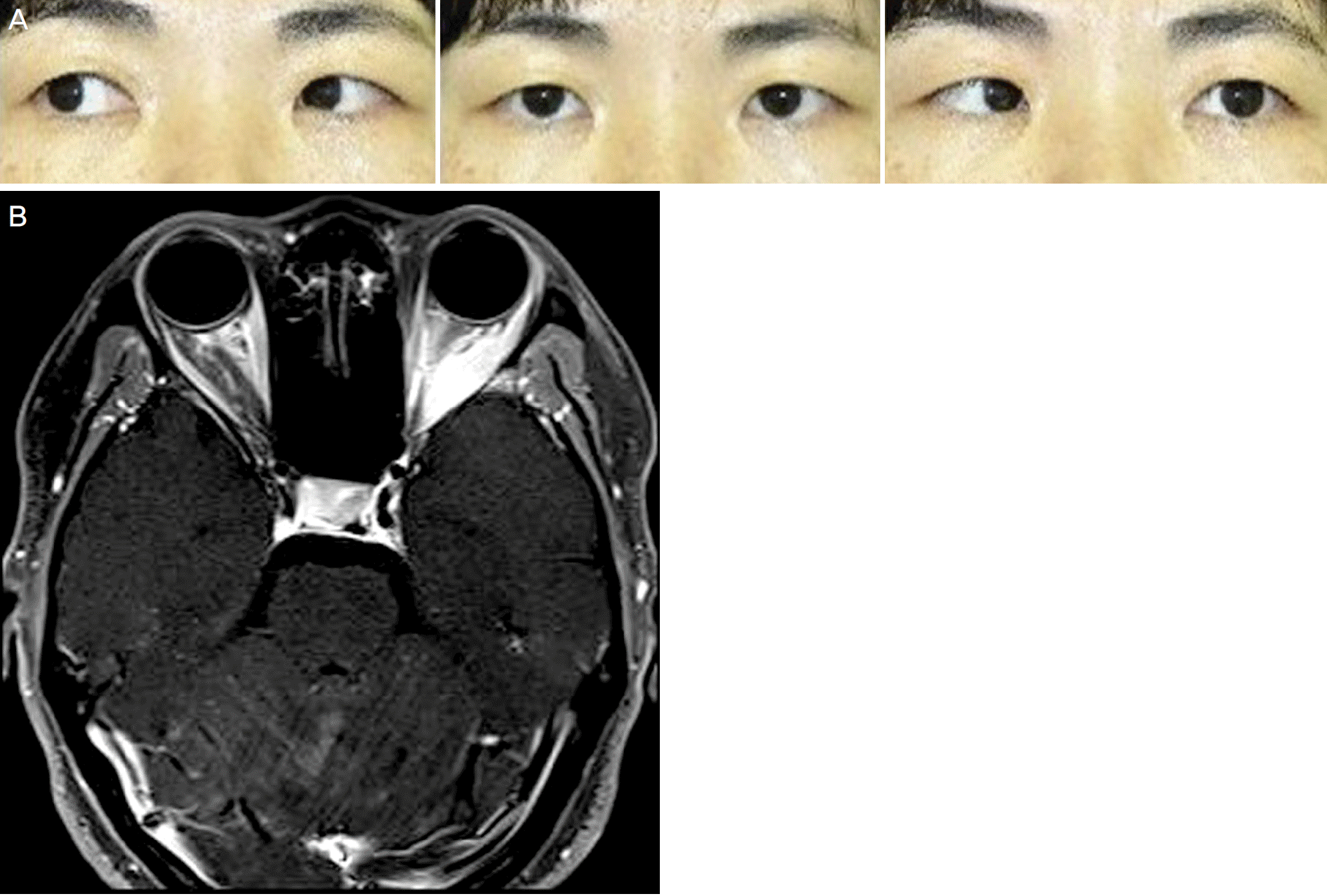
A 56-year-old woman presented with horizontal diplopia first noted 1 month earlier. She had a history of small cell lung cancer with brain and bone metastases. She had a −3 abduction deficit in the right eye and esotropia. The forced duction test showed no limitation in horizontal movement. Antibody tests for thyroid disease showed normal results. Brain magnetic resonance image showed multiple nodular enlargements of the right lateral and medial rectus muscles, al so multiple metastatic nodules in the brain. A 38-year-old woman presented with horizontal diplopia first noted 3 months previously. She had undergone breast cancer surgery 6 months earlier. The patient had a −4 abduction deficit in the left eye and esotropia. The forced duction test showed no limitation in horizontal movement. Antibody tests for thyroid disease showed normal results. Orbital magnetic resonance imaging showed nodular enlargement of left lateral rectus muscle including a tendon.
References
1. Capone A, Slamovits TL. Discrete metastasis of solid tumors to abdominal muscles. Arch Ophthalmol. 1990; 108:237–43.
2. Costa RM, Dumitrascu OM, Gordon LK. Orbital myositis: abdominal and management. Curr Allergy Asthma Rep. 2009; 9:316–23.
3. Ahmad SM, Esmaeli B. Metastatic tumors of the orbit and ocular adnexa. Curr Opin Ophthalmol. 2007; 18:405–13.

5. Park SY, Kim SY. A case of MALT lymphoma with left inferior rectus muscle invasion. J Korean Ophthalmol Soc. 2014; 55:947–51.

6. Shin JH, Lee JH, Lim HK, et al. Bilateral extraocular muscle abdominal of nasal rhabdomyosarcoma mimicking a thyroid associated orbitopathy: a case report. J Korean Soc Magn Reson Med. 2011; 15:176–80.
7. Scott AB, Kraft SP. Botulinum toxin injection in the management of lateral rectus paresis. Ophthalmology. 1985; 92:676–83.

8. Kim SD, Yang SW, Woo KI, et al. Ophthalmic Plastic and Reconstructive Surgery. 3rd ed.Goyang: Naewae haksool;2015. p. 384–434.
9. Char DH, Miller T, Kroll S. Orbital metastases: diagnosis and course. Br J Ophthalmol. 1997; 81:386–90.

10. Goldberg RA, Rootman J, Cline RA. Tumors metastatic to the abdominal: a changing picture. Surv Ophthalmol. 1990; 35:1–24.
11. Shields JA, Shields CL, Brotman HK, et al. Cancer metastatic to the orbit: the 2000 Robert M. Curts Lecture. Ophthal Plast Reconstr Surg. 2001; 17:346–54.
12. Goldberg RA, Rootman J. Clinical characteristics of metastatic abdominalal tumors. Ophthalmology. 1990; 97:620–4.
13. Patrinely JR, Osborn AG, Anderson RL, Whiting AS. Computed tomographic features of nonthyroid extraocular muscle enlargement. Ophthalmology. 1989; 96:1038–47.

14. Jiang H, Wang Z, Xian J, Ai L. Bilateral multiple extraocular abdominal metastasis from hepatocellular carcinoma. Acta Radiol Short Rep. 2012; 1:pii: arsr.2011.110002.
15. Bartalena L, Tanda ML. Clinical practice. Graves' ophthalmopathy. N Engl J Med. 2009; 360:994–1001.
Figure 1.
Photograph of case 1. (A) The patient had limitation of abduction in the right eye. (B) Axial image of T1-weighted contrast enhanced brain magnetic resonance showed nodular enlargement with heterogenous high signal intensities in the right lateral and medial rectus muscles.

Figure 2.
Photograph of case 2. (A) The patient had limitation of abduction in the left eye. (B) Axial image of T1-weighted contrast enhanced orbital magnetic resonance showed high signal intensities and large nodular enlargement in the left lateral rectus muscle including lateral rectus muscle tendon.


 PDF
PDF ePub
ePub Citation
 Print
Print


 XML Download