Abstract
Case summary
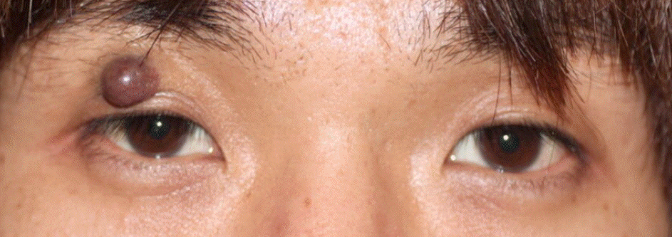
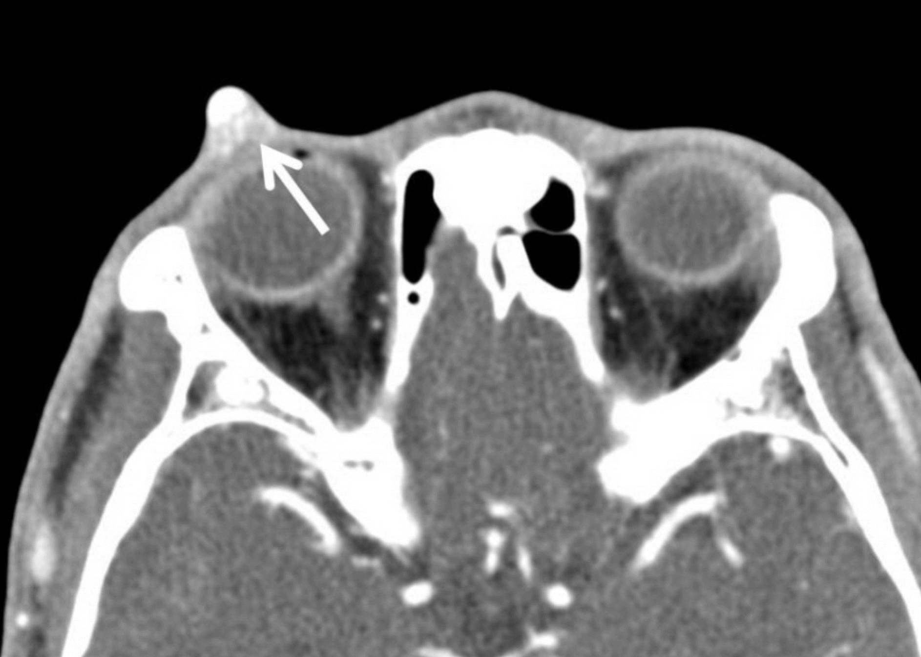
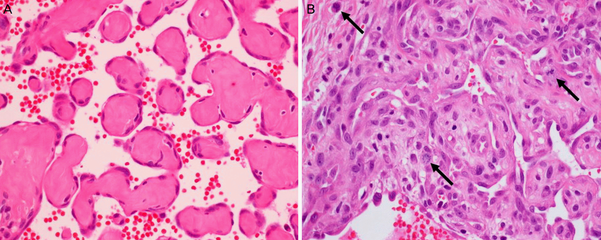
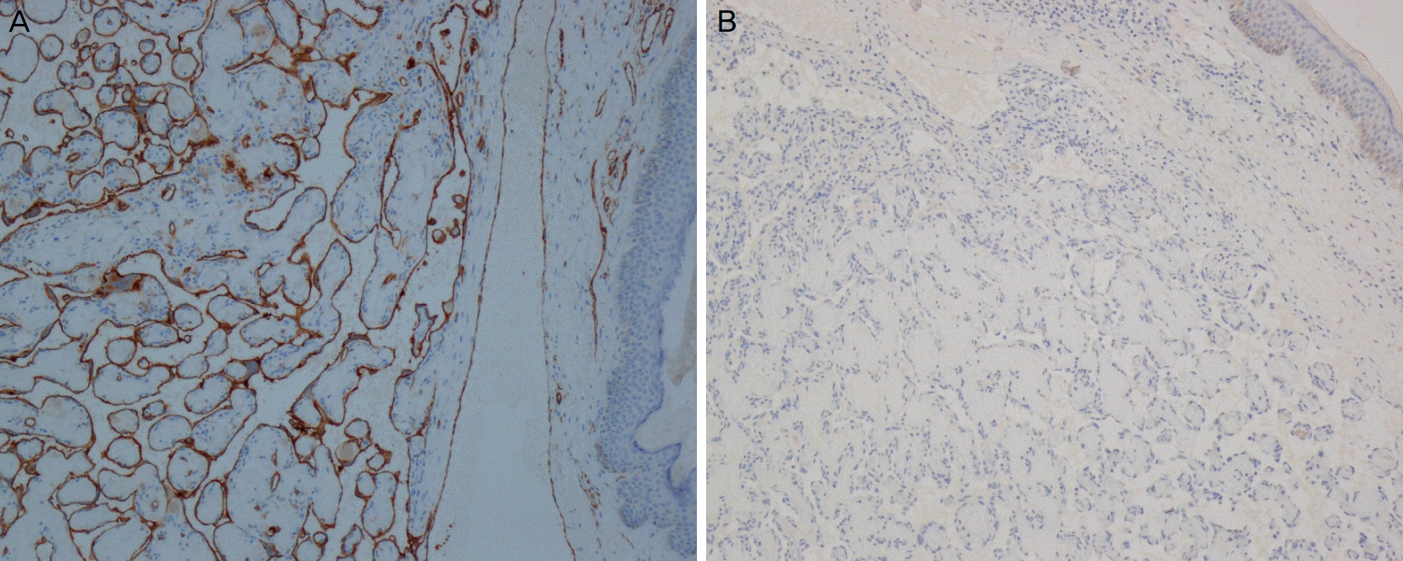
A 30-year-old male presented with a right upper eyelid mass, which had been growing in size for three years. The mass was about 7 mm in diameter, purplish, spherical, and cystic. Incision and drainage were performed, but the cystic mass instantly refilled with blood. Excisional biopsy was performed. On microscopic examination, a myriad of small delicate papillae projections into the dilated vascular lumen with organizing thrombus were noted. Each papilla was lined with a single layer of endothelial cells, surrounding a collagenized core. The endothelial cells were reactive for CD31 on immunohistochemical staining. There were focal areas of frequent mitoses, but neither cytological atypia nor necrosis was found. Hence, the lesion was diagnosed as intravascular papillary endothelial hyperplasia.
Conclusions
Intravascular papillary endothelial hyperplasia in the periorbital area is rarely reported, and it is important to distinguish it from hemangioma or angiosarcoma. Complete surgical excision is necessary to prevent recurrence. The authors report a case of intravascular papillary endothelial hyperplasia of the upper eyelid, which should be considered in the differential diagnosis of eyelid or orbital tumor.
Go to : 
References
1. Dutton JJ, Gayre GS, Proia AD. Diagnostic Atlas of Common Eyelid Diseases. 1st ed.New York: CRC Press;2007. p. 171–4.
2. Tosios K, Koutlas IG, Papanicolaou SI. Intravascular papillary abdominal hyperplasia of the oral soft tissues: report of 18 cases and review of the literature. J Oral Maxillofac Surg. 1994; 52:1263–8.
3. Shields JA, Shields CL, Eagle RC Jr, Diniz W. Intravascular abdominal endothelial hyperplasia with presumed bilateral orbital varices. Arch Ophthalmol. 1999; 117:1247–9.
4. Sorenson RL, Spencer WH, Stewart WB, et al. Intravascular abdominal endothelial hyperplasia of the eyelid. Arch Ophthalmol. 1983; 101:1728–30.
5. Wagh VB, Kyprianou I, Burns J, et al. Periorbital Masson's tumor: a case series. Ophthal Plast Reconstr Surg. 2011; 27:e55–7.

6. Font RL, Wheeler TM, Boniuk M. Intravascular papillary abdominal hyperplasia of the orbit and ocular adnexa. A report of five cases. Arch Ophthalmol. 1983; 101:1731–6.
7. Clearkin KP, Enzinger FM. Intravascular papillary endothelial hyperplasia. Arch Pathol Lab Med. 1976; 100:441–4.
8. Werner MS, Hornblass A, Reifler DM, et al. Intravascular papillary endothelial hyperplasia: collection of four cases and a review of the literature. Ophthal Plast Reconstr Surg. 1997; 13:48–56.
9. Hashimoto H, Daimaru Y, Enjoji M. Intravascular papillary abdominal hyperplasia. A clinicopathologic study of 91 cases. Am J Dermatopathol. 1983; 5:539–46.
10. Kuo T, Sayers CP, Rosai J. Masson's “vegetant intravascular he-mangioendothelioma:” a lesion often mistaken for angiosarcoma: study of seventeen cases located in the skin and soft tissues. Cancer. 1976; 38:1227–36.
Go to : 
 | Figure 1.External photograph of eyelid. Spherical bluish mass measured about 7mm in diameter on the right upper lid. |
 | Figure 2.Contrast enhanced oribtal computed tomography. It shows 7 × 8 sized, contrast enhancing round mass in right upper eyelid (arrow). |


 PDF
PDF ePub
ePub Citation
 Print
Print


 XML Download