Abstract
Purpose
To evaluate changes in subfoveal choroidal thickness (SFCT) after patterned panretinal photocoagulation (PRP) using pattern scan laser (PASCAL) in patients with diabetic retinopathy.
Methods
This study included 39 patients (50 eyes) treated with patterned PRP using PASCAL and who were followed for at least 3 months. Patients were classified into 2 groups according to severity: severe non-proliferative diabetic retinopathy and proliferative diabetic retinopathy. SFCT was measured by enhanced depth imaging of spectraldomain optical coherence tomography. The change in SFCT was analyzed at 1 and 3 months after PRP.
Results
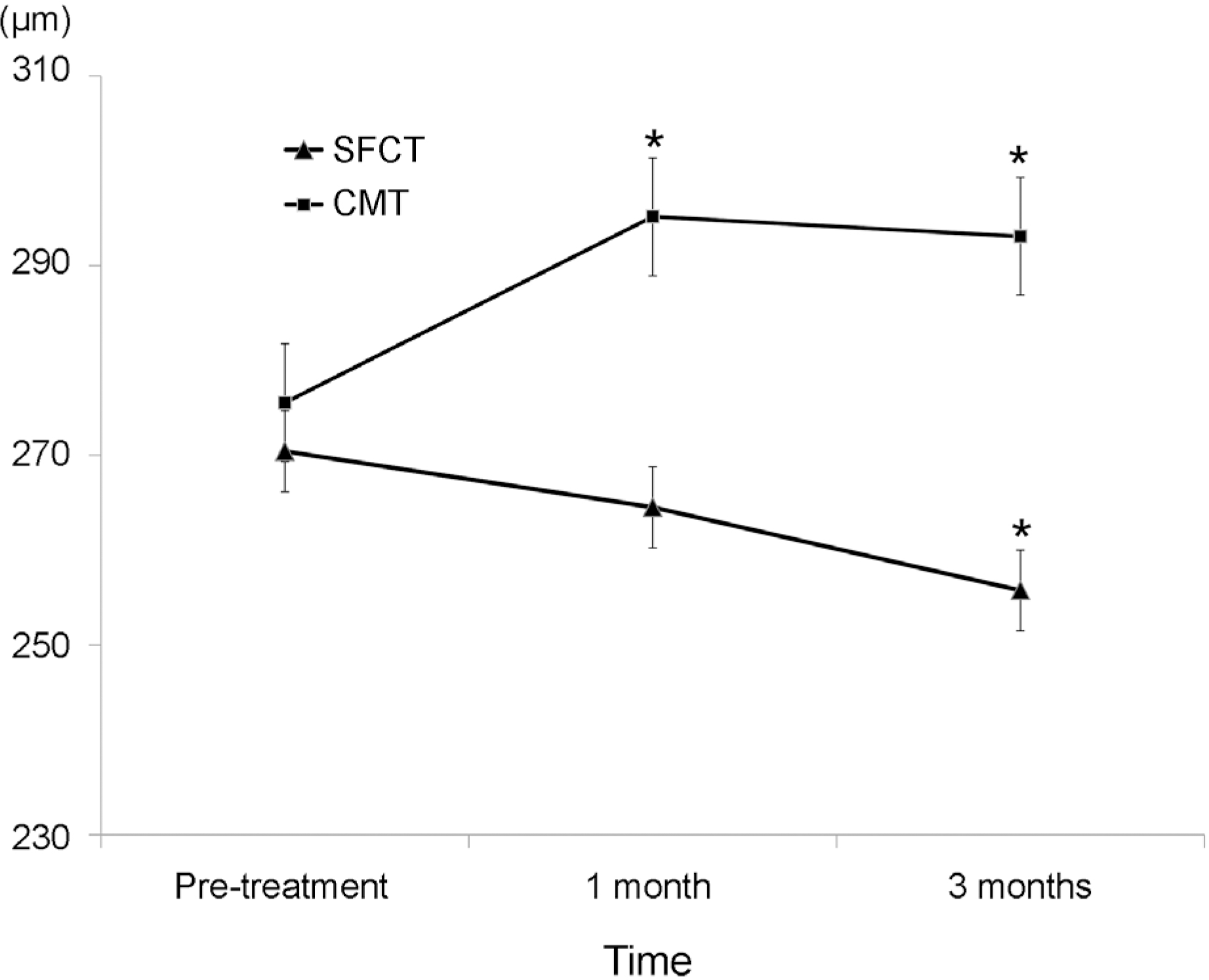
SFCT was 270.42 ± 61.44 μ m before PRP, 264.52 ± 60.78 μ m at 1 month, and 255.74 ± 56.89 μ m at 3 months after PRP. Significant change of SFCT was found at 3 months after PRP. Central macular thickness was 275.56 ± 50.61 μ m before PRP and increased to 295.18 ± 52.80 μ m and 293.10 ± 57.24 μ m at 1 and 3 months posttreatment, respectively. There were no significant differences between groups in SFCT at baseline or in the amount of change in SFCT after PRP.
Go to : 
References
1. Diabetic Retinopathy Study Research Group. Photocoagulation treatment of proliferative diabetic retinopathy. Clinical application of Diabetic Retinopathy Study (DRS) findings, DRS Report Number 8. The Diabetic Retinopathy Study Research Group. Ophthalmology. 1981; 88:583–600.
2. Early photocoagulation for diabetic retinopathy. ETDRS report number 9. Early Treatment Diabetic Retinopathy Study Research Group. Ophthalmology. 1991; 98(5 Suppl):766–85.
3. Weiter JJ, Zuckerman R. The influence of the photoreceptor-RPE complex on the inner retina. An explanation for the beneficial abdominals of photocoagulation. Ophthalmology. 1980; 87:1133–9.
4. Nonaka A, Kiryu J, Tsujikawa A, et al. Inflammatory response abdominal scatter laser photocoagulation in nonphotocoagulated retina. Invest Ophthalmol Vis Sci. 2002; 43:1204–9.
5. Spaide RF, Koizumi H, Pozzoni MC. Enhanced depth imaging spectraldomain optical coherence tomography. Am J Ophthalmol. 2008; 146:496–500.

6. Kim JT, Lee DH, Joe SG, et al. Changes in choroidal thickness in relation to the severity of retinopathy and macular edema in type 2 diabetic patients. Invest Ophthalmol Vis Sci. 2013; 54:3378–84.

7. Cho GE, Cho HY, Kim YT. Change in subfoveal choroidal abdominal after argon laser panretinal photocoagulation. Int J Ophthalmol. 2013; 6:505–9.
8. Zhu Y, Zhang T, Wang K, et al. Changes in choroidal thickness abdominal panretinal photocoagulation in patients with type 2 diabetes. Retina. 2015; 35:695–703.
9. Zhang Z, Meng X, Wu Z, et al. Changes in choroidal thickness after panretinal photocoagulation for diabetic retinopathy: a 12-week longitudinal study. Invest Ophthalmol Vis Sci. 2015; 56:2631–8.

10. Coleman DJ, Silverman RH, Chabi A, et al. High-resolution abdominal imaging of the posterior segment. Ophthalmology. 2004; 111:1344–51.
11. Hidayat AA, Fine BS. Diabetic choroidopathy. Light and electron microscopic observations of seven cases. Ophthalmology. 1985; 92:512–22.
12. Weinberger D, Kramer M, Priel E, et al. Indocyanine green abdominal findings in nonproliferative diabetic retinopathy. Am J Ophthalmol. 1998; 126:238–47.
13. Nagaoka T, Kitaya N, Sugawara R, et al. Alteration of choroidal circulation in the foveal region in patients with type 2 diabetes. Br J Ophthalmol. 2004; 88:1060–3.

14. Park BJ, Chung HW, Kim HC. Effects of diabetic retinopathy and intravitreal bevacizumab injection on choroidal thickness in abdominal patients. J Korean Ophthalmol Soc. 2013; 54:1520–5.
15. Yuki T, Kimura Y, Nanbu S, et al. Ciliary body and choroidal abdominal after laser photocoagulation for diabetic retinopathy. A high-frequency ultrasound study. Ophthalmology. 1997; 104:1259–64.
16. Gentile RC, Stegman Z, Liebmann JM, et al. Risk factors for abdominal effusion after panretinal photocoagulation. Ophthalmology. 1996; 103:827–32.
17. Yu S, Kim YI, Lee KW, Kang HG. Changes in choroidal thickness after panretinal photocoagulation in diabetic retinopathy patients. J Korean Ophthalmol Soc. 2016; 57:256–63.

18. Chappelow AV, Tan K, Waheed NK, Kaiser PK. Panretinal photo-coagulation for proliferative diabetic retinopathy: pattern scan abdominal versus argon laser. Am J Ophthalmol. 2012; 153:137–42.e2.
19. Paulus YM, Jain A, Gariano RF, et al. Healing of retinal photo-coagulation lesions. Invest Ophthalmol Vis Sci. 2008; 49:5540–5.

20. Yang JW, Lee YC. Comparison of the effects of patterned and abdominal panretinal photocoagulation on diabetic retinopathy. J Korean Ophthalmol Soc. 2010; 51:1590–7.
21. Arnarsson A, Stefánsson E. Laser treatment and the mechanism of edema reduction in branch retinal vein occlusion. Invest Ophthalmol Vis Sci. 2000; 41:877–9.
22. Mendivil A. Ocular blood flow velocities in patients with abdominal diabetic retinopathy after panretinal photocoagulation. Surv Ophthalmol. 1997; 42(Suppl 1):S89–95.
Go to : 
 | Figure 1.Consecutive changes of the mean subfoveal choroidal thickness (SFCT) and central macular thickness (CMT) after patterned panretinal photocoagulation. Data are expressed as the mean ± standard error of mean. * p < 0.05 by repeated measures analysis of variance (ANOVA). |
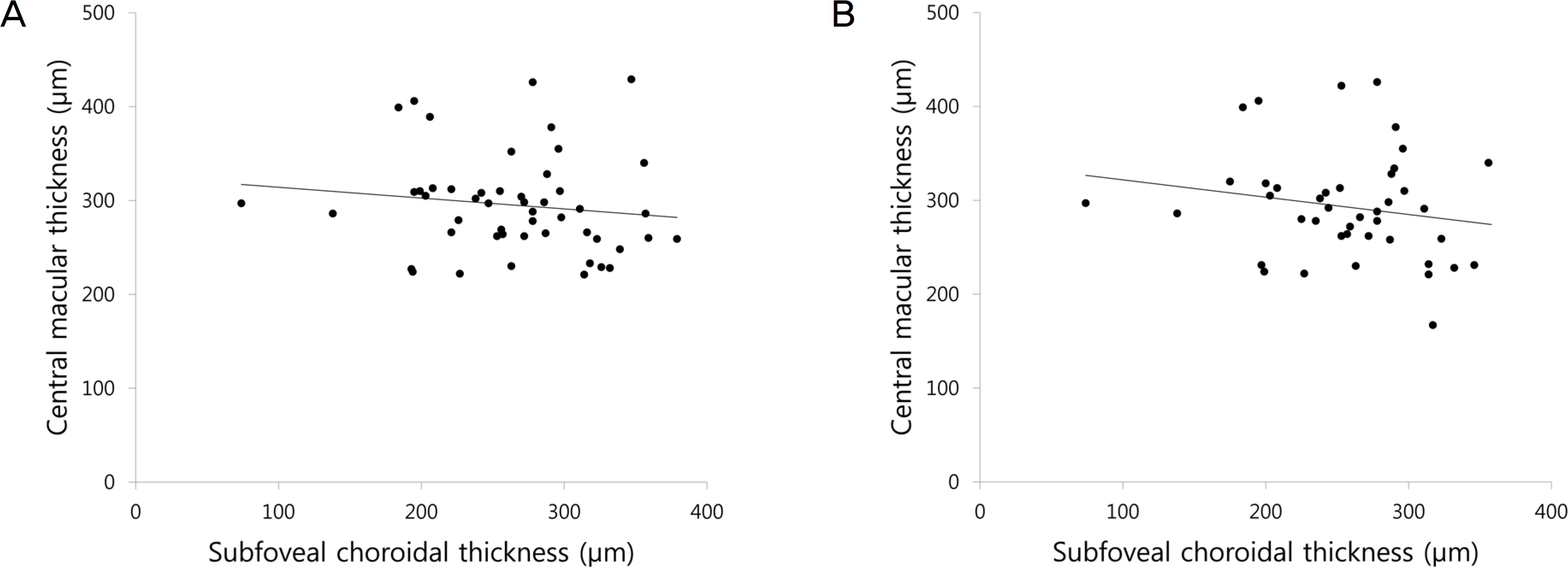
 | Figure 2.Scatter plots for correlation between subfoveal choroidal thickness (SFCT) and central macular thickness (CMT). (A) At 1 month after patterned panretinal photocoagulation (PRP), there was no significant correlation between SFCT and CMT (r = −0.133, p = 0.201). (B) At 3 months after patterned PRP, there was no significant correlation between SFCT and CMT (r = −0.184, p = 0.245). Compared by Pearson's correlation analysis. r = coefficient of correlation. |
Table 1.
Demographics of patients
Table 2.
Comparison of baseline and amount of change after patterned panretinal photocoagulation according to disease severity
| Severe NPDR | PDR | p-value* | |
|---|---|---|---|
| SFCT (μ m) | 290.05 ± 52.92 | 257.33 ± 64.03 | 0.064 |
| Δ† 1 month | –6.33 ± 20.67 | –5.25 ± 27.85 | 0.593 |
| Δ 3 months | –8.54 ± 20.60 | –13.36 ± 24.18 | 0.762 |
| CMT (μ m) | 277.53 ± 47.46 | 272.60 ± 56.15 | 0.739 |
| Δ 1 month | 20.47 ± 23.30 | 18.35 ± 57.83 | 0.308 |
| Δ 3 months | 20.79 ± 34.64 | 7.64 ± 42.70 | 0.290 |


 PDF
PDF ePub
ePub Citation
 Print
Print


 XML Download