Abstract
Purpose:
We report the first case in Korea of rapid bone formation on a subperiosteal orbital hematoma after trauma.
Case summary
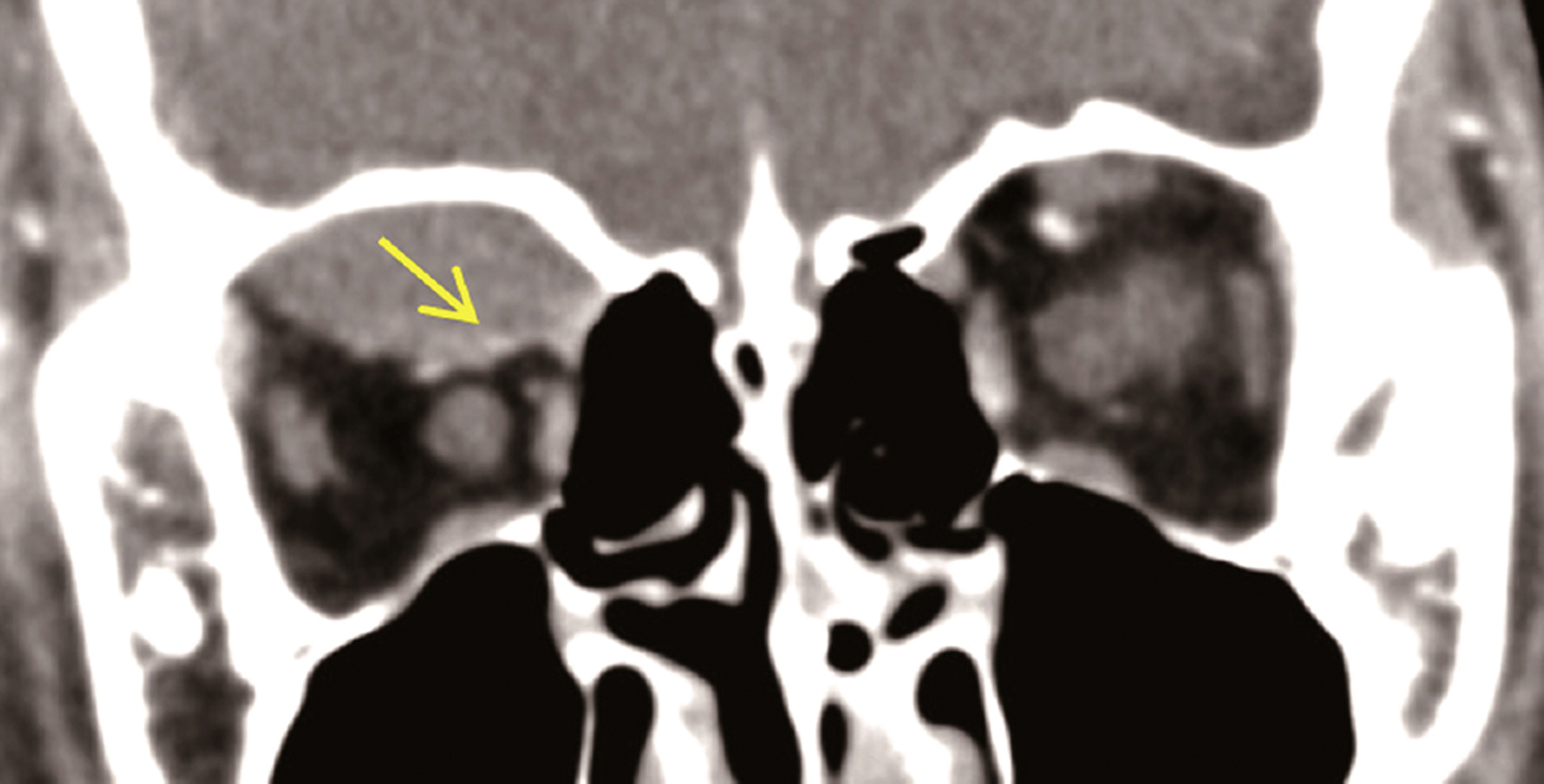
A 10-year-old boy who was in the intensive care unit after trauma showed proptosis and ocular movement limi-tation of the right eye associated with subperiosteal hematoma. On ocular examination, 3 mm of proptosis and limitation of right eye movement were observed; however, visual acuity was not decreased. At 1 month after the trauma, orbital computed tomog-raphy (CT) showed new bone formation at the margin of the hematoma border although the size of the hematoma decreased. The patient underwent hematoma and bony tissue removal using anterior orbitotomy approach. A new bone was formed be-tween the orbital border and hematoma from the anterior orbital margin to the orbital apex. During pathological examination, wo-ven bone tissue with fibrotic tissue was observed in the hematoma wall. One year after surgery, the patient’s proptosis and limi-tation of ocular movement disappeared without any evidence of new bone formation.
Conclusions
Waiting for spontaneous absorption of orbital subperiosteal hematoma is usually recommended unless there is significant functional impairment. However, as in our case, new bone formation could occur during a short period of less than 1 month; imaging follow-up is necessary in patients having intensive care or showing delayed absorption of a hematoma.
References
1. Hirakawa T, Tanaka A, Yoshinaga S. . Calcified chronic sub-dural hematoma with intracerebral rupture forming a subcortical hematoma. A case report. Surg Neurol. 1989; 32:51–5.
2. Chang JH, Choi JY, Chang JW. . Chronic epidural hematoma with rapid ossification. Childs Nerv Syst. 2002; 18:712–6.

3. Afra D. Ossification of subdural hematoma. Report of two cases. J Neurosurg. 1961; 18:393–7.
4. Erdogan B, Sen O, Bal N. . Rapidly calcifying and ossifying epidural hematoma. Pediatr Neurosurg. 2003; 39:208–11.

5. Niwa J, Nakamura T, Fujishige M. Hashi K. Removal of a large asymptomatic calcified chronic subdural hematoma. Surg Neurol. 1988; 30:135–9.

6. Sabet SJ, Tarbet KJ, Lemke BN. . Subperiosteal hematoma of the orbit with osteoneogenesis. Arch Ophthalmol. 2001; 119:301–3.
7. Wolter JR, Vanderveen GJ, Wacksman RL. Posttraumatic sub-galeal hematoma extending into the orbit as a cause of permanent blindness. J Pediatr Ophthalmol Strabismus. 1978; 15:151–3.

8. Seigel RS, Williams AG, Hutchison JW. . Subperiosteal hema-tomas of the orbit: angiographic and computed tomographic diagnosis. Radiology. 1982; 143:711–4.

9. Pope-Pegram LD, Hamill MB. Post-traumatic subgaleal hema-toma with subperiosteal orbital extension. Surv Ophthalmol. 1986; 30:258–62.

10. Kersten RC, Rice CD. Subperiosteal orbital hematoma: visual re-covery following delayed drainage. Ophthalmic Surg. 1987; 18:423–7.

11. Nakai K, Doi E, Kuriyama T. Tanaka Y. Spontaneous subperiosteal hematoma of the orbit. Surg Neurol. 1983; 20:100–2.

13. Carrion LT, Edwards WC, Perry LD. Spontaneous subperiosteal orbital hematoma. Ann Ophthalmol. 1979; 11:1754–7.
14. Wolter JR, Leenhouts JA, Coulthard SW. Clinical picture and man-agement of subperiosteal hematoma of the orbit. J Pediatr Ophthalmol. 1976; 13:136–8.
15. Ide M, Jimbo M, Yamamoto M. . Asymptomatic calcified chronic subdural hematoma-report of three cases. Neurol Med Chir (Tokyo). 1993; 33:559–63.
16. Chen NF, Wang YC, Shen CC. . Calcification and ossification of chronic encapsulated intracerebral haematoma: case report. J Clin Neurosci. 2004; 11:527–30.

17. Loh JK, Howng SL. Huge calcified chronic subdural hematoma in the elderly-report of a case. Kaohsiung J Med Sci. 1997; 13:272–6.
18. Nagane M, Oyama H, Shibui S. . Ossified and calcified epi-dural hematoma incidentally found 40 years after head injury: case report. Surg Neurol. 1994; 42:65–9.

19. McLaurin RL. McLaurin KS. Calcified subdural hematomas in childhood. J Neurosurg. 1966; 24:648–55.

20. Kaplan M, Akgün B. Seçer HI. Ossified chronic subdural hema-toma with armored brain. Turk Neurosurg. 2008; 18:420–4.
21. Katz B, Carmody R. Subperiosteal orbital hematoma induced by the valsalva maneuver. Am J Ophthalmol. 1985; 100:617–8.

22. Markovits AS. Evacuation of orbital hematoma by continuous suction. Ann Ophthalmol. 1977; 9:1255–8.
Figure 1.
Clinical photographs of the patients eyelids. The photographs shows hypoglobus (A) and exophthalmos (B) of the right eye.

Figure 2.
Orbit magnetic resonance imaging (MRI) taken 9 days after trauma. These pictures show subacute stage of hematoma. T1-weighted axial images (A) and coronal images (B) after gadolinium enhancement with fat suppression reveal a soft tissue mass at the superior portion of right orbit (arrows). Mass shows a high signal intensity on T1-weighted sagittal image with enhancement (C, arrow) and low and high signal intensity on T2-weighted axial image (D, arrow).

Figure 3.
Orbit CT taken at 2 weeks after trauma. Convex-shap-ed mass lesion with fine spots of calcifications at the superior or-bit (arrow). CT = computed tomography

Figure 4.
Orbit CT taken at 4 weeks after trauma. A convex le-sion was slightly decreased, but it accompanies a clear-cut cal-cification line at the inferior margin, in the coronal (A, arrow) and sagittal (B, arrow) CT scans. CT = computed tomography.


 PDF
PDF ePub
ePub Citation
 Print
Print


 XML Download