Abstract
Purpose
To introduce a case of a cyst containing a parasite misdiagnosed as a dermoid cyst, which is to the best of our knowledge, the first report in Korea of a parasite in a cyst located at the medial side of the orbit.
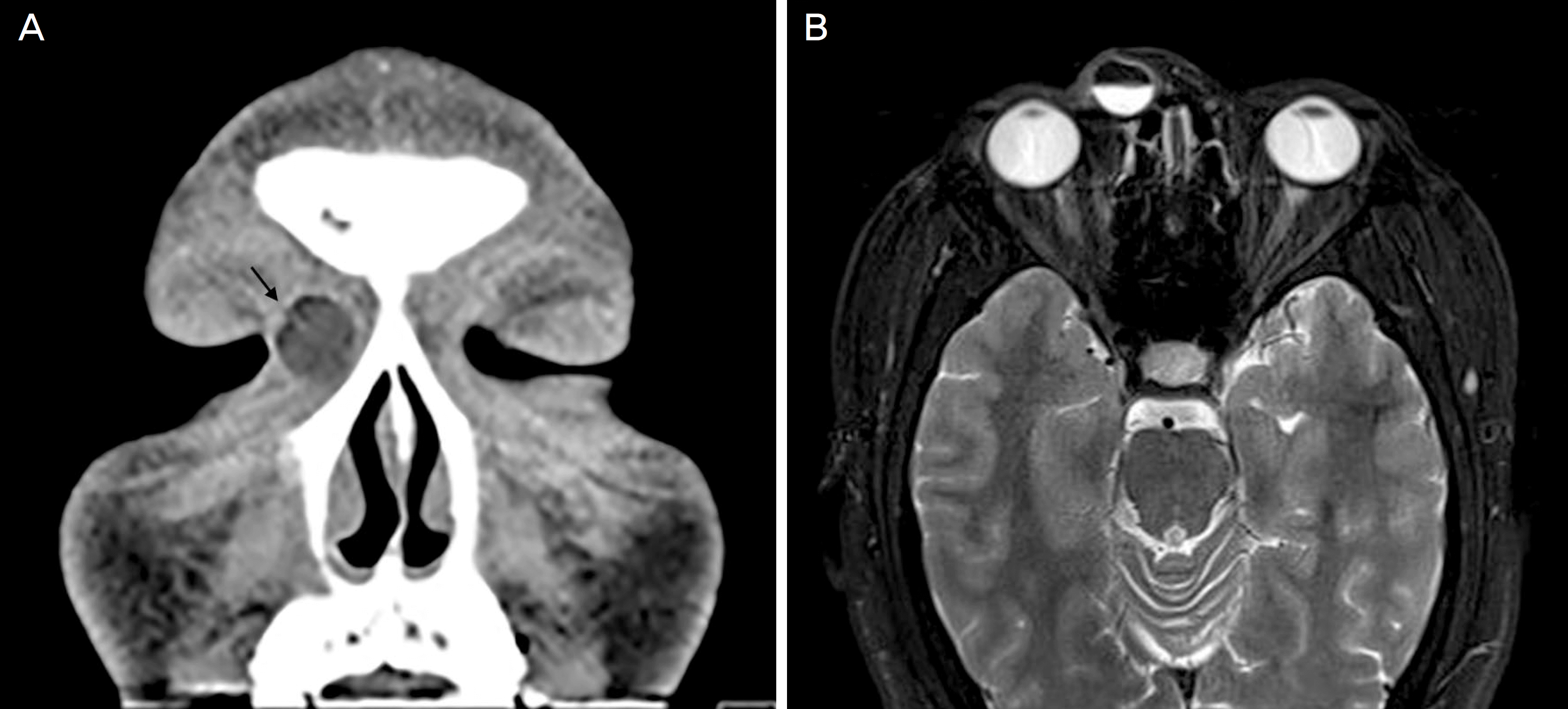
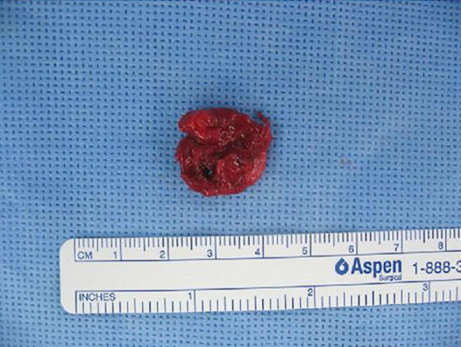
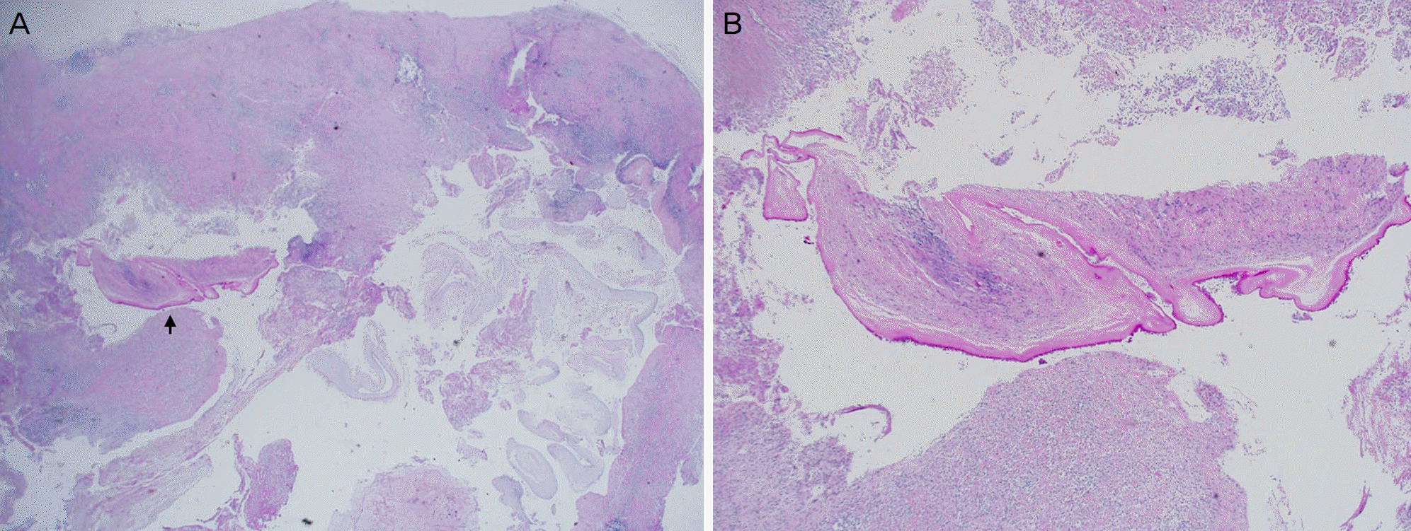
Case summary: A 31-year-old male visited the hospital with a 2-year history of a slowly growing mass at the medial side of the right orbit. The patient had a history of mass excision in the same location 18 years previously, however, biopsy was not performed at that time. Orbital computed tomography and magnetic resonance imaging revealed a 5.0 × 1.4 × 1.8 cm3 well-defined T1 high signal intensity unilocular cyst, thus our first impression was a dermoid cyst. The cyst was surgically removed with anterior orbitotomy. The cyst ruptured during the operation, and thus complete aspiration of the cystic fluid and in situ irrigation with antibiotics were performed. Histopathological examination revealed a fragmented adult parasite worm with chronic granulomatous change.
Conclusions
A differential diagnosis for orbital cyst based on clinical and radiological results is difficult. Thus, histopathological confirmation is required. A cyst containing a parasite located in the orbit has rarely been reported. A full examination of all infected patients must be conducted for parasite infection.
Go to : 
References
1. Pahwa S, Sharma S, Das CJ, et al. Intraorbital cystic lesions: an imaging spectrum. Curr Probl Diagn Radiol. 2015; 44:437–48.

3. Ziaei M, Elgohary M, Bremner FD. Orbital cysticercosis, case abdominal and review. Orbit. 2011; 30:230–5.
4. Gottstein B. Hydatid disease. Jonathan C, William GP, Steven MO, editors. Infectious Diseases. 3rd ed.China: Mosby;2010. 2:chap. 169.

5. Jack R. Diseases of the orbit: a multidisciplinary approach. 2nd ed.Philadelphia: Lippincott Williams and Wilkins;2003. p. 471–80.
6. Yoshino K. Studies on the post-embryonal development of Taenia solium: III. On the development of Cysticercus cellulosae within the definitive intermediate host. J Med Assoc Formosa. 1933; 32:166–9.
7. Ko JS, Kim GA, Shin JY, Byeon SH. A case of intravitreal cysticercosis with neovascular glaucoma. J Korean Ophthalmol Soc. 2013; 54:1610–3.

8. Kruger-Leite E, Jalkh AE, Quiroz H, Schepens CL. Intraocular cysticercosis. Am J Ophthalmol. 1985; 99:252–7.

9. Cardenas F, Quiroz H, Plancarte A, et al. Taenia solium ocular cysticercosis: findings in 30 cases. Ann Ophthalmol. 1992; 24:25–8.
10. Pushker N, Bajaj MS, Betharia SM. Orbital and adnexal cysticercosis. Clin Experiment Ophthalmol. 2002; 30:322–33.

11. Ryou JY, Kim KH, Kim SY. Hydatid cyst of the orbit. J Korean Ophthalmol Soc. 2006; 47:484–8.
12. Lerner SF, Gomez Morales A, Croxatto JO. Hydatid cyst of the orbit. Arch Ophthalmol. 1991; 109:285.

13. Gökçek C, Gökçek A, Akif Bayar M, et al. Orbital hydatid cyst: CT and MRI. Neuroradiology. 1997; 39:512–5.

14. Nugent RA, Lapointe JS, Rootman J, et al. Orbital dermoids: abdominals on CT. Radiology. 1987; 165:475–8.
Go to : 
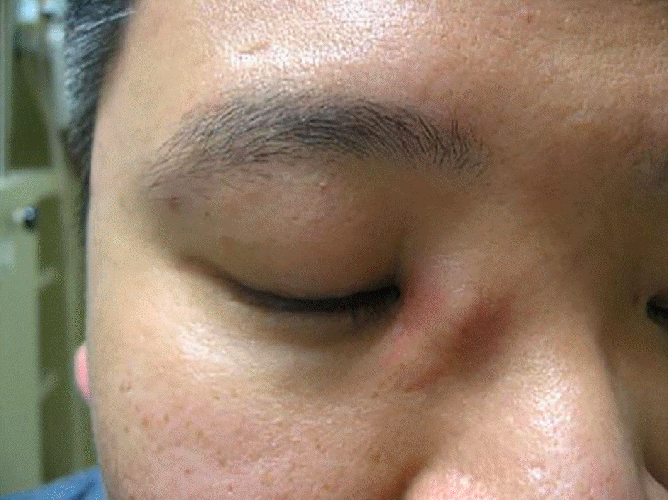
 | Figure 1.Clinical photograph. Slightly elevated cystic mass invading the medial side of the orbit. |
 | Figure 2.Radiologic images of the patient. (A) Computed tomography, coronal view image. The black arrow indicates a unilocular cystic mass with fluid-fluid level. (B) Magnetic resonance imaging, T2-enhanced image. The fluid inside the cyst had fluid-fluid level, showing high signal density on T1 and the same density with fat on T2. |


 PDF
PDF ePub
ePub Citation
 Print
Print


 XML Download