Abstract
Purpose
Hodgkin lymphoma involving the orbit and ocular adnexal tissue is very rare and usually appears in the form of a metastatic tumor at the end stage of the disease. Primary Hodgkin lymphoma in the orbit has not been previously reported, and herein we report a case of primary Hodgkin lymphoma occurring in the bilateral orbit.
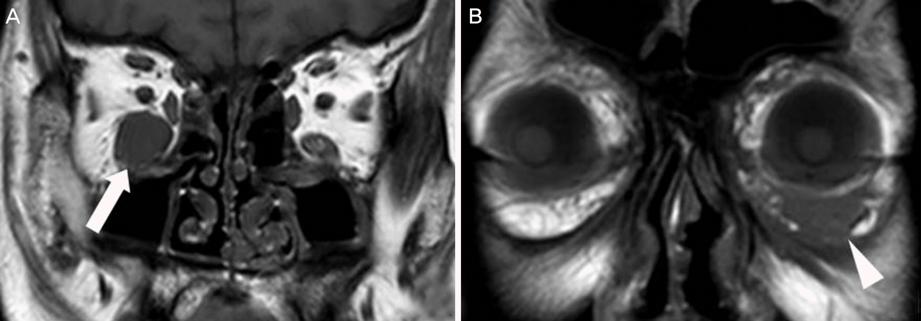
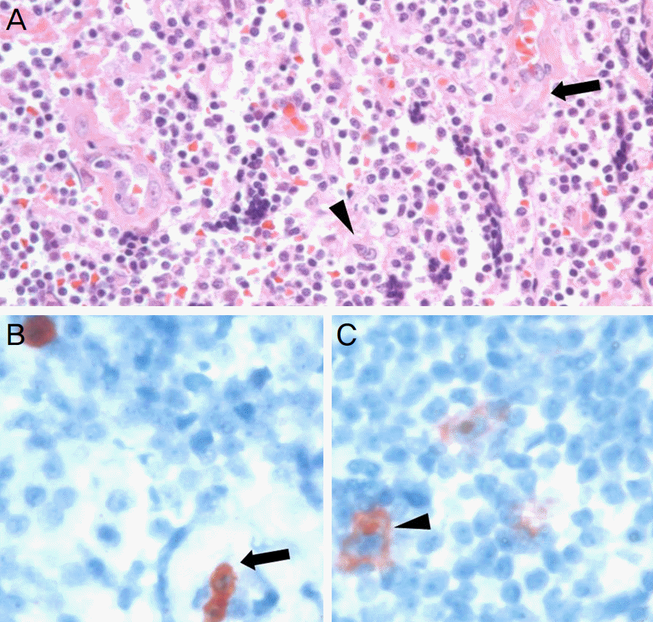
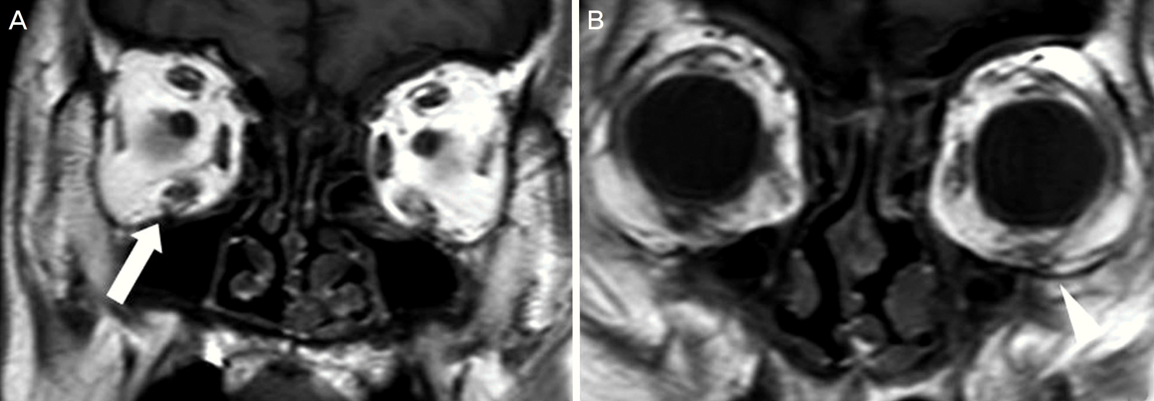
Case summary: A 64-year-old male presented with a left lower eyelid mass that increased in size over 2 years. The patient had no specific past medical history or family history except diabetes. During the physical examination, a fixed mass was gently palpated in the left lower eyelid. Mild upgaze limitation was observed during extraocular muscle movement examination in both eyes. Orbital computed tomography and magnetic resonance imaging showed soft tissue masses involving the bilateral inferior rectus muscle and left lower eyelid. The patient was diagnosed with nodular sclerosing Hodgkin lymphoma after pathological examination following incisional biopsy. The patient was transferred to the oncology department for tumor staging. Positron emission tomography showed no involvement of other organs except both orbits. After systemic chemotherapy and radiation therapy, the patient was under observation for 14 months without ophthalmic and systemic complications or recurrence.
Go to : 
References
2. Stefanovic A, Lossos IS. Extranodal marginal zone lymphoma of the ocular adnexa. Blood. 2009; 114:501–10.

3. Freeman C, Berg JW, Cutler SJ. Occurrence and prognosis of abdominal lymphomas. Cancer. 1972; 29:252–60.
5. Cho EY, Han JJ, Ree HJ, et al. Clinicopathologic analysis of ocular adnexal lymphomas: extranodal marginal zone b-cell lymphoma constitutes the vast majority of ocular lymphomas among Koreans and affects younger patients. Am J Hematol. 2003; 73:87–96.

6. Mani H, Jaffe ES. Hodgkin lymphoma: an update on its biology with new insights into classification. Clin Lymphoma Myeloma. 2009; 9:206–16.

7. Sengupta S, Pal R. Clinicopathological correlates of pediatric head and neck cancer. J Cancer Res Ther. 2009; 5:181–5.
8. Diehl V, Tesch H. Hodgkin's disease-environmental or genetic? N Engl J Med. 1995; 332:461–2.
9. Knowles DM 2nd, Jakobiec FA. Orbital lymphoid neoplasms: a clinicopathologic study of 60 patients. Cancer. 1980; 46:576–89.

11. Fratkin JD, Shammas HF, Miller SD. Disseminated Hodgkin's abdominal with bilateral orbital involvement. Arch Ophthalmol. 1978; 96:102–4.
Go to : 
 | Figure 1.A magnetic resonance imaging (MRI) of the orbit. T1-weighted orbital coronal MRI showing a diffuse mass in the right inferior rectus muscle (A, arrow) and left lower eyelid (B, arrowhead). |
 | Figure 2.Histopathological examination of nodular sclerosing Hodgkin lymphoma. (A) Dense collagen band (arrow) and the mixture of different cell types; small lymphocytes, plasma cells, eosinophils, binucleated owl-face Reed-Sternberg cell (arrowhead) (H&E stain, ×200). (B) CD30 stains Reed-Sternberg and Hodgkin cells (arrow) (CD30, ×400). (C) CD15 staining showing cell membrane positivity in the large cells (arrowhead) (CD15, ×400). |
 | Figure 3.A magnetic resonance imaging (MRI) of the orbit after chemotherapy and radiation therapy. T1-weighted orbital coronal MRI showing noticeably reduced mass in the right inferior rectus muscle (A, arrow) and normal left lower eyelid area (B, arrowhead). |
 | Figure 4.Preoperative and postoperative chemotherapy and radiation therapy photographs of the patient. In the preoperative photograph, left lower eyelid appeared protruding compared to the right lower eyelid (A), and postoperative photography showing both lower eyelid protruding lesions disappeared after chemotherapy and radiation therapy (B). |


 PDF
PDF ePub
ePub Citation
 Print
Print


 XML Download