

 PDF
PDF ePub
ePub Citation
 Print
Print


Abstract
Purpose
To evaluate long-term change in intraocular pressure (IOP) in eyes undergoing laser iridotomy (LI) and early phacoemulsification after LI in patients with acute angle-closure glaucoma (AACG).
Methods
The retrospective, comparative chart review included patients with AACG, Group A who underwent only LI and Group B who underwent early phacoemulsification within 1 month after LI. Patients were followed up on day 1; week 1; and months 1, 3, 6, and 12 after LI. IOP changes were studied.
Results
This study included a total 99 eyes from 99 patients, 37 in group A and 62 in group B. The mean IOP were not significantly different between the two groups at the initial visit or 1 month later. However, group B showed a consistently lower mean IOP that that of group A at 3, 6, and 12 months (p = 0.003, <0.001, <0.001, respectively). The prevalence of IOP increase to greater than 21 mmHg was 3 (8.11%), 5 (13.51%), and 5 patients (13.51%) in group A and 0, 2 (5.41%), and 1 patients (1.61%) in group B at 3, 6, and 12 months, respectively. Group B showed a significantly lower prevalence of IOP increase (p = 0.050, 0.038, 0.026).
Conclusions
We found that patients treated with early phacoemulsification after LI had better outcomes of well-maintained IOP compared to those undergoing LI alone. For AACG patients with coexisting cataract, early phacoemulsification after LI can be considered as a reasonable treatment to maintain IOP.
Go to : 
References
1. Kim YY, Lee JH, Ahn MD, et al. Angle closure in the Namil study in central South Korea. Arch Ophthalmol. 2012; 130:1177–83.

2. Kim CS, Seong GJ, Lee NH, et al. Prevalence of primary open-abdominal glaucoma in central South Korea the Namil study. Ophthalmology. 2011; 118:1024–30.
3. Quigley HA. Number of people with glaucoma worldwide. Br J Ophthalmol. 1996; 80:389–93.
4. Robin AL, Pollack IP. Argon laser peripheral iridotomies in the treatment of primary angleclosure glaucoma. abdominal follow-up. Arch Ophthalmol. 1982; 100:919–23.
5. Salmon JF. abdominal intraocular pressure control after Nd-YAG laser iridotomy in chronic abdominal glaucoma. J Glaucoma. 1993; 2:291–6.
6. Lam DS, Lai JS, Tham CC, et al. Argon laser peripheral iridoplasty versus conventional systemic medical therapy in treatment of acute primary abdominal glaucoma: a prospective, randomized, abdominalled trial. Ophthalmology. 2002; 109:1591–6.
7. Aung T, Ang LP, Chan SP, Chew PT. Acute primary abdominal: long-term intraocular pressure outcome in Asian eyes. Am J Ophthalmol. 2001; 131:7–12.
8. Ming Zhi Z, Lim AS, Yin Wong T. A pilot study of lens extraction in the management of acute primary abdominal glaucoma. Am J Ophthalmol. 2003; 135:534–6.
9. Tarongoy P, Ho CL, Walton DS. Angle-closure glaucoma: the role of the lens in the pathogenesis, prevention, and treatment. Surv Ophthalmol. 2009; 54:211–25.
10. Hayashi K, Hayashi H, Nakao F, Hayashi F. Effect of cataract abdominal on intraocular pressure control in glaucoma patients. J Cataract Refract Surg. 2001; 27:1779–86.
11. Jacobi PC, Dietlein TS, Lüke C, et al. Primary phacoemulsification and intraocular lens implantation for acute abdominal glaucoma. Ophthalmology. 2002; 109:1597–603.
12. Pereira FA, Cronemberger S. Ultrasound biomicroscopic study of anterior segment changes after phacoemulsification and foldable intraocular lens implantation. Ophthalmology. 2003; 110:1799–806.
13. Chen YY, Chen YY, Sheu SJ, Chou P. The biometric study in abdominal stages of primary abdominal glaucoma. Eye (Lond). 2013; 27:1070–6.
14. Shams PN, Foster PJ. Clinical outcomes after lens extraction for visually significant cataract in eyes with primary angle closure. J Glaucoma. 2012; 21:545–50.
15. Musch DC, Gillespie BW, Niziol LM, et al. Cataract extraction in the collaborative initial glaucoma treatment study: incidence, risk factors, and the effect of cataract progression and extraction on clinical and quality-of-life outcomes. Arch Ophthalmol. 2006; 124:1694–700.
16. Lowe RF. Aetiology of the anatomical basis for primary abdominal glaucoma. Biometrical comparisons between normal eyes and eyes with primary abdominal glaucoma. Br J Ophthalmol. 1970; 54:161–9.
17. Foster PJ, Buhrmann R, Quigley HA, Johnson GJ. The definition and classification of glaucoma in prevalence surveys. Br J Ophthalmol. 2002; 86:238–42.
18. Chen Y, Bao YZ, Pei XT. Morphologic changes in the anterior chamber in patients with cortical or nuclear age-related cataract. J Cataract Refract Surg. 2011; 37:77–82.
19. Erie JG, Hodge DO, Gray DT. The incidence of primary abdominal glaucoma in Olmsted County, Minnesota. Arch Ophthalmol. 1997; 115:177–81.
20. Ritch R, Lowe RF. Angle-closure glaucoma: therapeutic overview. Ritch R, Shields MB, Krupin T, editors. The Glaucomas: Glaucoma Therapy. 2nd ed.St Louis: Mosby;1996. p. 1521–31.
21. Strorey JK, Phillips CL. Ocular dimensions in angle closure glaucoma. Br J Physiol Optics. 1971; 26:228–42.
22. Nonaka A, Kondo T, Kikuchi M, et al. Cataract surgery for residual angle closure after peripheral laser iridotomy. Ophthalmology. 2005; 112:974–9.
23. Nonaka A, Kondo T, Kikuchi M, et al. Angle widening and abdominal of ciliary process configuration after cataract surgery for abdominal angle closure. Ophthalmology. 2006; 113:437–41.
24. Lam DS, Leung DY, Tham CC, et al. Randomized trial of early phacoemulsification versus peripheral iridotomy to prevent abdominal pressure rise after acute primary angle closure. Ophthalmology. 2008; 115:1134–40.
25. Dada T, Rathi A, Angmo D, et al. Clinical outcomes of clear lens extraction in eyes with primary angle closure. J Cataract Refract Surg. 2015; 41:1470–7.
26. Trikha S, Perera SA, Husain R, Aung T. The role of lens extraction in the current management of primary abdominal glaucoma. Curr Opin Ophthalmol. 2015; 26:128–34.
27. Azuara-Blanco A, Burr JM, Cochran C, et al. The effectiveness of early lens extraction with intraocular lens implantation for the treatment of primary abdominal glaucoma (EAGLE): study abdominal for a randomized controlled trial. Trials. 2011; 12:133.
28. Sihota R, Lakshmaiah NC, Walia KB, et al. The trabecular abdominal in acute and chronic angle closure glaucoma. Indian J Ophthalmol. 2001; 49:255–9.
29. Lee KM, Lee SH, Kim MS. Clinical results of phacoemulsification in eyes with acute abdominal glaucoma in the aspect of complications. J Korean Ophthalmol Soc. 2009; 50:44–50.
Go to :
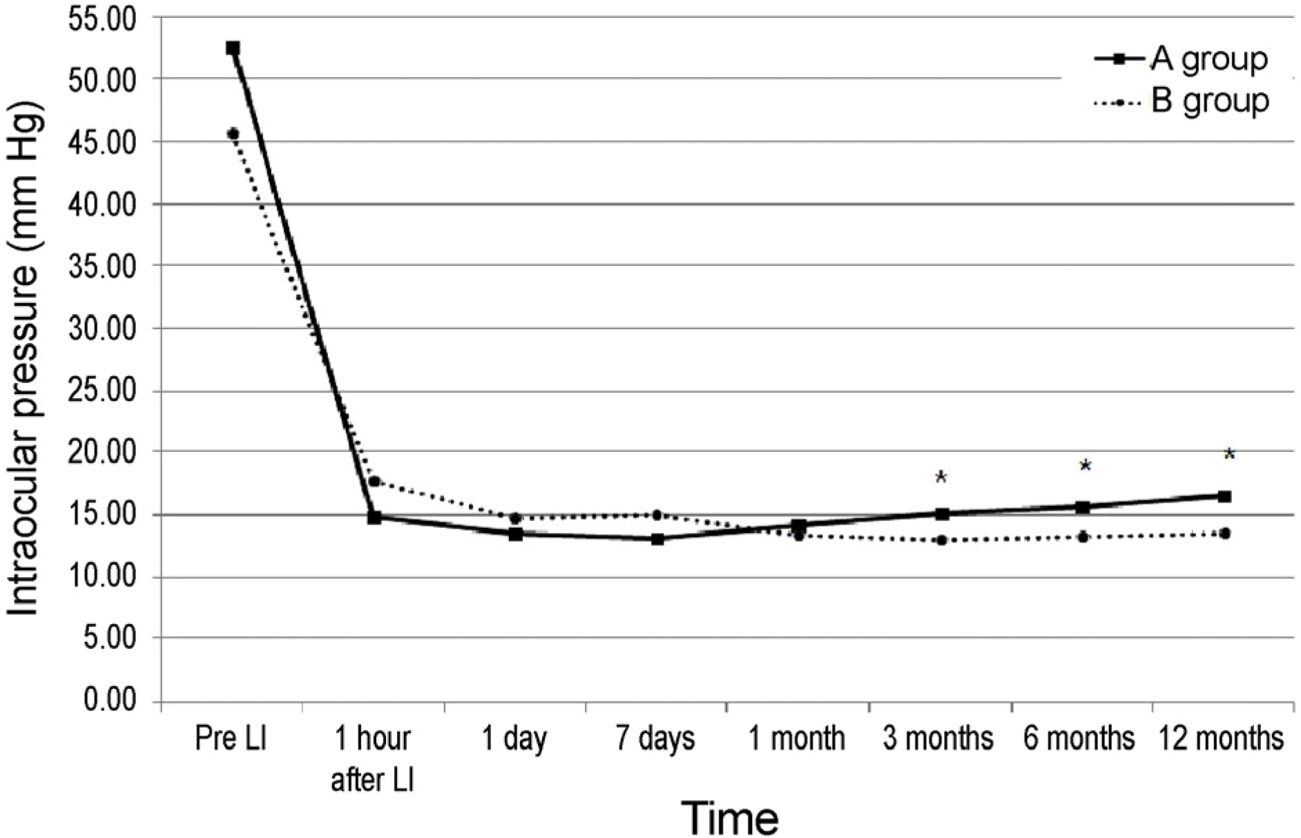 | Figure 1.Change of mean intraocular pressure for the Group A and B. Group A was 37 patients who underwent only laser iridotomy (LI) and Group B was 62 patients who underwent early phacoemulsification after LI within 1 month. The mean intraocular pressure (IOP) between two groups were not significantly different at initial visit and 1 month. But group B showed consistently lower mean IOP that that of group A for the follow-up at 3, 6, 12 months (p = 0.003, <0.001, <0.001). * p < 0.05 by independent samples t-test. |
Table 1.
Baseline demographics and presenting clinical feature of all subjects
| A group (N = 32) | B group (N = 67) | p-value | |
|---|---|---|---|
| Mean age (years) | 64.05 ± 7.40 | 69.32 ± 8.15 | 0.457* |
| Gender (male:female) | 7:30 | 11:51 | 0.883† |
| Laterality (right:left) | 24:13 | 33:29 | 0.254† |
| IOP at presentation (mm Hg) | 52.70 ± 10.13 | 45.56 ± 9.56 | 0.082* |
| IOP immediately before the intervention | 15.89 ± 9.56 | 14.84 ± 7.24 | 0.344* |
| Mean Shaffer gonoiscopy grading | 1.70 ± 0.46 | 1.80 ± 0.40 | 0.241* |
| Spherical equivalent (diopters) | 0.17 ± 1.12 | 0.88 ± 1.54 | 0.016* |
| Mean LOCS III scores | 2.08 ± 1.23 | 2.58 ± 0.80 | 0.037* |
Table 2.
Serial changes of mean intraocular pressure for the Group A and B
| A group (IOP, mm Hg) | B group (IOP, mm Hg) | p-value* | |
|---|---|---|---|
| Pre LI | 52.70 ± 10.13 | 45.56 ± 9.56 | 0.082 |
| 1 hour after LI | 14.89 ± 6.67 | 17.68 ± 9.07 | 0.108 |
| 1 day | 13.56 ± 7.49 | 14.76 ± 6.75 | 0.418 |
| 7 days | 13.11 ± 3.47 | 15.01 ± 9.51 | 0.244 |
| 1 month | 14.13 ± 3.64 | 13.35 ± 4.22 | 0.352 |
| 3 months | 15.16 ± 3.71 | 13.03 ± 3.18 | 0.003 |
| 6 months | 15.67 ± 2.75 | 13.27 ± 3.24 | <0.001 |
| 12 months | 16.54 ± 3.47 | 13.56 ± 3.02 | <0.001 |
Table 3.
Prevalence of intraocular pressure rise above 21 mm Hg for the Group A and B
| A group (N = 37) | B group (N = 62) | p-value* | |
|---|---|---|---|
| 1 month | 3 (8.11) | 5 (8.06) | 0.709 |
| 3 months | 3 (8.11) | 0 (0) | 0.050 |
| 6 months | 5 (13.51) | 2 (3.22) | 0.038 |
| 12 months | 5 (13.51) | 1 (1.61) | 0.026 |
Table 4.
Comparison of the clinical outcomes for the Group A and B
| A group (N = 32) | B group (N = 67) | p-value | |
|---|---|---|---|
| Initial BCVA (Snellen) | 0.24 ± 0.24 | 0.31 ± 0.30 | 0.260* |
| Final BCVA (Snellen) | 0.63 ± 0.26 | 0.83 ± 0.11 | <0.001* |
| PAS formation | 8 (21.62) | 0 (0) | <0.001† |
| Development of CACG | 11 (29.73) | 2 (3.22) | 0.001† |
 XML Download