

 PDF
PDF ePub
ePub Citation
 Print
Print


Abstract
Purpose
To compare neodymium-doped yttrium aluminum garnet (Nd:YAG) laser capsulotomy rates between hydrophobic and hydrophilic intraocular lenses.
Methods
The present retrospective study enrolled patients who received cataract surgery from a single surgeon between July 2006 to December 2009. Patients included in the study were implanted with SA60AT hydrophobic spherical intraocular lenses (Alcon, Fort Worth, TX, USA, 268 eyes) or I-FLEX hydrophilic spheric intraocular lenses (i-Medical®, Ophthalmic International Heidelberg GmbH, Mannheim, Germany, 331 eyes). The Nd:YAG capsulotomy rates and best-corrected visual acuity (BCVA) were compared between the two groups for 2 years after the operation.
Results
The mean follow-up period was 23.5 months and 22.6 months and the mean age was 68.6 years and 70.3 years in the SA60AT and I-FLEX groups, respectively. Follow-up periods were longer in the SA60AT group (p = 0.035), but ages were not significantly different between the two groups (p = 0.367). Nd:YAG laser capsulotomy rates were 6.3% in the SA60AT group and 11.2% in the I-FLEX group. Nd:YAG laser capsulotomy rates were significantly higher in the I-FLEX group (p = 0.020). BCVA before and after the Nd:YAG laser capsulotomy was not significantly different.
Conclusions
Nd:YAG laser capsulotomy rates were higher in the I-FLEX hydrophilic spheric intraocular lens group than in the SA60AT hydrophilic spheric intraocular lens group. Adhesion between capsular bag and intraocular lens by bioadhesive character of hydrophobic acryl intraocular lens may contribute to the prevention of lens epithelial migration and posterior capsule opacification.
Go to : 
References
1. Apple DJ, Solomon KD, Tetz MR, et al. Posterior capsule abdominal. Surv Ophthalmol. 1992; 37:73–116.
2. Schaumberg DA, Dana MR, Christen WG, Glynn RJ. A systematic overview of the incidence of posterior capsule opacification. Ophthalmology. 1998; 105:1213–21.

3. Marcantonio JM, Vrensen GF. Cell biology of posterior capsular opacification. Eye (Lond). 1999; 13(Pt 3b):484–8.
4. Meacock WR, Spalton DJ, Stanford MR. Role of cytokines in the pathogenesis of posterior capsule opacification. Br J Ophthalmol. 2000; 84:332–6.
5. Aslam TM, Devlin H, Dhillon B. Use of Nd:YAG laser abdominal. Surv Ophthalmol. 2003; 48:594–612.
6. Sterling S, Wood TO. Effect of intraocular lens convexity on abdominal capsule opacification. J Cataract Refract Surg. 1986; 12:655–7.
7. Born CP, Ryan DK. Effect of intraocular lens optic design on abdominal capsular opacification. J Cataract Refract Surg. 1990; 16:188–92.
8. Yamada K, Nagamoto T, Yozawa H, et al. Effect of intraocular lens design on posterior capsule opacification after continuous abdominal capsulorhexis. J Cataract Refract Surg. 1995; 21:697–700.
9. Kohnen T, Fabian E, Gerl R, et al. Optic edge design as long-term factor for posterior capsular opacification rates. Ophthalmology. 2008; 115:1308–14. 1314.e1–3.
10. Ursell PG, Spalton DJ, Pande MV, et al. Relationship between abdominal lens biomaterials and posterior capsule opacification. J Cataract Refract Surg. 1998; 24:352–60.
11. Hollick EJ, Spalton DJ, Ursell PG, et al. The effect of poly-methylmethacrylate, silicone, and polyacrylic intraocular lenses on posterior capsular opacification 3 years after cataract surgery. Ophthalmology. 1999; 106:49–54. discussion 54–5.
12. Hayashi K, Hayashi H, Nakao F, Hayashi F. Changes in posterior capsule opacification after poly(methyl methacrylate), silicone, and acrylic intraocular lens implantation. J Cataract Refract Surg. 2001; 27:817–24.
13. Kugelberg M, Wejde G, Jayaram H, Zetterström C. Posterior abdominal opacification after implantation of a hydrophilic or a abdominal acrylic intraocular lens: one-year follow-up. J Cataract Refract Surg. 2006; 32:1627–31.
14. Johansson B. Clinical consequences of acrylic intraocular lens abdominal and design: Nd:YAG-laser capsulotomy rates in 3 × 300 eyes 5 years after phacoemulsification. Br J Ophthalmol. 2010; 94:450–5.
15. Hollick EJ, Spalton DJ, Meacock WR. The effect of capsulorhexis size on posterior capsular opacification: one-year results of a randomized prospective trial. Am J Ophthalmol. 1999; 128:271–9.
16. Apple DJ, Peng Q, Visessook N, et al. Surgical prevention of abdominal capsule opacification. Part 1: Progress in eliminating this abdominal of cataract surgery. J Cataract Refract Surg. 2000; 26:180–7.
17. Peng Q, Apple DJ, Visessook N, et al. Surgical prevention of abdominal capsule opacification. Part 2: Enhancement of cortical cleanup by focusing on hydrodissection. J Cataract Refract Surg. 2000; 26:188–97.
18. Vasavada AR, Raj SM, Johar K, Nanavaty MA. Effect of abdominal alone and hydrodissection combined with rotation on lens epithelial cells: surgical approach for the prevention of abdominal capsule opacification. J Cataract Refract Surg. 2006; 32:145–50.
19. Werner L, Mamalis N, Pandey SK, et al. Posterior capsule abdominal in rabbit eyes implanted with hydrophilic acrylic intraocular lenses with enhanced square edge. J Cataract Refract Surg. 2004; 30:2403–9.
20. Nishi Y, Rabsilber TM, Limberger IJ, et al. Influence of 360-degree enhanced optic edge design of a hydrophilic acrylic intraocular lens on posterior capsule opacification. J Cataract Refract Surg. 2007; 33:227–31.
21. Vasavada AR, Raj SM, Shah A, et al. Comparison of posterior abdominal opacification with hydrophobic acrylic and hydrophilic abdominal intraocular lenses. J Cataract Refract Surg. 2011; 37:1050–9.
22. Bai L, Zhang J, Chen L, et al. Comparison of posterior capsule abdominal at 360-degree square edge hydrophilic and sharp edge hydrophobic acrylic intraocular lens in diabetic patients. Int J Ophthalmol. 2015; 8:725–9.
23. Kim SH, Park CY. Comparison of Nd: YAG laser capsulotomy rates between implantation of two different aspheric intraocular lenses. J Korean Ophthalmol Soc. 2015; 56:190–8.
24. Awasthi N, Guo S, Wagner BJ. Posterior capsular opacification: a problem reduced but not yet eradicated. Arch Ophthalmol. 2009; 127:555–62.
25. Buehl W, Findl O. Effect of intraocular lens design on posterior capsule opacification. J Cataract Refract Surg. 2008; 34:1976–85.
26. Nagamoto T, Fujiwara T. Inhibition of lens epithelial cell migration at the intraocular lens optic edge: role of capsule bending and abdominal pressure. J Cataract Refract Surg. 2003; 29:1605–12.
27. Nishi O, Yamamoto N, Nishi K, Nishi Y. Contact inhibition of mi-grating lens epithelial cells at the capsular bend created by a sharpedged intraocular lens after cataract surgery. J Cataract Refract Surg. 2007; 33:1065–70.
28. Nixon DR, Apple DJ. Evaluation of lens epithelial cell migration in vivo at the haptic-optic junction of a one-piece hydrophobic acrylic intraocular lens. Am J Ophthalmol. 2006; 142:557–62.
29. Katayama Y, Kobayakawa S, Yanagawa H, Tochikubo T. The relationship between the adhesion characteristics of acrylic intraocular lens materials and posterior capsule opacification. Ophthalmic Res. 2007; 39:276–81.
30. Lombardo M, Carbone G, Lombardo G, et al. Analysis of abdominal lens surface adhesiveness by atomic force microscopy. J Cataract Refract Surg. 2009; 35:1266–72.
31. Barbour W, Saika S, Miyamoto T, Ohnishi Y. Biological compati-bility of polymethyl methacrylate, hydrophilic acrylic and abdominal acrylic intraocular lenses. Ophthalmic Res. 2005; 37:255–61.
32. Leydolt C, Kriechbaum K, Schriefl S, et al. Posterior capsule abdominal and neodymium:YAG rates with 2 single-piece abdominal acrylic intraocular lenses: three-year results. J Cataract Refract Surg. 2013; 39:1886–92.
33. Chang A, Behndig A, Rønbeck M, Kugelberg M. Comparison of posterior capsule opacification and glistenings with 2 hydrophobic acrylic intraocular lenses: 5- to 7-year follow-up. J Cataract Refract Surg. 2013; 39:694–8.
34. Iwase T, Nishi Y, Oveson BC, Jo YJ. Hydrophobic versus dou-ble-square-edged hydrophilic foldable acrylic intraocular lens: abdominal on posterior capsule opacification. J Cataract Refract Surg. 2011; 37:1060–8.
35. Liu CS, Wormstone IM, Duncan G, et al. A study of human lens cell growth in vitro. A model for posterior capsule opacification. Invest Ophthalmol Vis Sci. 1996; 37:906–14.
36. Jamal SA, Solomon LD. Risk factors for posterior capsular pearling after uncomplicated extracapsular cataract extraction and pla-no-convex posterior chamber lens implantation. J Cataract Refract Surg. 1993; 19:333–8.
37. Elgohary MA, Dowler JG. Incidence and risk factors of Nd:YAG capsulotomy after phacoemulsification in nondiabetic and abdominal patients. Clin Experiment Ophthalmol. 2006; 34:526–34.
38. Kohnen S, Brauweiler P. First results of cataract surgery and abdominalation of negative power intraocular lenses in highly myopic eyes. J Cataract Refract Surg. 1996; 22:416–20.
39. Hayashi K, Yoshida M, Hayashi H. Posterior capsule opacification in myopic eyes. J Cataract Refract Surg. 2006; 32:634–8.
Go to :
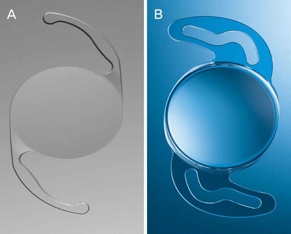 | Figure 1.The hydrophobic intraocular lens and hydrophilic intraocular lens used in this study. (A) AcrySof® SA60AT (Alcon Inc., Fort Worth, TX, USA). (B) I-FLEX (i-Medical® Ophthalmic International Heidelberg GmbH, Mannheim, Germany). |
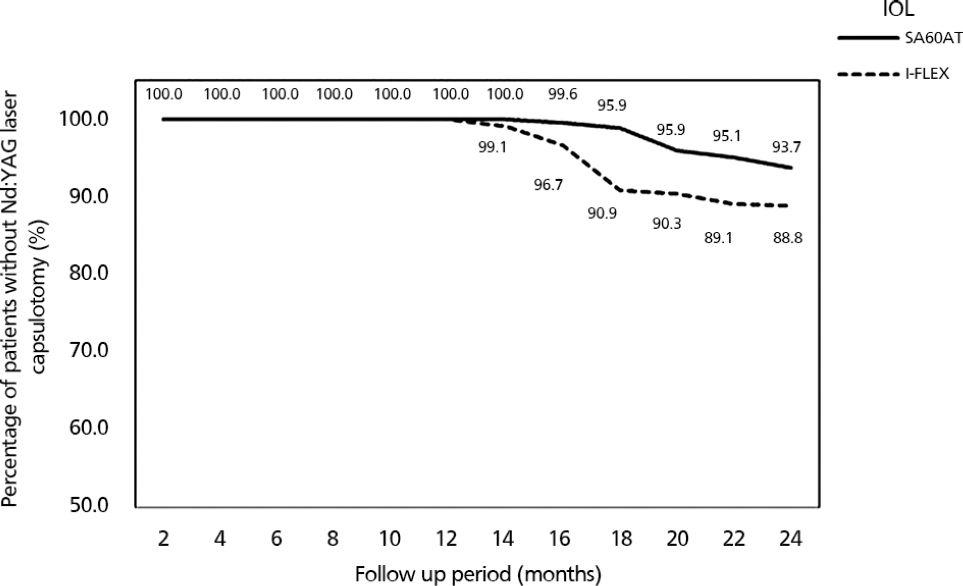 | Figure 2.Kaplan-Meier survival plots for eyes not requiring neodymium-doped yttrium aluminum garnet (Nd:YAG) laser posterior capsulotomy. Percentage of eyes without Nd:YAG laser capsulotomy after cataract surgery. Kaplan-Meier survival analysis showed that the rate of capsulotomy was statistically significantly higher in the hydrophilic group than in the hydrophobic group (p=0.02). At postoperative 24 months, Nd:YAG laser capsulotomy rates were 6.3% in eyes with SA60AT and 11.2% in eyes with I-FLEX. IOL = intraocular lens. |
Table 1.
Overview of intraocular lens characteristics
Table 2.
Patients' demographics
| SA60AT (n = 268) | I-FLEX (n = 331) | p-value | |
|---|---|---|---|
| Mean age (years, range) | 68.6 ± 12.9 (45–90) | 70.3 ± 12.2 (42–92) | 0.367* |
| Sex (male:female) | 95:173 | 116:215 | 0.525† |
| DM (n, %) | 21 (7.8) | 29 (8.8) | 0.317† |
| Right eye/Left eye (n) | 145/123 | 156/175 | 0.157† |
| Mean follow up time (months, range) | 23.5 ± 8.8 (6.3–36.2) | 22.6 ± 10.1 (6.2–34.5) | 0.035* |
Table 3.
The rate of Nd:YAG laser posterior capsulotomy and visual acuity in 2 intraocular lens groups (pre-laser & post-laser)
| SA60AT | I-FLEX | p-value | |
|---|---|---|---|
| Nd:YAG capsulotomy eye/total eye (%) | 17/268 (6.3) | 37/331 (11.2) | 0.020* |
| Mean time to Nd:YAG capsulotomy (months, range) | 20.1 ± 6.8 (15.8–30.5) | 18.3 ± 11.6 (13.5–32.0) | 0.045† |
| Pre-laser mean BCVA | 0.59 ± 0.36 | 0.61 ± 0.41 | 0.535† |
| post-laser mean BCVA | 0.10 ± 0.44 | 0.13 ± 0.46 | 0.122† |
Table 4.
Clinical characteristics of eyes with or without Nd:YAG laser capsulotomy
| Total | Non-capsulotomy | Nd:YAG capsulotomy | p-value | |
|---|---|---|---|---|
| Total No. | 599 | 545 | 54 | |
| Sex | ||||
| Male | 211 (35.2) | 191 (90.5) | 20 (9.5) | 0.165* |
| Female | 388 (64.8) | 354 (91.2) | 34 (8.8) | |
| Age (years) | 69.0 ± 12.6 (42–92) | 69.7 ± 11.9 (45–92) | 62.1 ± 16.9 (42–89) | 0.002† |
| DM | 50 (8.3) | 44 (88.0) | 6 (12.0) | 0.472* |
| Trauma history | 14 (2.3) | 10 (71.4) | 4 (28.6) | 0.312* |
| History of refractive surgery | 59 (9.8) | 45 (76.3) | 14 (23.7) | 0.121* |
| Axial length (mm) | ||||
| <22 | 9 (1.5) | 9 (100.0) | 0 (0) | |
| 22–26 | 384 (64.1) | 363 (94.5) | 21 (5.5) | <0.001* |
| >26 | 206 (34.4) | 173 (84.0) | 33 (16.0) | |
| IOL | ||||
| SA60AT | 268 (44.7) | 251 (93.7) | 17 (6.3) | |
| I-FLEX | 331 (55.3) | 294 (88.8) | 37 (11.2) | 0.020* |
 XML Download