

 PDF
PDF ePub
ePub Citation
 Print
Print


Abstract
Case summary
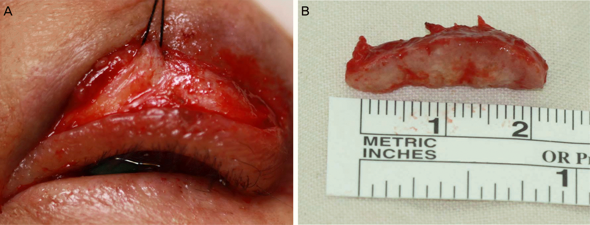
A 73-year-old male presented to our clinic with a palpable mass in his left upper eyelid that had appeared 10 days prior. The patient had a history of colon cancer that was diagnosed 2 years previously with liver and lung metastases, and he had underwent colon resection followed by chemotherapy. A 10.7 × 14.7 × 9.0 mm mass was observed on orbital computed tomography and a biopsy confirmed that the mass was a metastatic colonic adenocarcinoma. Because radical resection of the mass could not be performed, a debulking surgery was performed instead. The patient was followed up while receiving conservative treatment, but he died 3 weeks after surgery.
References
1. Kersten RC, Ewing-Chow D, Kulwin DR, Gallon M. Accuracy of clinical diagnosis of cutaneous eyelid lesions. Ophthalmology. 1997; 104:479–84.

2. Welch RB, Duke JR. Lesions of the lids; a statistical note. Am J Ophthalmol. 1958; 45:415–26.
3. Lee SB, Saw SM, Au Eong KG, et al. Incidence of eyelid cancers in Singapore from 1968 to 1995. Br J Ophthalmol. 1999; 83:595–7.
4. Lin HY, Cheng CY, Hsu WM, et al. Incidence of eyelid cancers in Taiwan: a 21-year review. Ophthalmology. 2006; 113:2101–7.
5. Aurora AL, Blodi FC. Lesions of the eyelids: a clinicopathological study. Surv Ophthalmol. 1970; 15:94–104.
6. Arnold AC, Bullock JD, Foos RY. Metastatic eyelid carcinoma. Ophthalmology. 1985; 92:114–9.
7. Wang JK, Liao SL, Jou JR, et al. Malignant eyelid tumours in Taiwan. Eye (Lond). 2003; 17:216–20.
8. Weiner JM, Henderson PN, Roche J. Metastatic eyelid carcinoma. AmJ Ophthalmol. 1986; 101:252–4.
9. Shields CL, Shields JA, Gross NE, et al. Survey of 520 eyes with uveal metastases. Ophthalmology. 1997; 104:1265–76.
10. Shields CL, Shields JA. Metastatic tumors to the orbit. Int Ophthalmol Clin. 1993; 33:189–202.
11. Shields JA, Shields CL, Brotman HK, et al. Cancer metastatic to the orbit: the 2000 Robert M. Curts lecture. Ophthal Plast Reconstr Surg. 2001; 17:346–54.
12. Kiratli H, Shields CL, Shields JA, DePotter P. Metastatic tumours to the conjunctiva: report of 10 cases. Br J Ophthalmol. 1996; 80:5–8.
13. Oltmans HJ. Carcinoma palpebrae metastaticum. Nederl T Geneesk. 1930; 74:1532.
14. Kim HJ, Kang JY. Metastatic gastric adenocarcinoma of the lower eyelid. J Korean Ophthalmol Soc. 1995; 36:1283–6.
15. Kong BD, Lee TW. Clinical analysis of metastatic intraocular malignancy. J Korean Ophthalmol Soc. 1999; 40:2928–34.
16. Park HN, Jung SK, Cho WK, et al. Clinicopathological characteristics of malignant eyelid tumor in Korea. J Korean Ophthalmol Soc. 2014; 55:348–53.
17. Ferry AP, Font RL. Carcinoma metastatic to the eye and orbit. I. A clinicopathologic study of 227 cases. Arch Ophthalmol. 1974; 92:276–86.
18. Bloch RS, Gartner S. The incidence of ocular metastatic carcinoma. Arch Ophthalmol. 1971; 85:673–5.
19. Bianciotto C, Demirci H, Shields CL, et al. Metastatic tumors to the eyelid: report of 20 cases and review of the literature. Arch Ophthalmol. 2009; 127:999–1005.
20. Mansour AM, Hidayat AA. Metastatic eyelid disease. Ophthalmology. 1987; 94:667–70.
21. Riley FC. Metastatic tumors of the eyelids. Am J Ophthalmol. 1970; 69:259–64.
22. Hogan MJ, Zimmerman LE. Ophthalmic pathology, an atlas and textbook. 2nd ed.Philadelphia: WB Saunders Company;1962. p. 413–49.
23. Albert DM, Jakobiec FA. Principles and practice of ophthalmology: basic sciences. 1st ed.Vol. 1. Philadelphia: WB Saunders Company;1994. p. 1819.
24. Lee TY, Cho JY, Kang BN. A case of metastatic conjunctival tumor. J Korean Ophthalmol Soc. 1986; 27:949–53.
25. Kim JH, Yu HG. Clinical characteristics of metastatic choroidal tumors in Korean patients. J Korean Ophthalmol Soc. 2008; 49:1785–93.
26. Ostriker PJ. Metastasis of adenocarcinoma of colon to conjunctival surface of lid. AMA Arch Ophthalmol. 1957; 57:279–81.
Figure 1.
(A) A photograph taken at initial presentation shows a diffuse erythematous and edematous lesion. The mass was palpable in the left upper eyelid. (B) A photograph taken on the day of debulking surgery, 1 month after initial presentation, shows a rapidly growing mass and near total ptosis of the left upper eyelid.
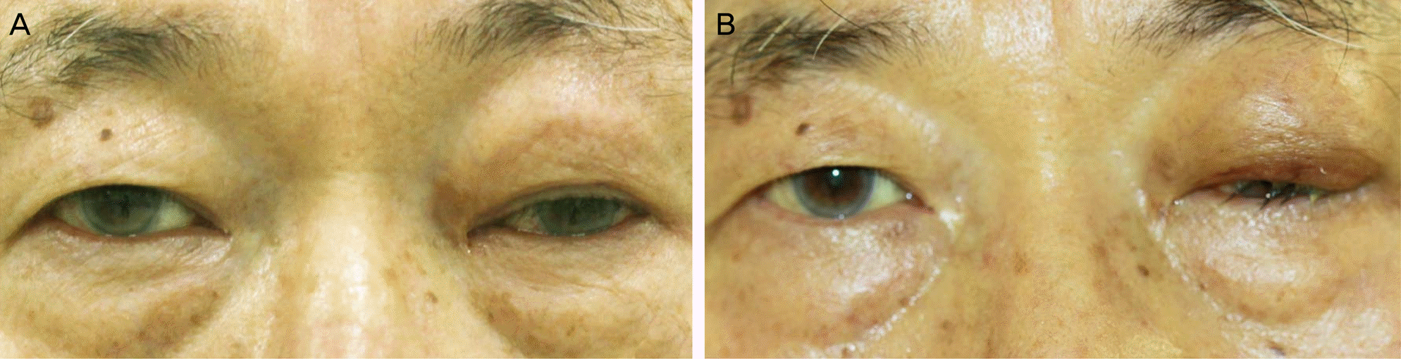
Figure 2.
(A) Coronal and (B) axial computed tomography images of the orbit showing an enhanced 10.7 × 14.7 × 9.0 mm mass in the left upper eyelid (white arrow). (C, D) One month later, orbital magnetic resonance imaging was performed; T2-weighted (C) axial and (D) coronal images show an approximately 28.0 × 11.0 × 10.0 mm well-defined enhancing mass (white arrow). The mass showed rapid growth and its size was increased compared to that observed 1 month previously.

Figure 3.
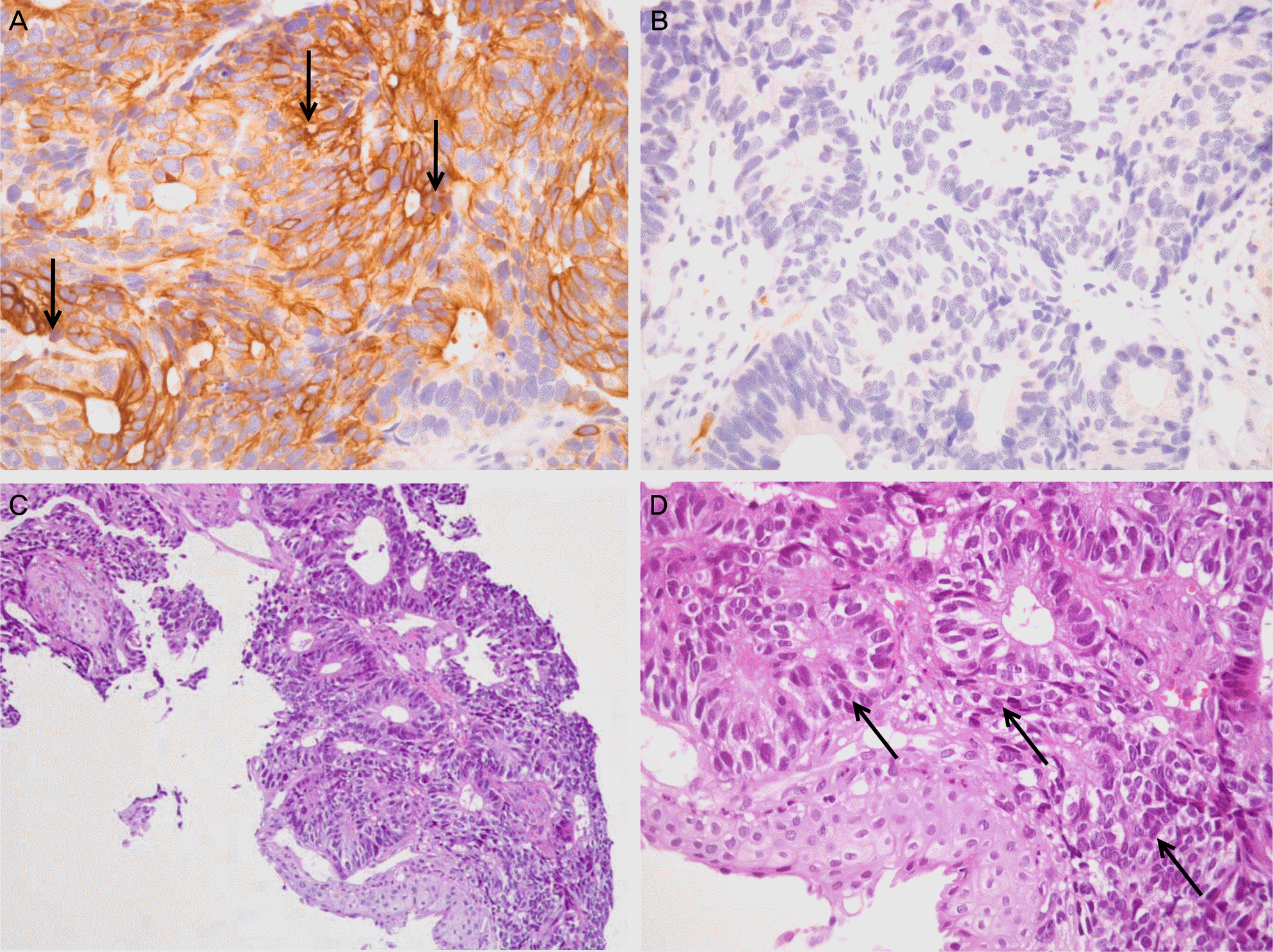
(A) Immunohistochemistry for CK20 shows brown positive staining of the tumor section (×400) (black arrows). (B) Immunohistochemistry for CK7 is negative (×400). (C, D) Moderately differentiated metastatic colonic adenocarcinoma shows an irregular, glandular structure (C, hematoxylin and eosin staining, ×200, D, hematoxylin and eosin staining, ×400) (D: black arrows).
 XML Download